IMAGE ARTICLE | VOLUME 2, ISSUE 1 | OPEN ACCESS DOI: 10.23937/2474-3682/1510021

Coiled Nasogastric Tube in Oesophagus

Sameer Sethi

Department of Anaesthesia & Intensive Care, Post Graduate Institute of Medical Education and Research, India

*Corresponding author: Sameer Sethi, Associate Professor, Department of Anaesthesia & Intensive Care, Post Graduate Institute of Medical Education and Research (PGIMER), Sector 12, Chandigarh, India, PIN: 160012, Tel: 9317851002, 7087008491, E-mail: sameersethi29@gmail.com

Published: January 06, 2016

Citation: Sethi S (2016) Coiled Nasogastric Tube in Oesophagus. Clin Med Img Lib 2:021. doi.org/10.23937/2474-3682/1510021

Copyright: © 2016 Sethi S. This is an open-access content distributed under the terms of the Creative Commons Attribution License, which permits unrestricted use, distribution, and reproduction in any medium, provided the original author and source are credited.

This is a image of an plain X-Ray Chest PA view of a 32 year old female postoperative patient of liver transplant. In this X- ray a nasogastric tube (NG) is seen coiled in the oesophagus which is a rare event. It must be noted that nasogastric tube is coiled as a single U-shaped structure. The tip of NG tube is seen in the upper oesophagus. Normally it gets coiled in the mouth while being inserted through the nose.


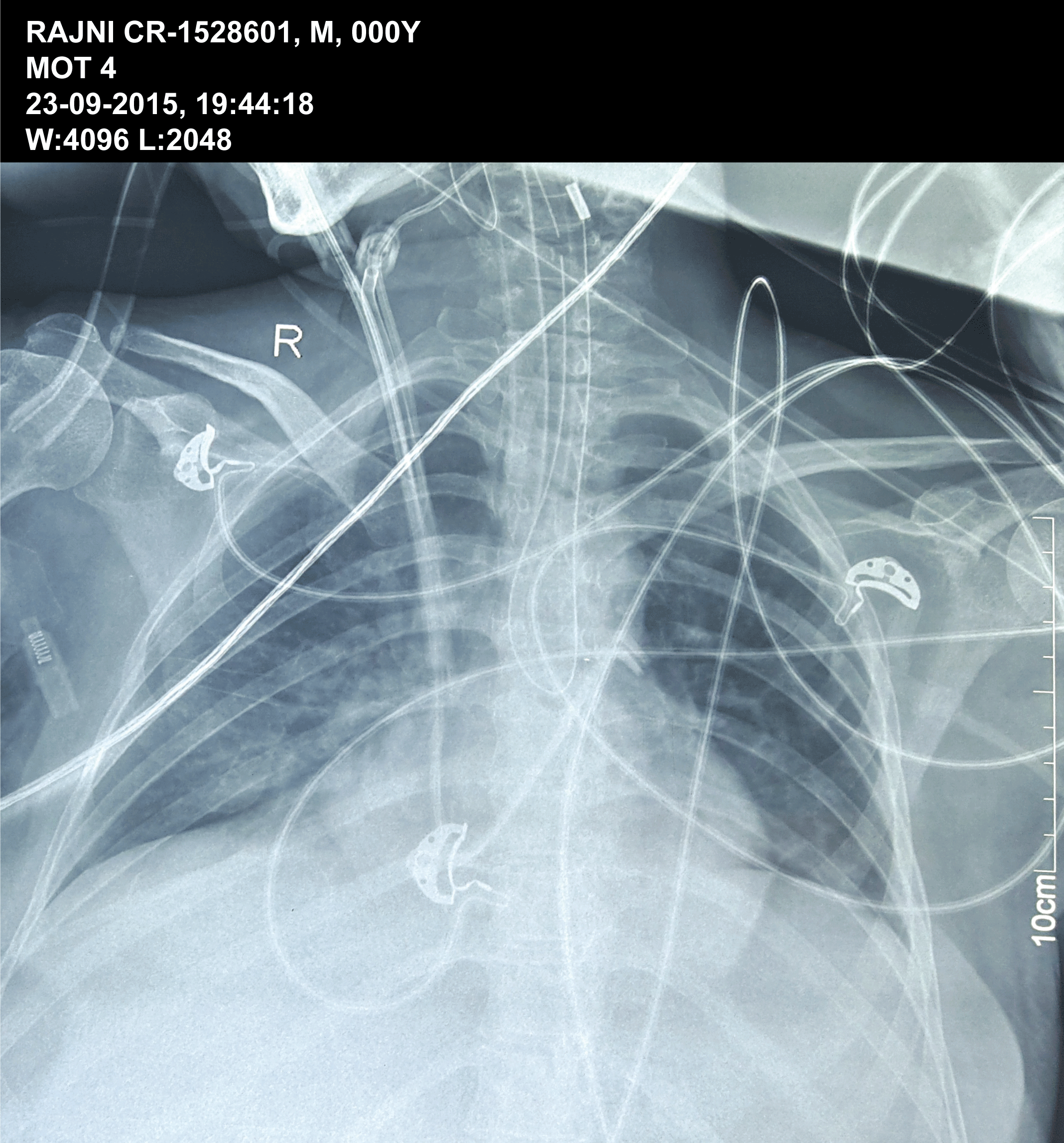
Figure 1: This is a image of an plain X-Ray Chest PA view of a 32 year old female postoperative patient of liver transplant. In this X- ray a nasogastric tube (NG) is seen coiled in the oesophagus which is a rare event. It must be noted that nasogastric tube is coiled as a single U-shaped structure. The tip of NG tube is seen in the upper oesophagus. Normally it gets coiled in the mouth while being inserted through the nose.