Condylar hyperplasia is a pathologic condition due to progressive overgrowth of one or both mandibular condyles, causing either secondary facial asymmetry or mandibular prognathism. Although it is self-limiting, the overgrowth may continue even after skeletal growth stops. The most common kind of hyperplasia is hemi-mandibular elongation. The diagnostic is based on clinical correlation of facial characteristics, intraoral occlusal characteristics and radiographic or tomographic findings. Once established the clinical diagnostic, it is necessary to check the active or passive state of overgrowth by Single Photon Emission Computerized Tomography (SPECT). If the active state of hyperplasia is confirmed, the therapeutic treatment includes high condylectomy and orthodontics to stop the advance of the disease and reduce its facial and functional sequelae (Figure 1, Figure 2, Figure 3, Figure 4 and Fgiure 5).
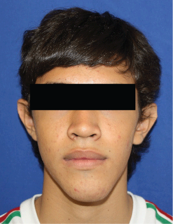
Figure 1: Facial asymmetry due to mandibular lateral deviation to the left side (Mandibular levognathism).
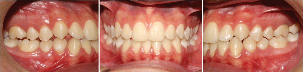
Figure 2: The occlusal characteristics include. Right side molar and canine class III dental malocclusion, mandibular midline deviation to the left and tendency to edge to edge bite or left side cross bite.areas, suggesting fat containing lesion. Doppler (b) showed no flow and elastography (c) demonstrated soft stiffness.
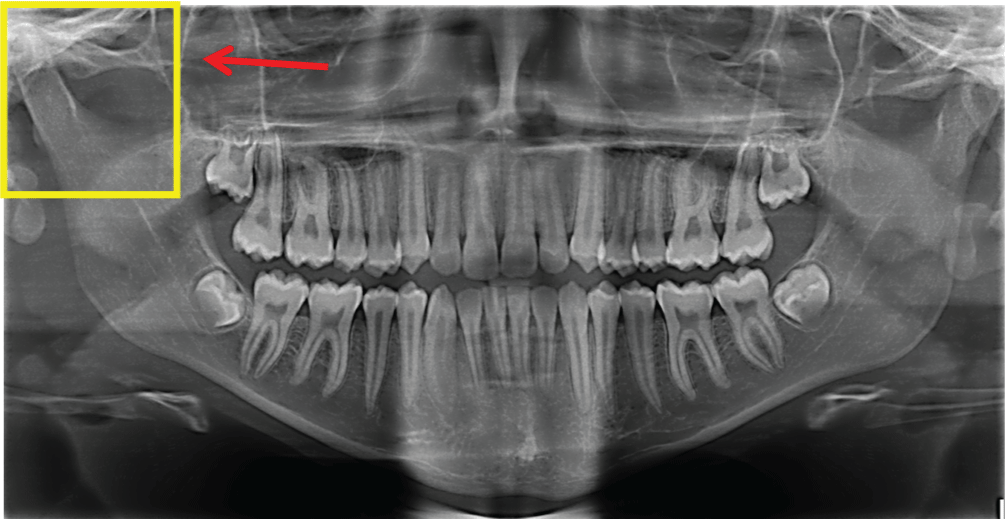
Figure 3: Panoramic X-ray film. The arrow indicates the evidence of right condyle neck longer and narrower than in the contralateral side.
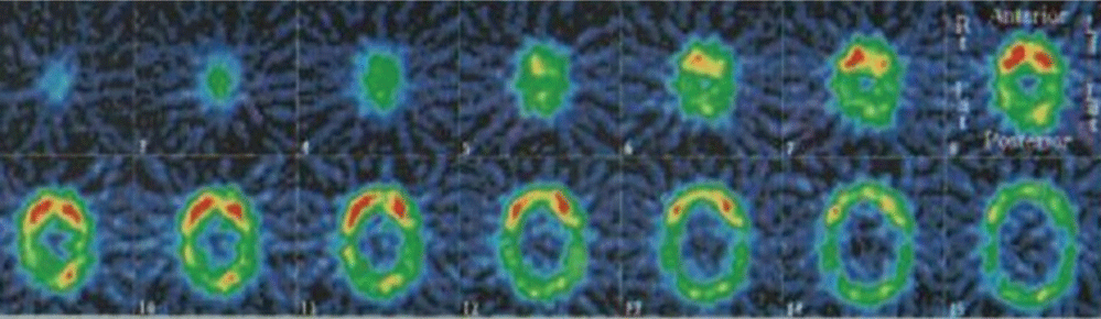
Figure 4: TMJ bone scintigraphy indicates more uptake of the radiopharmaceutical m-Technetium 99 methyl diphosphonate in the right condyle, which indicates hyperplasia active state in that side.

Figure 5: Pre-auricular access for right side high condylectomy of the hyperplasic condyle. Six mm length of condyle head removed from medial to lateral pole.