IMAGE ARTICLE | VOLUME 3, ISSUE 2 | OPEN ACCESS DOI: 10.23937/2474-3682/1510065
Dehiscent High Jugular Bulb
Hasan Erdogan , Serdar Arslan, Fatma Zeynep Arslan, Mehmet Sedat Durmaz and Arzu Cengiz
Department of Radiology, University of Health Sciences, Turkey
*Corresponding author:
Hasan Erdogan, MD, Specialist, Department of Radiology, Konya Education and Research Hospital, University of Health Sciences, 42090, Meram, Konya, Turkey, Tel: +90-506-473-0225, E-mail: dr.hasanerdogan@gmail.com
Received: April 03, 2017 | Accepted: June 12, 2017 | Published: June 14, 2017
Citation: Erdogan H, Arslan S, Arslan FZ, Durmaz MS, Cengiz A (2017) Dehiscent High Jugular Bulb. Clin Med Img Lib 3:065. doi.org/10.23937/2474-3682/1510065
Copyright: © 2017 Erdogan H, et al. This is an open-access article distributed under the terms of the Creative Commons Attribution License, which permits unrestricted use, distribution, and reproduction in any medium, provided the original author and source are credited.
Keywords
Dehiscent jugular bulb, CT, Otoscopy, Pulsatile mass
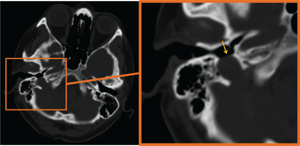
A 41-year-old female patient with 2-year history of tinnitus in right ear admitted to our clinic. The patient underwent temporal bone CT imaging. CT demonstrated right dehiscent high riding jugular bulb with absence of the right sigmoid plate (Figure 1).
A dehiscent jugular bulb is defined as a normal venous variant with superior and lateral extension of the jugular bulb into the middle-ear cavity through a dehiscent sigmoid plate [1]. Despite its rare frequency, dehiscent jugular bulb is one of the common cause of pulsatile tinnitus. In a study, high resolution CT scans of 700 temporal bones of 350 patients were retrospectively examined for the incidence of vascular variations showed that dehiscent jugular bulb was seen in 27 (3.9%) temporal bones [2]. Patients with dehiscent jugular bulb can be completely asymptomatic or may suffer from dizziness, pulsatile tinnitus and hear loss [3]. Otoscopy may show a bluish, pulsatile mass. CT has a major role in outlining the defect of sigmoid plate [4]. During the middle ear surgery jugular bulb may injure; consequently, it is important to warn surgeons about this pitfall before the operation.
Source of Support
None.
Conflict of Interest
None.
References
-
Harnsberger R, Hudgens P, Wiggins R, Davidson C (2006) Dehis-cent jugular bulb. In: Harnsberger R, Diagnostic Imaging: Head and Neck. (3rd edn), Salt Lake City, Amirsys/Elsevier Saunders, Utah, 18-22.
-
Ball M, Elloy M, Vaidhyanath R, Pau H (2010) Beware the silent presentation of a high and dehiscent jugular bulb in the external ear canal. J Laryngol Otol 124: 790-792.
-
Weissman JL, Hirsch BE (2000) Imaging of Tinnitus: A Review. Radiology 216: 342-349.
-
Atilla S, Akpek S, Uslu S, Ilgit ET, Isik S (1995) Computed tomographic evaluation of surgically significant vascular variations related with the temporal bone. Eur J Radiol 20: 52-56.