Diabetes insipidus, Polyuria, Polydipsia, Magnetic resonance imaging
An 18-year-old male patient presented to our hospital with polyuria, polydipsia and nocturia. He reports drinking 4-5 gallons of water every day due to extreme thirst. Her laboratory tests revealed serum sodium in the range of 140-145 mEq/L, serum osmolality of 295 mOsm/kg, with a concomitant urine osmolality of 279 mOsm/kg. The patient underwent pituitary Magnetic Resonance Imaging (MRI). MRI studies showed pituitary gland height is 4.5 mm and adenohypophyse signal in within normal limits. However, the hyperintense signal of the posterior pituitary lobe is absent in T1 weighted images (Figure 1).
Central Diabetes Insipidus (CDI) is a disorder of excessive renal water loss caused by a deficiency of antidiuretic hormone. CDI is characterized by polyuria, polydipsia, and the inability to concentrate urine [1]. Dysfunction of the system that maintains water homeostasis may result in life-threatening hypernatremia, seizures, dehydration, and failure to thrive [2]. CDI is the end result of a number of conditions that affect the hypothalamo hypophyseal region [1,2]. MR images allow us to clearly delineate the clinoids, the clivus, and the two components of the pituitary gland. The posterior lobe has been described as a high-intensity structure in the posteroinferior margin of the pituitary gland on T1-weighted sagittal images [3]. MRI should be performed in the diagnostic workup of central diabetes insipidus in childhood to search the hypothalamohypophyseal region abnormalities.
None.
None.
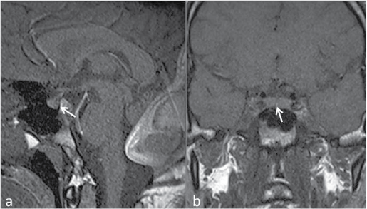
Figure 1: Sagittal T1-weighted. (a) and coronal T1-weighted; (b) MR images show the absence of normally high signal of posterior hypophysis (arrows).