Calcification or ossification of the auricular cartilage, also known as "petrified ears", was first described by Bochdalek in 1866. In our article, a seventy-two years-old male was evaluated for stiffed left ear over few years. No obvious cause was demonstrated on laboratory tests. Calcification or ossification of the left external auricular cartilage was demonstrated on non-contrast temporal bone Computed Tomography (CT) scan and therefore a rare case of idiopathic petrified left ear was diagnosed.
Calcification or ossification of the auricular cartilage, also known as "petrified ears" was first described by Bochdalek in 1866. In our article, a seventy-two years-old male was evaluated for stiffed left ear over few years. He has no history of trauma or medical disorder of persistent hypercalcemia. The left pinna was rigid and non-foldable. The right pinna was normal and foldable. Hearing was normal in both ears. Laboratory testings including complete metabolic panels, S. calcium, S. phosphorus, parathyroid hormone, uric acid, s. vitamin D and thyroid function tests were within the normal range.
Calcification or ossification of the left external auricular cartilage was demonstrated on non-contrast temporal bone Computed Tomography (CT) scan (Figure 1A-Axial, Figure 1B-Sagital, Figure 1C-Coronal image). Volume Rendered Technique (VRT) shows three dimensional images of calcified auricular cartilage (Figure 1D). Based on above clinical and radiographic findings, a rare diagnosis of idiopathic petrified left ear was made.
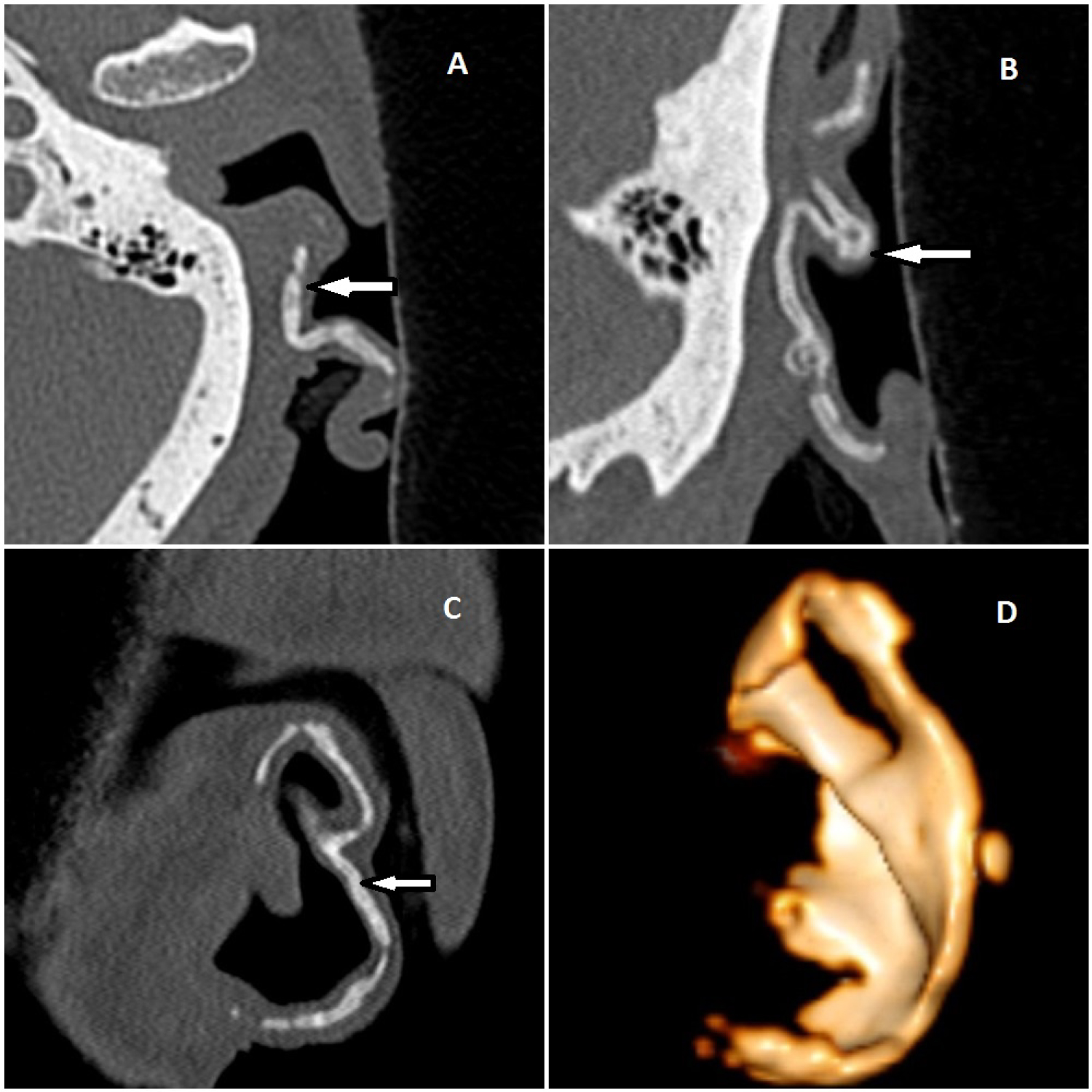
Figure 1: (A) Axial; (B) Sagital and; (C) Coronal reformated images on non-contrast temporal bone Computed Tomography (CT) scan demonstrated calcification or ossification of the left external auricular cartilage. Volume rendered technique; (D) Reveals three dimensional images of calcified auricular cartilage.