Myxoma, Tumor plop, Presyncope, Echocardiography
A 45-year-old male was referred to the out-patient clinic of Cardiology department with complaints of recurrent lightheadedness and dizziness on standing and changing postures in bed for past 3 months. He was normotensive, and non-diabetic person with no history of chest pain, dyspnea, palpitations, TIA or stroke. His ECG was unremarkable. CVS examination revealed a low pitch, early diastolic sound along with a soft systolic murmur at the apex. 2D-Echocardiographic study revealed a large, pedunculated mass in the left atrium with stalk attached to fossa ovalis region (Figure 1, Figure 3, Video 1 and Video 2). M-mode echocardiography showed a mass 'plopping' out through LA producing a characteristic multiple diastolic echoes within the mitral orifice as well as systolic echoes within the left atrium (Figure 2). Patient underwent emergency operation for resection of LA mass (Figure 4). Histopathological findings were consistent with myxoma. There was no recurrence of myxoma 6 months post-excision.
Myxomas are the most frequent benign tumors of the heart, with > 80% of myxomas localized to left atrium, with a peak incidence of cardiac myxoma at 40 to 60 years of age, with a female-to-male ratio of approximately 3:1. Patients are commonly asymptomatic and the tumor is found as an incidental finding on two-dimensional echocardiography. Positional dyspnea that is worse while lying on the left side, should alert an astute physician to the presumptive diagnosis of a myxoma. Most signs and symptoms related to myxoma can be attributed to obstruction (syncope, dyspnea, and pulmonary edema), embolism or constitutional symptoms (fatigue, cough, low-grade fever, arthralgia, myalgia, weight loss, and erythematous rash. Findings on physical examination can reveal a systolic murmur caused by damage to the valve or failure of the leaflets to coapt, or a diastolic murmur due to obstruction of the mitral valve orifice by the myxoma. Detection of "tumor plop" - a low pitched, early diastolic sound heard as the tumor prolapses into the left ventricle, helps to identify such patients and expedites them for urgent or emergent surgery.
None.
None.
We, hereby declare that all authors contributed equally to the scientific content, designing and writing of this image article.
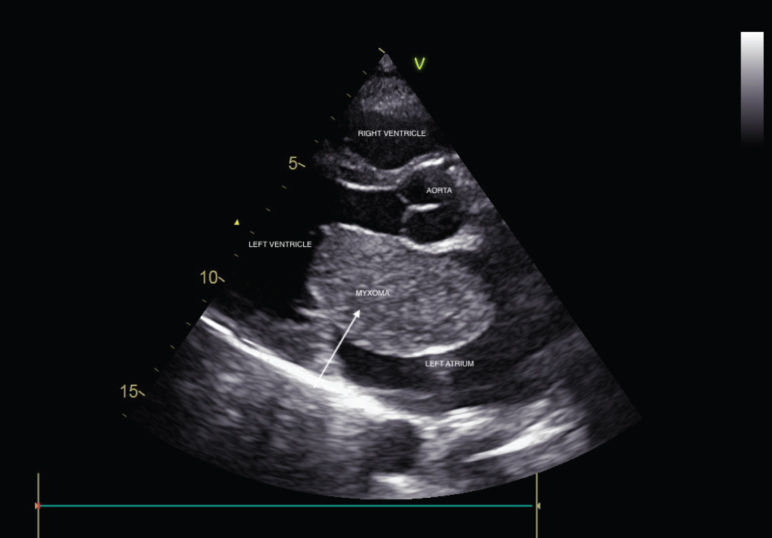
Figure 1: Parasternal long axis view on two-dimensional echocardiography showing large mass inside left atrium.
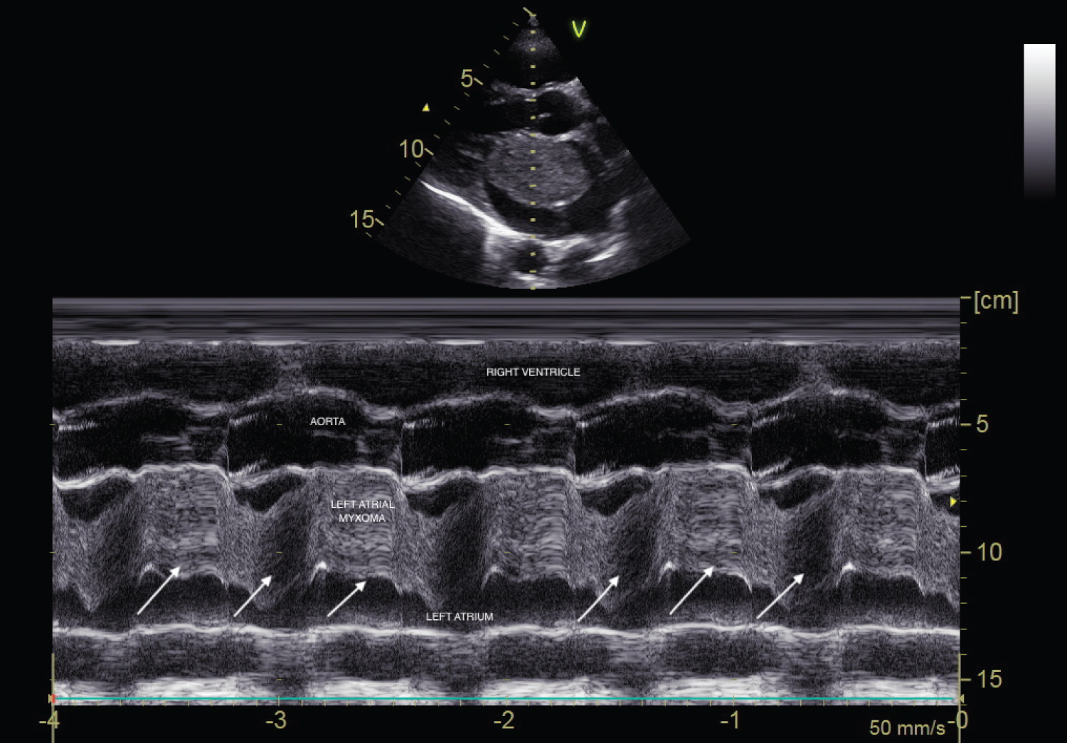
Figure 2: M-mode echocardiography in parasternal long axis view showing systolic and diastolic echoes produced in left atrium as a result of plopping of tumour across mitral orifice.
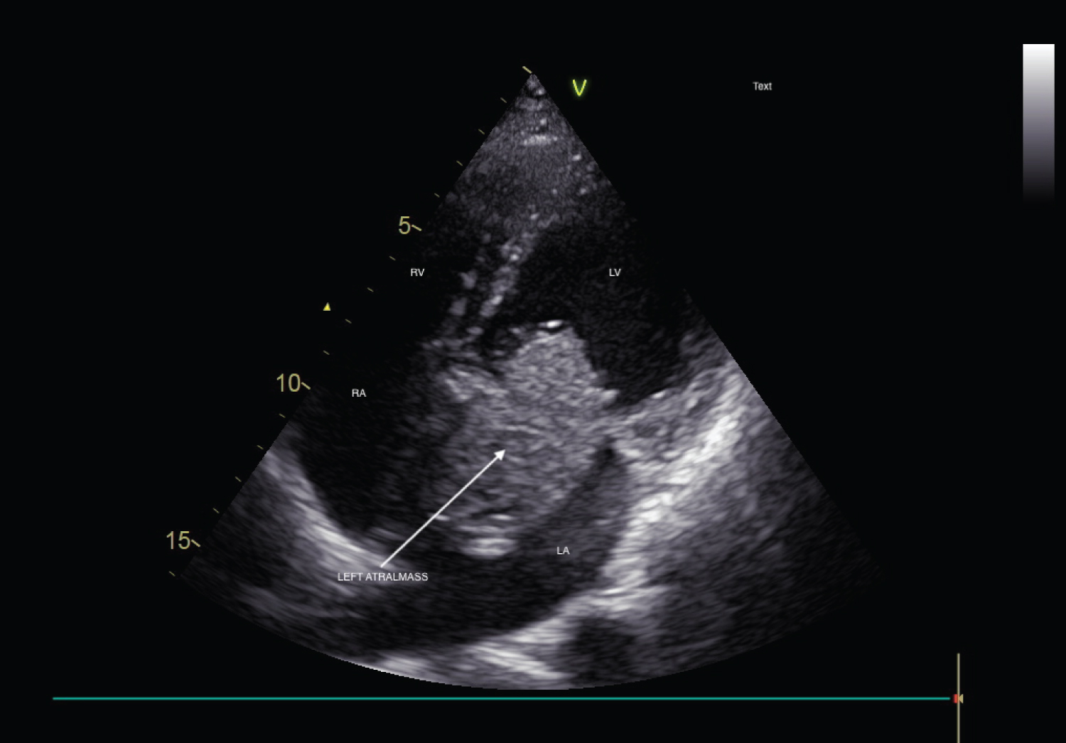
Figure 3: Apical 4 chamber view showing large left atrium mass with peduncle attached to fossa ovalis region.
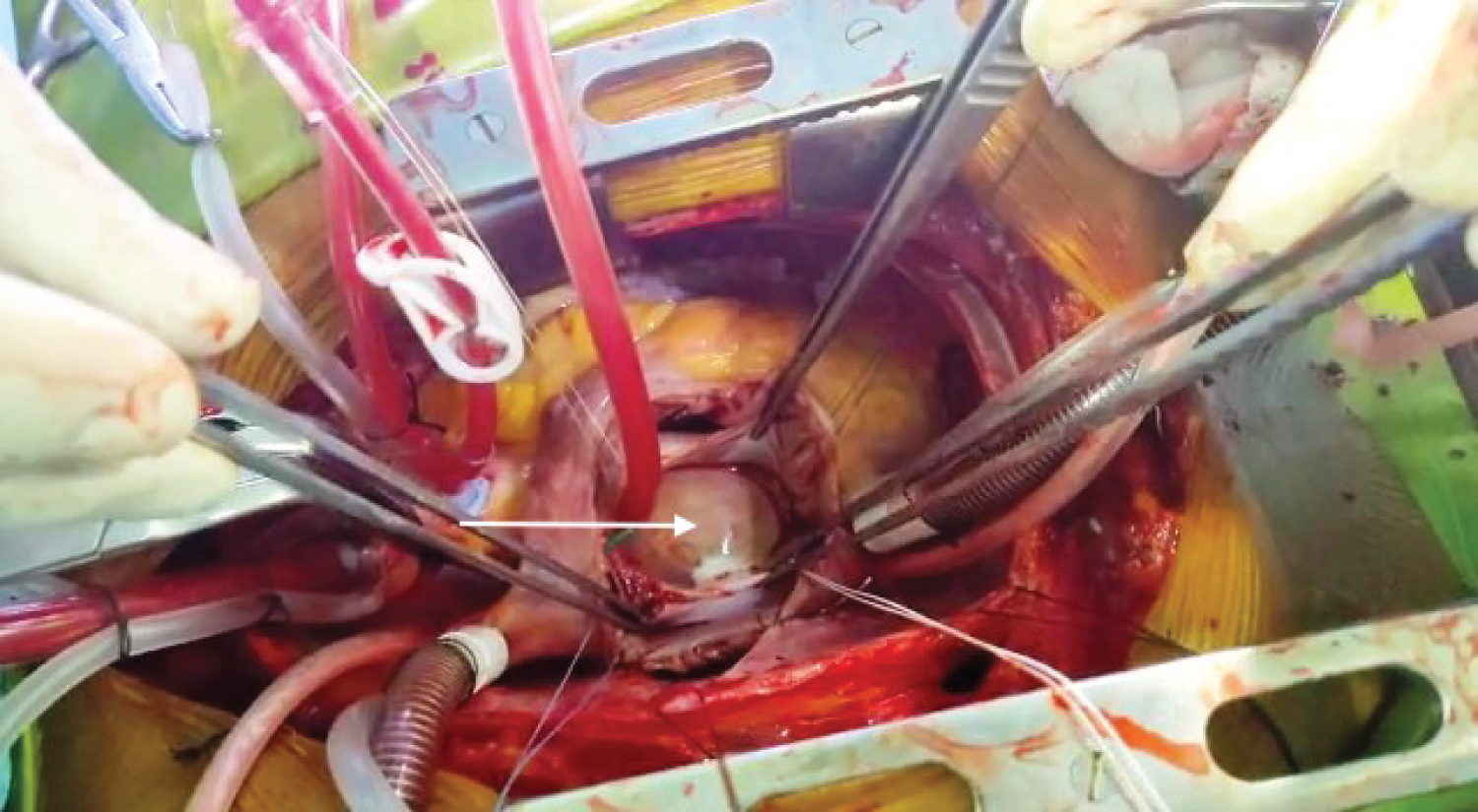
Figure 4: LA myxoma's gross morphological appearance as the mass is being excised during cardiac surgery.
Video 1: Parasternal long axis view showing LA myxoma as it protrudes through mitral orifice in to LV during diastole.
Video 2: Apical 4 chamber view showing LA myxoma moving to- and -fro inside left atrium with peduncle attached to inter-atrial septum near fossa ovalis.