Giant urinary bladder stone, Renal failure, Cystolithotomy
Bladder stones are an uncommon condition in women and according to the literature, less than 2% of all bladder stones occur in women [1,2]. This condition is often overlooked because of its rarity, hence the diagnostic and therapeutic delay which can lead to potentially serious complications. Its treatment is essentially surgical and the choice between endoscopic, laparoscopic or open surgery methods depends mainly on the size of the stones and its etiology.
In women, the occurrence of urinary bladder stones is often secondary to predisposing factor like an intra-vesical foreign body, neurogenic bladder, repeated urinary tract infections, history of surgical treatment for stress urinary incontinence.
However, bladder stones without obvious causes, called idiopathic stones, can be found. We report the case of a 33-years-old female patient with a large bladder stone that affected the upper urinary tract leading to acute renal failure.
The Kidney, Ureter, and Bladder (KUB) X-ray showed a pelvic projection opacity (Figure 1A).
A renal ultrasound was performed showing a large, bilateral ureterohydronephrosis, upstream of a voluminous calcification at the pelvic level measuring 8 cm in diameter with a posterior enhancement.
The CT scan (Figure 1B) showed a bilateral uretero-pyelocalyceal dilation upstream of a large bladder stone measuring 9 cm in diameter.
The patient initially underwent emergency drainage of the upper urinary tract by bilateral nephrostomy and then underwent an open cystolithotomy (Figure 1C).
None declared.
Obtained.
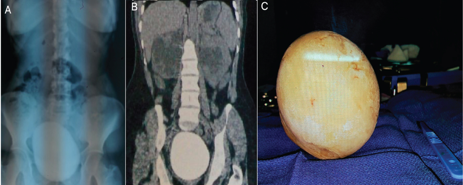
Figure 1: (A) (KUB) X-ray showing a pelvic projection calcification; (B) CT scan showing a bilateral uretero-pyelocalyceal dilatation upstream of a large bladder stone measuring 9 cm in diameter; (C) Lithiasis after extraction via open cystolithotomy.