

Clinical Medical
Reviews and Case Reports
Reactive Edema after Endoscopic Polipectomy Simulating a Polypoid Lesion: A New Pitfall in CT Colonography
Francesco Lassandro1*, Luigi Urciuoli2, Marilina D'Amora2, Tullio Valente1, Giulia Lassandro3 and Elvira Guerriero2
1Department of Radiology, Monaldi Hospital, Naples, Italy
2Second University of Naples, Naples, Italy
3University Federico II, Naples, Italy
*Corresponding author: Dr. Francesco Lassandro, MD, Department of Radiology, Monaldi Hospital, Naples, Italy, Tel: +39-335-6760716, Fax: +39-081-5786114, E-mail: f.lassandro@gmail.com
Clin Med Rev Case Rep, CMRCR-2-074, (Volume 2, Issue 12), Case Report; ISSN: 2378-3656
Received: September 15, 2015 | Accepted: December 04, 2015 | Published: December 08, 2015
Citation: Lassandro F, Urciuoli L, D'Amora M, Valente T, Lassandro G, et al. (2015) Reactive Edema after Endoscopic Polipectomy Simulating a Polypoid Lesion: A New Pitfall in CT Colonography. Clin Med Rev Case Rep 2:074. 10.23937/2378-3656/1410074
Copyright: © 2015 Lassandro F, et al. This is an open-access article distributed under the terms of the Creative Commons Attribution License, which permits unrestricted use, distribution, and reproduction in any medium, provided the original author and source are credited.
Abstract
Computed Tomography Colonography (CTC) is a feasible and minimally invasive technique widely accepted both for screening and diagnosis of colon cancer. The major limit of CTC compared to optical endoscopy resides in its minor specificity due to false positive findings. Several non-pathologic aspects mimicking true polyps are described in medical literature and are well known to radiologists. In this study we report seven pathologically documented cases of reactive edema appearing after a previous endoscopic polipectomy. This represents another possible and relatively common source of false positive examination of which radiologists should be aware when interpreting CTC after previous endoscopic removal of polyps.
Keywords
CT colonography, Reactive edema, Pitfall, Polypoid lesion, Polyp, Endoscopic polypectomy, Virtual colonoscopy
Introduction
Computed Tomography Colonography (CTC), also called virtual colonoscopy, is a feasible and minimally invasive technique for full exploration of the colon; it has greatly improved over the years and is now a widely accepted technique both for screening and diagnosis of colon cancer. CTC uses data obtained from CT imaging to generate two-dimensional (2D) and three-dimensional (3D) images of the entire colon [1].
CTC is a rapid, non invasive imaging method suitable for colon and rectum investigation [1].
The role of CTC has progressively increased, allowing detection of primary cancer or its recurrence, metachronous disease, lesions located proximal to a tight stenosis and distant metastases in one single study [2-4].
The primary target lesion in colorectal screening is the adenomatous polyp, since detection and removal of these lesions can potentially prevent approximately 95% or more of all colon cancers. However, a variety of other findings may be encountered that can mimic true polyps [5-7].
CTC is often used to complete partial endoscopic examinations due to stricture that impede a complete study of the colon [2-4]. In some cases of neoplastic strictures a polyp distal to stenosis could be found and removed endoscopically.
So far we found just one description in literature of CTC appearance of colonic mucosa at the site of previous polypectomy [8]. These authors describe a flat mucosal relevance mimicking a flat polyp.Our purpose is to investigate if any mucosal change after endoscopic removal of polyps, how it appears in CTC eventually arising a false-positive finding observable in CTC.
Methods
From 2005 to 2014 we performed 1257 CTC exams at Monaldi Hospital. We searched on our computed database for the presence of the word "polyp" in the radiologic report. Thereafter we matched our data with the Service of Digestive Endoscopy of our Hospital to find patients who had previous optical endoscopy with polyp removal.
We retrospectively analyzed the radiologic characters of the polyps described in the radiologic report and compared it with surgical and pathologic data of patients that underwent successive operation.
Bowel preparation
All patients started a low residue diet 3 days before CTC and fasted the day of examination. They received a cathartic bowel preparation, consisting of polyethylene glycol (PEG) (Selg-Esse, Promefarm Srl), 4 sachets of 70 g each of powder dissolved in 4 L of water the day before oral administration. Fecal tagging was obtained through ingestion of 10 ml of a 50% aqueous solution of an iodinate contrast media consisting of Amidotrizoato Sodium and Meglumine Amidotrizoato (Gastrografin, Bayer) at morning, afternoon and evening.
The anticholinergic drug Hyoscine-N-butylbromide 20 mg (Buscopan - Ingelheim Italia) was injected per venam just before CTC examination to improve colonic distension and patient tolerance and to reduce intestinal spasms.
Colonic distention
Before the examination, the colon was gently distended with air by means of a standard hand-held enema bulb attached to a small flexible rectal catheter positioned in the rectum, according to the patient's tolerance. In order to prevent air escaping from the anus, the enema catheter was clamped and left in situ during CT acquisition. Before each CT acquisition, adequate distension of the colon was ensured by a scout anteroposterior CT scan. Additional air was insufflated each time one or more colonic segments were not clearly identified [3].
CT Scanning protocol
CTC was performed using a 16-section multi-detector row CT scanner (Philips Brilliance 16) with the following settings: collimation 16 × 0.75 mm; thickness 2 mm; increment 1 mm; gantry rotation time 0.5 s; kVp 120; mA 70; pitch 0,938; filter smooth (A). Scanning was performed in cranio-caudal direction during a single breath-hold (scanning time 4-9 s), both in prone and supine position [3].
Interpretation of CTC images
Images were visualized on a commercially available workstation (Philips Medical Systems Nederland B.V. Extended Brilliance TM Workspace Release 2.1.1), which allows analysis of both 3D endoluminal surface-rendered images and 2D multiplanar reformations.
In all cases were reviewed primarily the 3D endoluminal CTC images followed analysis of the two-dimensional images. CT scan without intravenous contrast medium was used to evaluate the extracolonic compartment, with particular attention to abdominal lymph nodes.
Results
We identified seven patients (four females and three males; mean age 69.85 years range 56-78) who had a colonic stenosis due to adenocarcinoma that were submitted to endoscopic polipectomy, in four cases for tubular adenoma and in three cases for adenoma, located distally to the site of the stenosis and that required CTC to have a complete evaluation of the unexplored colon with optical endoscopy.
.
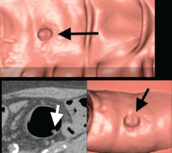
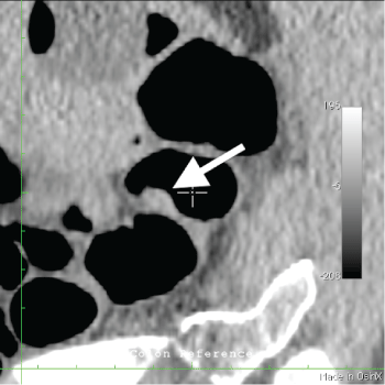
Figure 1: Polyp-like image visible on 3D and confirmed in 2D reconstruction (arrows). Tissue projecting from colonic mucosa is not distinguishable from true colonic polyps
View Figure 1
On CTC a polyp-like lesion indistinguishable from a true polyp was found in all these patients at the site of previous polyp endoscopic removal [Figure1, Figure 2a, Figure 2b, Figure 3a, Figure 3b, Figure 3c, Figure 3d and Figure 3e]. There weren't other associated colonic lesions.
.
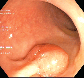
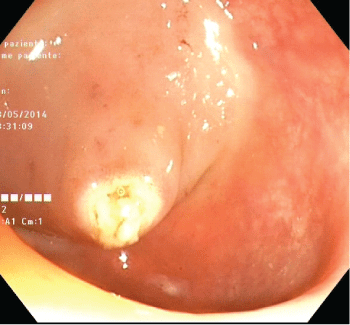
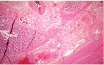
Figure 3A: Endoscopic view of a colonic polyp just before removal with diathermic loop
View Figure 3A
Time from the polyp endoscopic removal and successive CTC was 8-21 days.
.
Figure 3B: Endoscopic view of colonic mucosa after polyp removal with diathermic loop
View Figure 3B
.
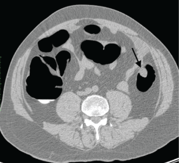
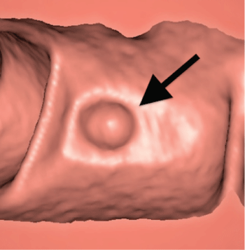
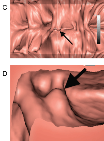
Figure 3C and 3D: CTC, 3D reconstruction: Colonic pseudopolyp 21 days after endoscopic removal of a true polyp at the same site (arrow)
View Figure 3C and 3D
All these seven patients were submitted to surgery to remove the neoplastic stenosis of the colon and had pathologic analysis of the colon encompassing the tract in which the removed polyp was located.
.
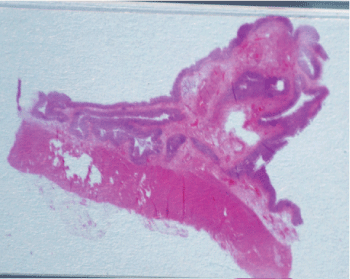
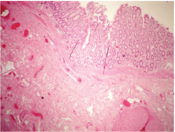
Figure 4A: Anatomic examination of the corresponding polyp-like lesion visible in CTC reconstructions in figure 1. Colonic eversion due to a fibrovascular axis with congested vesselscovered by a regular epithelial layer without adenomatous proliferation
View Figure 4A
Microscopical examination showed reactive swelling of mucosa [Figure 4a, Figure 4b and Figure 4c]. No neoplastic cell was found at the site of pseudopolyp.
.
Figure 4B: Histologic aspect of the pseudopolyp showing a normal epithelial layer
View Figure 4B
.
Figure 4C: Histologic aspect of the pseudopolyp disclosing a normal fibrovascular axis
View Figure 4C
Discussion
Computed Tomography Colonography (CTC) is widely used for screening of colorectal cancer and to study colon proximal to cancer stricture not permitting optical endoscopic evaluation [5-11].
A great variety of pitfalls can mimic true polyps at CTC, most of which should be recognized as "don't touch" lesions [9,10]. The most important of these pitfalls responsible of false-positive findings at CTC is residual stool [11]. Thus, fecal tagging is useful strategy to improve CTC specificity differentiating true soft-tissue polyps from adherent stool on 2D images processed with soft-tissue window settings [12,13].
Diverticula also may arise some concern when 3D endoluminal views are used alone for data evaluation [14]. In fact, while diverticula may simulate polyps on 3D virtual images [15], they are correctly recognized as air-filled outpouchings of the colonic wall on two-dimension CTC, whereas polyps appear as round intraluminal filling defects with soft-tissue attenuation [16].
Sometimes thickened, complex and bulbous haustral folds may mimic polyps on CTC, particularly when they are profile seen on 2D axial images [17,18]. They represent a diagnostic problem particularly in patients with diverticular disease, where muscular hypertrophy may cause distortion of the overlying folds [19].
In these cases endoluminal 3D images provide a view of the colonic mucosa very useful to avoid misinterpretation of the finding [9].
Pneumatosis cystoides intestinalis may appear as a polypoid lesion on endoscopy or 3D CTC, but on axial CT images the characteristic air content of these cysts allows a correct diagnosis [20,21].
Submucosal lipomas may appear sessile or peduncolate on 3D virtual images, but fat attenuation on 2D images usually allows the recognition of the exact nature of these lesions [12,13].
When advanced or thrombosed, internal hemorrhoids may also appear as mass-like lesions at CTC, usually recognized on the basis of morphology on 2D images [12,13].
External compression of colon by abnormal structures may mimic a polypoid structure on 3D CTC images [9]. Careful correlation with the corresponding axial 2D images may be essential to detect the correct origin of the protrusion observed on endoluminal images [22].
Endoluminal foreign bodies, such as pill fragments, capsules and undigested food may be occasionally found in the colon [22], but their mobility, internal heterogeneity and peculiar attenuation values usually furnish sufficient data to distinguish these findings from a soft-tissue polyp [12].
Based on our research in literature the CTC aspect of colonic mucosa after endoscopic removal of polyps has poorly been analyzed. Some authors suggest that after polypectomy the colonic mucosa remains edematous, simulating flat lesions on CTC [8]. They define this mucosal change as "scar after polypctomy".
We found in 7 patients a post-polypectomy reactive edema that produced a bump simulating a polyp on both 2D and 3D endoluminal views; in our cases the correct interpretation of the finding was possible only referring to the clinic history of the patient and by sharing information with the endoscopist who performed colonoscopy.
All these patients had CTC as a complementary diagnostic procedure after failure of optical endoscopy to perform a complete colon examination up to cecum, these patients subsequently underwent surgery with removal of the segment of the colon with the polyp-like lesion; histology of the specimen confirmed the inflammatory nature of the CTC finding. In fact, the diathermic loop excision procedure caused an inflammatory reaction that manifested on CTC as a protrusion, as confirmed by histology of the tissue specimen obtained after resection.
We suppose that this aspect on CTC may be more common than observed so far. Even though further analysis is needed to assess the real incidence of this radiological finding, we believe that it is important to be aware of this aspect that underlines the importance of cooperation between radiologists and endoscopists for correct patient management.
Conclusion
CTC is nowadays a widely accepted technique both for screening and diagnosis of colon cancer. Radiologists have to be aware of non-pathologic aspects that can mimic polyps to minimize false positive exams and improve accuracy of the method. We identified in the reactive edema that appears after an endoscopic polipectomy a possible pitfall that could be not unusual since not rarely CTC is used to complete colon examination in cancer patients with partial optical examination due to colonic stenosis.
References
-
Ganeshan D, Elsayes KM, Vining D (2013) Virtual colonoscopy: Utility, impact and overview. World J Radiol 5: 61-67.
-
Morrin MM, Farrell RJ, Raptopoulos V, McGee JB, Bleday R, et al. (2000) Role of virtual computed tomographic colonography in patients with colorectal cancers and obstructing colorectal lesions. Dis Colon Rectum 43: 303-311.
-
Neri E, Vagli P, Turini F, Cerri F, Faggioni L, et al. (2010) Post-surgical follow-up of colorectal cancer: role of contrast-enhanced CT colonography. Abdom Imaging 35: 669-675.
-
Ferrucci JT (2001) Colon cancer screening with virtual colonoscopy: promise, polyps, politics. AJR Am J Roentgenol 177: 975-988.
-
Aschoff AJ, Ernst AS, Brambs HJ, Juchems MS (2008) CT colonography: an update. Eur Radiol 18: 429-437.
-
Guerrisi A, Marin D, Laghi A, Di Martino M, Iafrate F, et al. (2010) Diagnostic accuracy of translucency rendering to differentiate polyps from pseudopolyps at 3D endoluminal CT colonography: a feasibility study. Radiol Med 115: 758-770.
-
Halligan S, Altman DG, Taylor SA, Mallett S, Deeks JJ, et al. (2005) CT colonography in the detection of colorectal polyps and cancer: systematic review, meta-analysis and proposed minimum data set for study level reporting. Radiology 237: 893-904.
-
Lefere P, Gryspeerdt S (2011) Virtual colonoscopy. A practical Guide 2nd Revised Edition. Springer.
-
Macari M, Megibow AJ (2001) Pitfalls of using three-dimensional CT colonography with two-dimensional imaging correlation. AJR Am J Roentgenol 176: 137-143.
-
Pickhardt PJ and Jong-Ho Richard Choi (2003) Electronic cleansing and stool tagging in CT colonography: advantages and pitfalls with primary three-dimensional evaluation. Am J Roentgenol 181: 799-805.
-
Pickhardt PJ (2003) Three-dimensional endoluminal CT colonography (virtual colonoscopy): comparison of three commercially available systems. American Journal of Roentgenology 181: 1599-1606.
-
Pickhardt PJ (2004) Differential diagnosis of polypoid lesions seen at CT colonography (virtual colonoscopy). Radiographics 24: 1535-1556.
-
Pickhardt PJ, Hassan C, Laghi A, Zullo A, Kim DH, et al. (2008) Small and diminutive polyps detected at screening CT colonography: A decision analysis for referral to colonoscopy. AJR Am J Roentgenol 190: 136-344.
-
Mang T, Maier A, Plank C, Mueller-Mang C, Herold C, et al. (2007) Pitfalls in multi-detector row CT colonography: a systematic approach. Radiographics 27: 431-454.
-
Fenlon HM, Clarke PD, Ferrucci JT (1998) Virtual colonoscopy: imaging features with colonoscopic correlation. AJR Am J Roentgenol 170: 1303-1309.
-
Hara AK, Johnson CD, Reed JE (1997) Colorectal lesions: evaluation with CT colography. Radiographics 17: 1157-1167.
-
Liedenbaum MH, de Vries AH, Halligan S, Bossuyt PM, Dachman AH, et al. (2009) CT colonography polyp matching: differences between experienced readers. Eur Radiol 19: 1723-1730.
-
Fenlon HM (2002) CT colonography: pitfalls and interpretation. Abdom Imaging 27: 284-291.
-
Lefere P, Gryspeerdt S (2011) CT colonography: avoiding traps and pitfalls. Insights Imaging 2: 57-68.
-
Pickhardt PJ, Kim DH, Taylor AJ (2008) Asymptomatic pneumatosis at CT colonography: a benign self-limited imaging finding distinct from perforation. AJR Am J Roentgenol 190: W112-117.
-
Pickhardt PJ (2004) Translucency rendering in 3D endoluminal CT colonography: a useful tool for increasing polyp specificity and decreasing interpretation time. AJR Am J Roentgenol 183: 429-436.
-
Macari M, Bini EJ, Jacobs SL, Lange N, Lui YW (2003) Filling defects at CT colonography: pseudo- and diminutive lesions (the good), polyps (the bad), flat lesions, masses, and carcinomas (the ugly). Radiographics 23: 1073-1091.
