Radicular cysts are considered rare in primary dentition. The aim of this article was to present two cases of radicular cysts associated to primary molars with different characteristics. Two children sought care at a dental clinic with complaints of a painless swelling and the absence of a premolar, respectively. The first case was a seven-year-old boy who complained of an increased volume in the region of the mandibular right primary second molar. The second case was a 12-year-old girl, who complained of the absence of the mandibular right second premolar and gingival inflammation. In both cases, surgery was performed to remove the lesion and the elements involved. Histological examinations confirmed the radicular cysts. After six months, complete regression of the lesions was reported, and the patients were referred for orthodontic treatment. Radicular cysts can cause damage to the elements involved, adjacent teeth and the occlusion of the patient.
Radicular cyst, Primary tooth, Child
Radicular cysts represent 60% of odontogenic cysts but are very rare in primary dentition [1]. According to a study by Mass, et al. in 1995, the mean age of all the cases studied was 7.7-years-old, with an almost equal distribution between genders [2]. In relation to the location in the arcades, the mandibular primary teeth are affected more frequently than the maxillary teeth [3]. In most cases they are asymptomatic lesions with slow development; however, these cysts can become large and lead to tooth mobility and displacement of adjacent teeth [4].
The etiology is related to pulp injuries or pulp necrosis caused by deep dental caries or dental trauma [2]. According to Bernardi, et al. [5], the mechanism to develop radicular cyst includes pulp necrosis, colonization and proliferation of microorganisms within the root canal system, release of bacteria toxins and inflammatory mediators into the periapical region and a combination of factors involving epithelial-stromal interaction. The periradicular inflammation leads to proliferation of epithelial cell rests [5].
A radicular cyst can be found via a routine radiography but the definite diagnosis can only be made by histopathologic examination. Radiographically, they appear as round or pear-shaped unilocular radiolucent lesions in the periapical region [6] and are bordered by a thin rim of cortical bone [7]. Radicular cysts and periapical granulomas have a similar radiographic appearance; however, radicular cysts are less common and often larger [8]. Various treatment options include root canal therapy, cyst enucleation, extraction of the affected tooth and marsupialization for the decompression of larger cysts [4,5]. This paper related two cases of children with radicular cysts associated to primary molars, with different characteristics.
A 7-year-old male patient was brought by his parents to the Pediatric Dental Clinic of the Federal University of Rio de Janeiro with the chief complaint of a painless swelling in the lower right side of his face. Anamnesis revealed no important systemic diseases. The extra-oral examination indicated a well-defined, firm swelling on the right side of the face. The intra-oral examination revealed mobility of the mandibular right primary second molar (#T) which presented a temporary restoration (Figure 1A). The patient also presented caries lesions in the primary maxillary incisors (#D, #E, #F, #G), maxillary right primary second molar (#B), mandibular right primary first molar (#S), maxillary left primary second molar (#J) and mandibular left primary second molar (#K). Cone-beam CT examination showed a rounded radiolucent unilocular lesion below the mandibular right primary second molar (#T), displacing the permanent second premolar successor (#29) to the lower mandible border (Figure 1B) and a cortical expansion (Figure 1C), suggesting a radicular cyst. Treatment plan included removal of the elements #T and #S, followed by surgical enucleation of the lesion. The affect teeth were extracted under local anesthesia. Analgesics and antibiotics were prescribed and post-operative orientations were given.
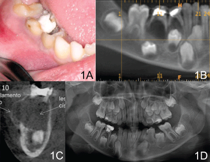 Figure 1: Clinical and radiographic exams of Case 1. (A) Initial intraoral examination with an extensive provisory restoration and edema; (B) Tomography showed a radiolucent unilocular lesion below the mandibular right primary second molar displacing the permanent second premolar successor; (C) Image showed a cortical expansion; (D) Radiographic follow-up after six months. View Figure 1
At the one-week follow up consultation, the suture was removed and there was good healing of the gingival tissue. Restorative treatments in the maxillary primary incisors and the maxillary primary second molars were then carried out. It was not possible to perform the space maintainer because the first permanent molar was not erupted. The patient is in orthodontics follow-up for space management. After six months a periodical clinical follow-up is still ongoing; however, the loss of space was noticeable. Radiographically no changes have been observed in the successor tooth or the presence of any further injuries (Figure 1D).
A 12-year-old female patient was seen at the Pediatric Dental Clinic for her first consultation with complaints of the absence of her right lower second premolar (#29) and gingival inflammation. Anamnesis revealed that the patient was having nutritional counseling since she was overweight, but no systemic diseases were reported. The intra-oral examination revealed that the patient was in her permanent dentition, but the right lower second premolar (#29) was absent and she presented gingival hyperplasia (Figure 2A). Carious lesions were observed in the following elements: left lower first permanent molar (#18) and right lower first permanent molar (#30); and generally there was poor oral hygiene. A panoramic exam suggested a radicular fragment of the right lower second primary molar (#T) with an extensive radicular radiolucent lesion and the absence of the right lower second premolar (#29) (Figure 2B). The tooth fragment and lesion were removed under local anesthesia. The wound was sutured and post-operative orientations were given. After one week, the patient returned for suture removal and cementation of a space maintainer. Finally, restorative treatment of the other tooth elements was performed. Clinical and radiographic follow-ups are ongoing for six months (Figure 2C).
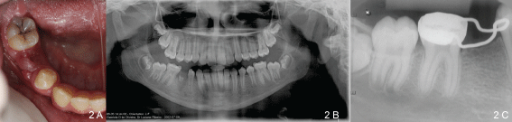 Figure 2: Clinical and radiographic exams of Case 2 (A) Initial clinical view with absence of the right lower second premolar and gingival hyperplasia; (B) Initial panoramic radiograph; (C) Radiographic follow-up after six months. View Figure 2
The curettage material was sent for histopathological analysis that showed a predominant presence of acute inflammatory cells, stratified squamous epithelium with underlying connective tissue and confirmed the diagnostic of radicular cysts (Figure 3).
 Figure 3: (A) Histopathological analysis of case 1; (B) Histopathological analysis of case 2; both cases showed a predominant presence of acute inflammatory cells. View Figure 3
Radicular cysts are considered rare in primary dentition, compared with permanent dentition [9]. This prevalence may be underestimated, as often a histopathological diagnosis is not performed when primary teeth are involved [10]. The most affected teeth in primary dentition are mandibular molars with endodontic involvement. On the other hand, in the permanent dentition, the teeth most affected are the incisors [11]. In both cases here, the dental elements involved were primary lower molars, which is in agreement with the observations in the literature.
Dental caries is the most common etiological factor of radicular cysts in primary teeth [12]. In this report, the patient in Case 1 had an extensive provisional restoration due to previous caries, an experience which may explain the appearance of the radicular cyst. In Case 2, no previous history relating to the element #T was found, and therefore no etiological factor for the development of the cyst was identified.
Usually radicular cysts are asymptomatic, have slow growth and affect a permanent dentition. A study by Mass, et al. presenting 32 children with radicular cysts, reported that all cysts were related to primary molars. The clinical feature of most of the patients was swelling and pain [5]. In this report, each case presented different clinical features and radiographic characteristics. In the first case, there was a bony swelling, tooth mobility of element #T, a radiolucent lesion around and below the affected tooth and displacement of the successor tooth #29. While in the second case, there was gingival hyperplasia, absence of any bony swelling and absence of tooth #29. However, there were no complaints of pain in either case.
The treatment of choice for both cases was to remove the affected teeth, enucleation of the lesion, space management of the involved region and protection against infection. The surgical technique used in both cases was enucleation, in which the involved teeth and lesion are removed. This technique is best suited in order to avoid the permanence of any infected tissue and to obtain samples for histopathological analyzes. On the other hand marsupialization, which is a more conservative technique, does not obtain the best material for histopathological examinations [13]. The two cases are presently in clinical and radiographic monitoring for six months. Case 1 was referred to orthodontics to recover adequate space for the successor tooth to erupt; while in Case 2, a space was preserved for a future dental implant. Both cases had a common purpose to allow the tooth to perform its physiological functions.
Routine clinical and radiographic examinations to find any asymptomatic injuries in pediatric patients are required since patients often do not have any complaints, especially since undiagnosed injuries can bring problems, as could have happened in these two cases. In the first case the germ of the permanent successor could have been affected and in the second case a loss of space would have made future treatment more complex.