Pulmonary hyalinizing granuloma (PHG) which should be considered in the differential diagnosis of pulmonary nodules of unknown origin is a distinct fibrosing lesion of lung that occurs in middle-aged patients. It has been usually reported as case reports in the literature. Patients are usually symptomatic. We present the case which recognized during operation and determined rarely.
Bronchoscopy, Pulmonary hyalinizing granuloma, Lung cancer
Pulmonary hyalinizing granuloma (PHG) which should be considered in the differential diagnosis of pulmonary nodules of unknown origin is a distinct fibrosing lesion of lung that occurs in middle-aged patients [1,2]. The radiological appearance of the lesion is that of multiple, bilateral nodules which sometimes mimic metastatic carcinoma [1-3]. Although the etiopathogenesis of PHG is unknown, it has been postulated that it represents an exaggerated immune response, possibly due to chronic granulomatous infections such as tuberculosis or histoplasmosis or antigen-antibody complexes so that this immune response may result in deposition of immunoglobulins or immune complexes in the lung [1-8]. A patient with suspected lung cancer was diagnosed pulmonary hyalinizing granuloma by transthoracic fine needle aspiration (TTFNA). We present the case which usually recognized during operation and determined rarely.
A 62-year-old female presented to the clinic with complaints of dyspnea, cough and sputum. In her history; she had cholecystectomy, hypertension and biomass exposure. Family history was unremarkable. On physical examination, blood pressure 125/75, pulse 95 and fingertip saturation was 97%. Other system examinations were normal. Laboratory examination revealed hemoglobin 11.5 g/dL, hematocrit 34.2%, WBC 7,730/mm3 and CRP of 0.48 mg/dl. Chest X ray showed homogeneous densities in the right middle zone (Figure 1). The thorax Computed Tomography (CT) showed 27 mm lesions in the right lower lobe and approximately 30 mm in diameter left hilar lesions with irregular margins were detected (Figure 2). Positron Emission Tomography (PET) CT Standardized Uptakes Values (SUV) max of the relevant lesions came in 5.2 (Figure 3).
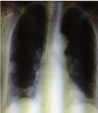 Figure 1: Image from chest X-ray.
View Figure 1
Figure 1: Image from chest X-ray.
View Figure 1
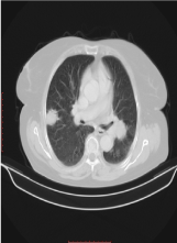 Figure 2: Image from Thorax CT.
View Figure 2
Figure 2: Image from Thorax CT.
View Figure 2
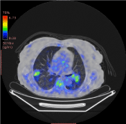 Figure 3: Image from PET CT.
View Figure 3
Figure 3: Image from PET CT.
View Figure 3
Endobronchial lesions were not observed. The left upper lobe bronchial lavage was reported as benign. TTFNA was performed to the lesion in the right lower lobe.
On microscopic examination; thick lamellar structure around small vessels, hyaline collagen fibers and a small number of kappa and lambda positive polyclonal lymphocytic inflammation was observed. Masson Trichrome, Congo Red and Crystal Violet used for differential diagnosis and hyaline connective tissue staining pattern was observed (Figure 4). The patient was diagnosed as 'Pulmonary hyalinizing granuloma'.
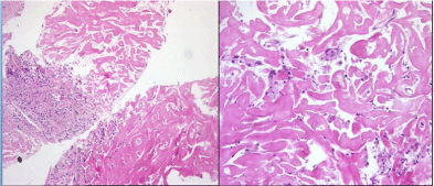 Figure 4: Microscopic view of lesion.
View Figure 4
Figure 4: Microscopic view of lesion.
View Figure 4
PHG is a rare, benign process which occurs as a result of inflammatory or post-inflammatory changes and consists of hyalinizing dense connective tissue. More than half of the patients have autoimmune phenomena or previous exposures to mycobacterial or fungal antigens [3]. It was first described in literature by Engleman, et al. in 1977. It has been usually reported as case reports in the literature. It's frequently seen in the 4th-5th decades, and the male/female ratio is approximately equal [1-4].
Patients are usually symptomatic, as in our case, although it can also be detected in asymptomatic cases. The clinical symptoms are nonspecific, including cough, fever, fatigue, dyspnea, pleuritic chest pain, sinusitis, and pharyngitis. However the most frequent symptoms are cough, shortness of breath and pain [4,5,7,9,10].
Radiography and CT findings showed randomly distributed nodules and masses with well-defined borders, some with and some with-out calcium. Radiologically single or multiple nodules between 0.2-15 cm are often, may be unilateral or bilateral and any lobe of the lung could be involved [2,4,6,11,12]. Therefore PHG should not only be considered in patients showing multiple pulmonary nodules but also in patients showing solitary nodules. In many cases, multiple lesions are present, simulating metastatic tumors. Spicules extensions, calcification, cavitation or necrosis may contain. Usually grows slowly [11].
F-18 fluordeoxyglucose positron emission tomography (FDG-PET/CT) can reveal increased metabolic activity in PHG lesions [6]. However, an accurate diagnosis of PHG can only be made with histopathological examination. Diagnosis is usually made by open lung biopsy. Bronchoscopic procedure and TTFNA are usually insufficient to diagnose, but we diagnosed our case with TTFNA [12].
The etiology of PHG is unknown. There is theoretical consideration of possible strong antigenic challenge that produce a chronic immune response, which provokes the development of PHG nodules. For pathological differential diagnosis pseudotumor, tuberculosis, sarcoidosis, Wegener's granulomatosis and silicosis should be considered. Case reports in the literature, which may be accompanied by some other entities have been reported. Among them; sclerosing mediastinitis, retroperitoneal fibrosis, rheumatoid arthritis, uveitis, ocular papillitis and neoplastic diseases (abdominal lymphoma, multiple myeloma, Paget's disease of the breast) can be observed [1,2,4,13]. It has been hypothesized that all of these conditions may essentially represent the same reactive response of an immunologic mechanism triggered by infectious agents [6]. However, in our case there was no additional pathology. As noted in the literature, in our case we suppose that an unusual immune response induced by inflammation cause the lesion. The prognosis for patients with PHG is generally excellent with no significant impact on longevity. Single lesions tend to be stable and resection is often curative.
In conclusion, Pulmonary hyalinising granuloma, a usually benign condition, should be kept in mind when encountered with patients presenting with nonspecific chest symptoms and bilateral pulmonary nodules on chest radiographs. Pulmonary hyalinizing granulomas are rare conditions which should be considered patients with pulmonary tumors.