

International Journal of Clinical Cardiology
Validation and Simplification of a Scoring Model Derived for Prediction of Poor Coronary Collateral Circulation in Acute Non-St Elevation Myocardial Infarction
Mehmet İleri1*, Koray Gürsel1, Funda Başyiğit1, Pınar Türker Bayır1, Deniz Şahin1, Ümit Güray1, Özgül Uçar1, Burak Acer2 and Yahya Büyükaşık3
1Department of Cardiology, Ankara Numune Education and Research Hospital, Ankara, Turkey
2Department of Cardiology, Türkiye Yüksek İhtisas Education and Research Hospital, Ankara, Turkey
3Department of Hematology, Faculty of Medicine, Hacettepe University, Ankara, Turkey
*Corresponding author: Mehmet İleri, Department of Cardiology, Ankara Numune Education and Research Hospital, Ankara, Turkey, Tel: +90 505 485 9803, E-mail: maraspoli2020@gmail.com
Int J Clin Cardiol, IJCC-3-087, (Volume 3, Issue 2), Research Article; ISSN: 2378-2951
Received: April 15, 2016 | Accepted: September 17, 2016 | Published: September 20, 2016
Citation: İleri M, Gürsel K, Başyiğit F, Bayır PT, Şahin D, et al. (2016) Validation and Simplification of a Scoring Model Derived for Prediction of Poor Coronary Collateral Circulation in Acute Non-St Elevation Myocardial Infarction. Int J Clin Cardiol 3:087. 10.23937/2378-2951/1410087
Copyright: © 2016 İleri M, et al. This is an open-access article distributed under the terms of the Creative Commons Attribution License, which permits unrestricted use, distribution, and reproduction in any medium, provided the original author and source are credited.
Abstract
Introduction and objective: The aim of this study was to provide validation and simplification of the scoring model derived for prediction of poor coronary collateral circulation (CCC) in acute non-ST elevation myocardial infarction (NSTEMI) in a population-based prospective cohort.
Patient and methods: The validation cohort consisted of 136 consecutive patients with NSTEMI admitted to coronary care units of three referral hospitals within 24 hours of symptom onset and scheduled to undergo coronary angiography within 48 hours of hospitalization. Coronary collateral development was graded according to the Cohen-Rentrop method. Presence of diabetes mellitus (DM), ≥ 7.85 × 10³/μL WBC and ≥ 6.25 × 10³/μL neutrophil count were assigned with 2 points; high NLR (≥ 4.5) with 1 point and older age (≥ 70 years old) with -1 point as defined in the derivation cohort. These individual points were then added together to provide a total risk score for every patient.
Results: In our validation cohort, the global score was well predictive for poor CCC (AUC [95% confidence interval]: 0.859 [0.797-0.922], p < 0.001). Eliminations of age, NLR or both did not impair predictivity of the score. The simplified score including only WBC, neutrophil and DM had 45/51 (88.2%) positive and 40/45 (88.9%) negative predictivities for total scores of ≥ 4 (2 or 3 risk factors) and 0 (no risk factor), respectively. The model was informative in 96 of 136 (70.5%) patients.
Conclusion: This study represents successful validation and simplification of a scoring model derived for prediction of PCC in patients with acute NSTEMI in a prospective cohort.
Keywords
Coronary collateral circulation, Risk scoring, Validation
Introduction
Determinants of coronary collateral circulation (CCC) in human heart are still incompletely identified. In acute non-ST elevation myocardial infarction (NSTEMI), abrupt vessel occlusion results in myocardial necrosis in the jeopardized area. Angiographic collaterals to myocardial tissue distal to an acutely occluded coronary vessel can reduce infarct size and risk for post-infarct complications as well as infarct related mortality. These prognostic implications of collateral function, makes it necessary to have a better understanding of the factors promoting collateral development in acute setting of myocardial infarction. We recently developed a risk scoring model for predicting poor collateralization in patients with NSTEMI at hospital admission [1]. The derivation cohort included 224 consecutive patients with NSTEMI admitted to coronary care unit within 24 hours of symptom onset and scheduled to undergo coronary angiography within 48 hours of hospitalization. The simplicity of the score and relatively limited number of variables included make it an attractive choice. Details on development of the model can be found in our previous article [1]. The aim of this study was to provide validation of this score in a population-based prospective cohort.
Patients and Methods
This was a prospective study conducted from 15th April 2015 to 25th September 2015 at Ankara Numune Training and Research Hospital (ANTRH), Türkiye Yüksek Ihtisas Training and Research Hospital (TYITRH) and Ankara Umut Hospital (AUH). ANTRH and AYITRH were tertiary care referral centers whereas AUH was a secondary care non-governmental hospital. The validation cohort consisted of 136 consecutive patients with NSTEMI admitted to coronary care units of these three hospitals (72 from ANTRH, 30 from AYITRH and 34 from AUH) within 24 hours of symptom onset and scheduled to undergo coronary angiography within 48 hours of hospitalization. Patients who did not have a significant stenosis (≥ 70%) in at least one of the major epicardial coronary arteries in coronary angiograms were excluded. Left main coronary artery narrowing of ≥ 50% was considered as significant. Non ST elevation myocardial infarction was diagnosed in the presence of two following criteria: (1) an accelerating pattern of or prolonged (lasting ˃ 20 minutes) angina or recurrent episodes of angina either at rest or during minimal exertion within the 48 hours; and (2) levels of cardiac biomarkers (troponin or creatine kinase MB isoenzymes) those were above the upper limit of the normal range. Patients who had overt congestive heart failure, idiopathic dilated or hypertrophic cardiomyopathy, chronic active pulmonary disease, history of renal or hepatic dysfunction, inflammatory rheumatic disease, recent infection, cancer, pregnancy, any hematological or immune system disorders and treatment with immunosuppressive medications including steroids were excluded. Those with a history of coronary artery bypass grafting or stenting within the prior 3 months were also not included. All subjects in the study group gave informed consent. The ethical committee of our hospital approved the study protocol.
Quantitative coronary angiography was performed in all patients within 48 hours after admission in multiple orthogonal projections using the Judkins technique by two experienced independent interventional cardiologists. Coronary collateral grading was carried out by two experienced cardiologists who are not informed of the clinical characteristics and biochemical results of the study patients. Collateral development was graded according to the Cohen-Rentrop method [2]: grade 0 (no filling of any collateral vessels), grade 1 (filling of side branches of the artery to be perfused by collateral vessels without visualization of the epicardial segment), grade 2 (partial filling of the epicardial segment by collateral vessels), grade 3 complete filling of the epicardial artery by collateral vessels). Patients were then divided into two groups according to their collateral grades, with the first group having poorly developed CCC (grade 0 and 1) and the second group having well-developed CCC (grade 2 and 3).
Blood samples at hospital admission were drawn in the emergency room from the antecubital vein by careful venipuncture using a 21-guage needle attached to a sterile syringe without stasis. Hematological parameters such as red blood cells, platelets, white blood cells (WBC) and their subtypes were measured in blood collected in dipotassium ethylenediaminetetraacetic acid (EDTA) containing tubes by flow cytometry in an automated blood cell counter (Sysmex, XT-2000i) immediately within 30 minutes after sampling. Neutrophil to lymphocyte ratio (NLR) was calculated as the mean value of the ratio of neutrophils to lymphocytes, both obtained from the same blood sample.
Statistical Analysis
Statistical Package for Social Sciences (SPSS) version 17.0 (SPSS Inc., Chicago, Illinois, USA) was used for all statistical calculations. A two tailed P value lower than 0.05 was considered to be statistically significant. The categorical variables were shown as numbers of cases with percentages. Continuous variables were defined as mean ± standard deviation for parametric; and median with minimum and maximum levels for nonparametric variables. Derivation of the risk score has been described in the original publication. Presence of diabetes mellitus (DM), ≥ 7.85 × 10³/μL WBC and ≥ 6.25 × 10³/μL neutrophil count were assigned with 2 points; high NLR (≥ 4.5) with 1 point and older age (≥ 70 years old) with -1 point. These individual points were then added together to provide a total risk score for every patient. Receiver operating characteristic (ROC) curve analysis was performed in order to determine success of the model. An area under the curve (AUC) value of 0.5 denotes random predictions and a value of 1 indicates perfect prediction of poor collateralization. Positive and negative predictivity were calculated using score-collateralization cross tabulations. Effects of eliminations of the parameters assigning lowest scores (i.e. age and NLR) on the model predictivity were also evaluated using ROC curve analyses.
.
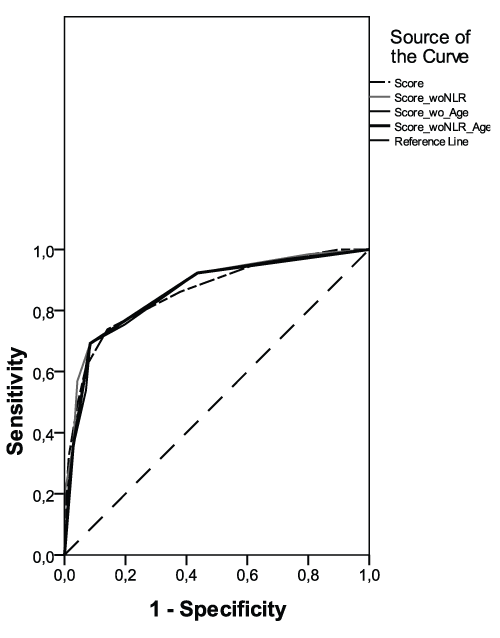
Figure 1: Receiver operating characteristic curves for the original model and various models excluding age and/or neutrophil to lymphocyte ratio (NLR).
View Figure 1
![]()
Table 1: Baseline clinical and laboratory findings in validation cohort.
View Table 1
Results
Baseline clinical characteristics and laboratory findings of patients in validation cohort at hospital admission were summarized in table 1. A total of 136 patients (81 male and 55 female, mean age 65.9 ± 9.5 years old) were enrolled in this study. Rentrop coronary grade was distributed as followed among subjects: 28 (20.6%) with grade 0, 36 (%26.5) with grade 1, 41 (30.1%) with grade 2 and 31 (47.1%) with grade 3. There were 64 (47.1%) patients in poor CCC group and 72 (52.9%) patients in good CCC group. In our validation cohort, the global score was well predictive for poor CCC (AUC [95% confidence interval]: 0.859 [0.797-0.922], p < 0.001). Eliminations of age, NLR or both did not impair predictivity of the score as shown in table 2 and figure 1. The simplified score including only WBC, neutrophil and DM had 45/51 (88.2%) positive and 40/45 (88.9%) negative predictivities for total scores of ≥ 4 (2 or 3 risk factors) and 0 (no risk factor), respectively. As all 3 variables in the model were assigned with 2 points, it is easier to consider count of the risk factors instead of scoring. The model was informative in 96 of 136 (70.5%) patients.
![]()
Table 2: Area under the curve and related statistics for the original model and various models excluding age and/or NLR.
View Table 2
Discussion
Recent trends in the incidence of acute myocardial infarction show that NSTEMI have increased by nearly 25% over the past decade while rates of ST elevation myocardial infarction have decreased [3]. While ST elevation myocardial infarction and NSTEMI have similar cardiac risk factors, their clinical presentations and epidemiologic features are distinct and warrant different management strategies. Thus, identifying clinical and angiographic factors that affect prognosis in this growing patient population is important. Collateral flow can provide an alternative blood supply to myocardium jeopardized by stenosis or occlusion of a coronary artery in NSTEMI. A well developed collateral circulation in acute setting of infarction have various protective effects including a reduction in ischemic damage and prevention of fatal arrhythmias and aneurysm formation [4]. Residual blood flow carried by collateral vessels at the time of acute occlusion is also associated with positively influenced post infarct re-modeling and preserved myocardial function [5]. Therefore, we interested in developing a model to assess the degree of collateral circulation in the setting of an acute NSTEMI. Prior to our derivation study, no scoring model for predicting poor collateralization in acute infarction had been established.
In this study, we successfully validated our model in a prospective cohort. Validation study was conducted during different time periods in 3 different cardiac centers that regularly admit high numbers of patients with acute NSTEMI. This score provided good discrimination of poor collateralization risk both in the derivation and validation sets with an excellent predictive power. Poor CCC can be easily and exactly identified from simple clinical variables on hospital admission of initial presentation of the patient in the emergency room. The risk parameters are all routinely measured and do not require expensive or complicated technology to investigate. Therefore this model is widely applicable and clinically relevant. An advantage of using routinely collected data is that with the advent of electronic patients’ records, this information can be used easily to flag patients at high risk to individual clinicians. Determination of NSTEMI patients with poor collateralization using this score may be useful for identifying high risk patients who require aggressive medical and invasive treatment. The original model was based on five variables which were significant at the P ˂ 0.05 level following multivariable logistic regression analysis. Presence of diabetes mellitus (DM), ≥ 7.85 × 10³/μL WBC and ≥ 6.25 × 10³/μL neutrophil count were assigned with 2 points; high NLR (≥ 4.5) with 1 point and older age (≥ 70 years old) with -1 point as defined in the derivation cohort. These individual points were then added together to provide a total risk score for every patient. The global risk score was well predictive for poor CCC (AUC [95% confidence interval]: 0.859 [0.797 - 0.922], p < 0.001) in our validation group. Interestingly, we observed that the model can be simplified by excluding the 2 variables which were assigned with the lowest scores (i.e. age and NLR). Eliminations of age, NLR or both did not impair predictivity of the score as shown in table 2 and figure 1. The simplified score including only WBC, neutrophil and DM had 88.2% (45/51) positive and 88.9% (40/45) negative predictivities for total scores of ≥ 4 (2 or 3 risk factors) and 0 (no risk factor), respectively. Adequacy of statistical predictivity values depends on a specific clinical scenario. We think that 88% approx. positive and negative predictivities are quite good values for prediction of poor CCC in acute NSTEMI.
In the original risk model, presence of diabetes mellitus, elevated WBC and neutrophil counts and NLR on admission were independent predictors of poor CCC in patients with acute NSTEMI, whereas older age emerged as a negative indicator. Previous data have clearly shown that poor collateralization in coronary artery disease was related to a low grade inflammation [6-9]. White blood cells and its subtypes play a major role in modulating this inflammatory response. In the case of acute infarction, leukocytosis is a common finding that reflects the infiltration of WBCs into damaged tissue in response to ischemia and reperfusion. In particular, neutrophils are the first leukocytes to be found in jeopardized myocardium. Then, monocytes migrate from capillaries to the extravascular space, transformed into macrophages and outnumber neutrophils 2-3 days after the acute event. Absolute and relative lymphocyte levels are considerably lower in acute cardiac events due to the physiological stress of such conditions. In the derivation cohort, patients with poor CCC had significantly higher WBC and neutrophil counts compared to those with good CCC. Similarly, the association between NLR and poor collateralization may also be explained by increased inflammatory activity and endothelial dysfunction.
The risk model was consistent with previous data indicating that collateral growth is impaired in type-2 diabetes mellitus [10,11]. One of the major features of diabetes is the persistent baseline inflammatory state characterized by elevated oxidative stress and endothelial dysfunction. In diabetic patients, synthesis of vasodilators including nitric oxide, prostacyclin and endothelial hyperpolarizing factors is decreased and synthesis of endothelial derived vasoconstrictors is increased. Since endothelial cell function is essential for good collateralization, diabetes severely impairs coronary collateral growth. The original model confirmed a negative association between older age and poor collateralization in contrast to some previous reports. Evidence from experimental studies suggests that aging negatively affects collateral growth via impaired endothelial nitric oxide synthase pathways and by increased oxidative stress [12]. In the setting of an acute infarction, the human collateral function can be a direct indicator of coronary artery disease severity. Elderly patients probably have more severe disease which itself positively effects collateralization. Interestingly, elimination of age in the original model did not itself impair predictivity of the score.
Several issues about this validation and the risk scoring model should be highlighted. First in derivation and validation cohorts, since collateral flow index was not used, collateral function in NSTEMI is likely to be underestimated by only angiographic visualization alone. Angiography demonstrates only vessels of which the luminal diameter is 100 μm or larger. In addition angiography shows epicardial collateral vessels, whereas the majority of human collaterals are subendocardial. Angiographic visualization of collateral circulation may be too insensitive to exclude their presence or potential improvement over time. Pre-existing high grade stenosis and the duration of acute ischemia influence the development of visible collaterals. Thus in the acute infarction, the timing of angiographic evaluation may be particularly important because collaterals may rapidly develop during ongoing ischemia. Second, we only performed laboratory tests at one point in time. A single measurement of blood cells may not reflect lifetime status. However, since we try to explore the predictive value of them measured at one point in time to detect the relation with the presence of CCC, our study design reflects routine daily practice in the majority of cardiology clinics. Third, further testing of the risk model is required to prove its accuracy in other, more geographically and temporally distinct populations.
Conclusion
This study represents successful validation and simplification of a scoring model derived for prediction of poor CCC in patients with acute NSTEMI in a prospective cohort. The robust performance of the score in both derivation and validation cohorts provides assurance of its utility in both clinical settings and research investigations in the future.
Conflict of Interest
The authors declare no conflict of interest.
References
-
Ileri M, Güray Ü, Yetkin E, Gürsoy HT, Bayır PT, et al. (2016) A new risk scoring model for prediction of poor coronary collateral circulation in acute non-ST elevation myocardial infarction. Cardiol J 23: 107-113.
-
Rentrop KP, Cohen M, Blanke H, Phillips RA (1985) Changes in collateral channel filling immediately after controlled coronary artery occlusion by an angioplasty balloon in human subjects. J Am Coll Cardiol 5: 587-592.
-
Giugliano RP, Braunwald E (2011) The year in non-ST-segment elevation acute coronary syndrome. J Am Coll Cardiol 58: 2342-2354.
-
Seiler C, Stoller M, Pitt B, Meier P (2013) The human coronary collateral circulation: development and clinical importance. Eur Heart J 34: 2674-2682.
-
Seiler C (2010) The human coronary collateral circulation. Eur J Clin Invest 40: 465-476.
-
Kerner A, Gruberg L, Goldberg A, Roguin A, Lavie P, et al. (2007) Relation of C-reactive protein to coronary collaterals in patients with stable angina pectoris and coronary artery disease. Am J Cardiol 99: 509-512.
-
Kadı H, Ceyhan K, Karayakalı M, Koç F, Celik A, et al. (2011) The relationship between coronary collateral circulation and blood high-sensitivity C-reactive protein levels. Turk Kardiyol Dern Ars 39: 23-28.
-
Seiler C, Pohl T, Billinger M, Meier B (2003) Tumor necrosis factor alpha concentration and collateral flow in patients with coronary artery disease and normal systolic left ventricular function. Heart 89: 96-97.
-
Guray U, Erbay AR, Guray Y, Yilmaz MB, Boyaci AA, et al. (2004) Pooor coronary collateral circulation is associated with higher concentrations of soluble adhesion molecules in patients with single-vessel disease. Coron Artery Dis 15: 413-417.
-
Sun Z, Shen Y, Lu L, Zhang RY, Pu LJ, et al. (2013) Clinical and angiographic features associated with coronary collateralization in stable angina patients with chronic total occlusion. J Zhejiang Univ Sci B 14: 705-712.
-
Rocic P (2012) Why is coronary collateral growth impaired in type II diabetes and the metabolic syndrome? Vascul Pharmacol 57: 179-186.
-
Epstein SE, Lassance-Soares RM, Faber JE, Burnett MS (2012) Effects of aging on the collateral circulation, and therapeutic implications. Circulation 125: 3211-3219.
