The standard first-line therapy for severe asthma exacerbation is bronchodilators and corticosteroids. Any patients not responding to this standard therapy often needs invasive ventilation, which can promote numerous related complications and mortality. Recent studies conclude that ECMO can be adopted as an alternative approach as it significantly enhances the survival rate in severe respiratory failure compared to standard invasive ventilation. In contrast, ECMO effectiveness in near-fatal asthma is not well studied, possibly due to unpredictable asthma death before reaching the hospital and lack of current evidence-based studies that support the usage and benefits of ECMO. Herein, we present 39-year-old male as a case of near-fatal asthma with severe persistent hypercapnic respiratory failure refractory to conventional ventilation. Our early application of VV-ECMO for continuous 58 hours improved the patient hemodynamics and respiratory mechanics along with a rapid reduction of carbon dioxide tension. Thus it reduces the further complications and the length of the intensive care unit. Our case demonstrates the significance of the early hours of ECMO as promising surrogate therapy when conventional treatment fails. Long-term research urged to validate VV ECMO effectiveness in this group of patients.
ECMO: Extracorporeal membrane oxygenation, NFA: Near fatal asthma, ABG: Arterial blood gas
Asthma is a chronic inflammatory disorder of the airways associated with variable expiratory flow, airway wall thickening, respiratory symptoms, and exacerbations (flare-ups), possibly require hospitalizations and may be fatal [1]. It is the most prevalent chronic global disorder with an incidence of 235 million people in the world community [2]. Despite the succession of modern pharmacological therapy in asthma, hospitalization for asthma exacerbation is significant until the present, and mortality after admission to critical care is increasing [3,4]. The near fatal asthma (NFA) is a life-threatening condition of asthma associated with characteristics of hypoxemia, hypercapnia, altered mental status, not responding to conventional therapy, and often requiring mechanical ventilation. Mechanical ventilation can generate harmful outcomes in NFA due to the presence of worsening dynamic hyperinflation and increase intrathoracic pressure, which may result in hemodynamic instability and sudden cardiovascular arrest. The reported mortality rate in patients who are in ventilator assistance is 30%. Also, Mechanical ventilation might lead to several complications attributed to the related mortality of 7-8% [5]. The ECMO is an alternative approach of cardio-pulmonary support in which oxygen supplemented and carbon dioxide (CO2) is expelled through an extracorporeal membrane using arteriovenous or venovenous cannulation. The goal of this therapy is to minimize ventilator-induced lung injury and to provide adequate time for the lung to heal from the inflammatory process. Several present studies conclude that ECMO enhances survival in adults with reversible respiratory failure compared to mechanical ventilation. Although NFA usually occurs in healthy individuals, and it is potentially reversible, its benefits from ECMO is limited and confined to a few case reports [6,7]. We present a case of successful early use of ECMO for 58 hours to correct the persistent severe hypercapnic reversible respiratory failure that was refractory to conventional ventilation.
A 39-year-old male known smoker history of childhood bronchial asthma on irregular asthmatic medications was reported to the emergency room (ER) with acute respiratory distress and shortness of breath refractory to inhaled SABA and productive cough for one day. The past clinical history summarized one episode of emergency admission to the hospital before 14 months with similar clinical symptoms and responded well to standard first-line therapy without the need for noninvasive or invasive ventilation. Presently, on examination, the patient is in severe respiratory exhaustion using accessory muscles, his vitals are blood pressure 145/84 mmHg, regular heart rate 125/min, respiratory rate 43/min, temperature 37 °C, oxygen saturation was 84% on room air. His Physical examination: Weight of 82 kg, height of 174 cm, BMI 28 kg/m2, mild jugular distension, Glasgow Coma Scale (GCS) score was 15/15. On auscultation, diffuse wheezing with insignificant bilateral breath sounds. Anteroposterior chest X-ray revealed bilateral increased Bronchovascular markings, and hyperinflated lungs (Figure 1) and electrocardiogram showed sinus tachycardia. In an emergency room, the patient treated with O2 15 L/min via a non-breathing mask, and the oxygen saturation rose to 90%-91%. His initial blood biochemistry was within normal limits. His treatment in the emergency room included intravenous fluids of normal saline 500 ml, hydrocortisone 100 mg TID, methylprednisolone 125 mg intravenous stat, 2 gram of magnesium sulphate, continuous inhaled ipratropium bromide, inhaled salbutamol. Arterial blood gas analysis (ABG) obtained, nasal swab for merscov and sputum for cultures collected. His initial ABG showed respiratory acidosis Ph 7.23 pCO2 75.3 pO2 72.9 HCO3 24.6 mmol/L. The diagnosis at admission was severe acute asthma exacerbation. His condition deteriorated subsequently with desaturation to 79% with 15 L/min of oxygen. A trial of noninvasive ventilation (NIV) via BIPAP was initiated to reduce the work of breathing to promote oxygenation and ventilation. Nevertheless, he cannot endure the inspiratory pressure of 10 cmH2O and expiratory pressure of 4 cmH2O. Repeated ABG revealed increasing respiratory acidosis: pH 7.14, pCO2 96.7 mmHg, pO2 63 mmHg, HCO3 23.4 mmol/, and low GCS (11/15). Therefore, he transferred to the intensive care unit (ICU) for close observation and he continued to deteriorate over the next 60 minutes from admission with an altered mental status (GCS 9/15) and hypopnea with worsening respiratory acidosis: pH 7.09, pCO2 106 pO2 61 HCO3 21.4. He was electively intubated in a rapid sequence manner, and post-intubations X-ray illustrated hyperinflated lungs. Started on volume control mode ventilation with FiO2 of 1.0, respiratory rate of 18, tidal volume 5 ml/kg, I:E ratio of 1:4, positive end-expiratory pressure (PEEP) 3 cmH2O. Furthermore, to bridge synchrony between the patient and ventilator. The patient was well sedated with propofol 70 mg/hr, midazolam 7 mg/hr, fentanyl 100 mcg/hr, ketamine 50 mg/hr, atracurium 40 mg/hr. In addition, high dose of inhaled bronchodilators (salbutamol 5 mg/hr, adrenalin 1 mg/hr, ipratropium 0.5 mg/hr), intravenous corticosteroid (methylprednisolone 80 mg/8h) and magnesium sulfate 12 g/24h were included. Despite standard measures, peak airway and plateau pressure are at 57 and 40 cmH2O, dynamic compliance is 13 ml/cmH2O, and total PEEP of 18 cmH2O. His continuous air trapping, high Peak, and high plateau pressure are in the high chance to promote barotrauma. Hence, the tidal volume, peep are reduced to 3.0 ml/kg and 0 cmH2O, respectively. Despite adequate manipulation of mechanical ventilation, the outcome of respiratory mechanics is unsatisfactory. Therefore, pressure control ventilation was initiated at 6th hour of post-intubation with FiO2 1.0, RR 18, PEEP 0, PC 30. Thereby Peak alveolar pressure and the risk of barotrauma were minimized. However, the hypercapnic respiratory failure persisted in the patient's serials of blood gases with no improvements along with high plateau pressure > 37 cmH2O. The arterial blood gas values at 8th hours of post-intubation were: pH 7.06, pCO2 111 mmHg, PO2 63 mmHg, HCO3- 20 mmol/L. At this moment, our team decides to initiate veno-venous extracorporeal membranous oxygenation (VV ECMO) based on the following clinical presentation; paO2/FiO2 which remained < 100 mmHg after 10 hours of mechanical ventilation, respiratory acidosis with a pH < 7.2, high plateau pressure ≥ 35 m bar with tidal volumes > 3.5 ml/kg ideal body weight (IBW), invasive ventilation for less than 3 days, Murray lung injury score ≥ 3, and to prevent the risk of ventilation-induced lung injury.
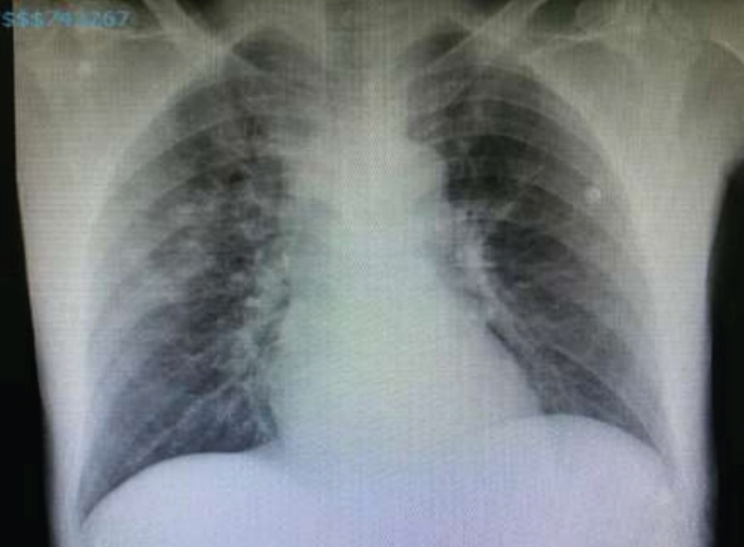 Figure 1: X-ray admission.
View Figure 1
Figure 1: X-ray admission.
View Figure 1
VV-ECMO Cannulation is performed with complete sterile measures of seldinger technique under the guidance of ultrasonography. A draining cannula of 25 fr is inserted into the right femoral vein and advanced into the inferior vena cava. Likewise, a returning cannula of 19 fr is inserted into the right internal jugular vein and advanced into the superior vena cava (Figure 2). A centrifugal pump used was Getinge/Maquet®, Rastatt, Germany, with an initial flow of 2 L/min, sweep gas 2 L/m. Heparin 5000 IU at cannulation. Then 1200 IU per hour to keep the target APTT ratio of 1.5-2.5 Immediate after ECMO cannulation, the blood gas value rapidly normalized. The first set of BGA Values at 60 minutes after starting ECMO were: pH 7.143, pCO2 86.2 mmHg, pO2 74.9 mmHg, HCO3- 21.5 mmol/L. There was no complication associated with VV ECMO cannulation. Adequate gas exchanges achieved with pressure control ventilation at PIP of 22 cmH2O, rate 12/min, FiO2 40%, and PEEP 3 cmH2O and decide to maintain the same MV settings for next 60 hours in adjunct with VV ECMO. The patient monitored closely with an hourly recording of all vitals; ABGs were repeated at every 2-hour interval for first 24 hours and every 6th hourly for the next 34 hours of ECMO therapy.
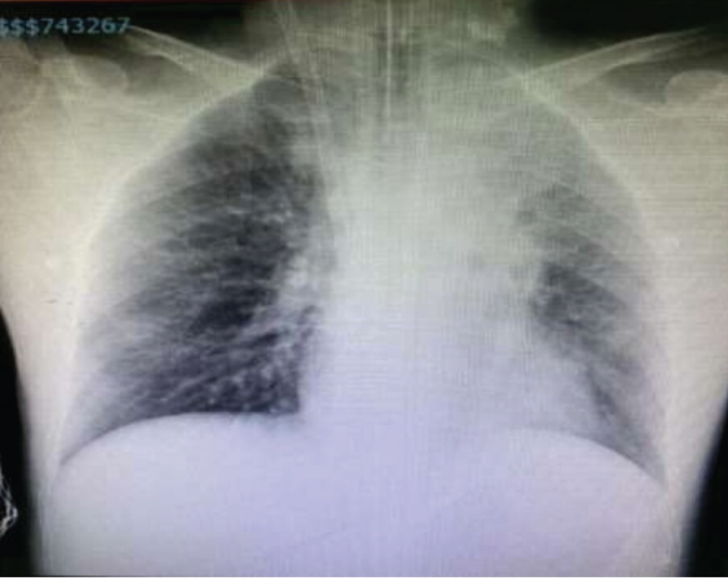 Figure 2: X-ray immediate after VV-ECMO cannulation.
View Figure 2
Figure 2: X-ray immediate after VV-ECMO cannulation.
View Figure 2
After 58 hours of continuous ECMO, the patient's clinical condition improved significantly, along with a reduction in PCO2 to 39.6 mmHg. Meanwhile, during 58 hours of ECMO, the patient was kept well sedated and ventilated; he continued on antibiotics, nebulization, steroids, and IV salbutamol, heparin infusion along with atracurium 40 mg per hour. By day 3, ECMO was weaned off by gradual flow and discontinued without any associated complications (Figure 3). On day 4, he was on a spontaneous breathing trial, respiratory mechanics, and ABG is ph 7.47 pCO2 34.2 pO2 108 HCO3 26 satisfactory. Therefore, he was extubated and transferred to the ward on day 6 and discharged home on day 16th without any complications. The serials of high ligtened PCO2 reduction in over 58 hours of ECMO therapy are shown in the Table 1. He turned to be influenzas B infection from nasal swab result exacerbating his near fatal asthma episode and it has been treated with Tamiflu 75 mg BID for 5 days.
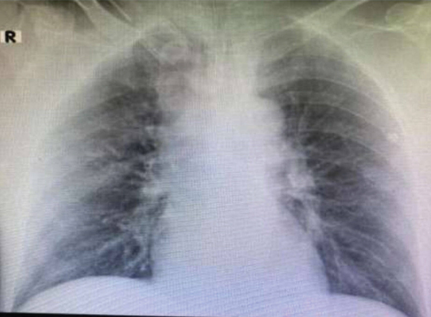 Figure 3: X-ray after decannulation of VV-ECMO.
View Figure 3
Figure 3: X-ray after decannulation of VV-ECMO.
View Figure 3
Table 1: Blood gas parameters before ECMO and during and after ECMO. View Table 1
The key factors that differentiate the patient of near-fatal asthma from severe asthma are: If there is no response to repeated sessions of Beta 2-agonist therapy, which results in respiratory failure necessitating hypercapnia (> 6.5 kPa), with or without altered consciousness for ICU admission for the possible requirement of mechanical ventilation, or history of the previous hospitalization [8]. The MV in NFA patients is a lifesaving emergency act as well; it requires a high dosage of sedation, analgesics, and muscle relaxants for good MV synchrony. At the same time, high sedation can increase further bronchospasms, air trapping, hyperinflation, which results in low functional residual capacity and increased intrathoracic pressures affecting the gas exchange [9,10]. While recent days asthma mortality is declining but remains significant. Substantial mortality and morbidity seen in asthma may be associated with MV itself in comparison to disease progression. In this case, our main objective is to reduce alveolar hyperinflation, avoid barotrauma, and maintain adequate oxygenation and ventilation by acknowledging some degree of permissive hypercapnia, until bronchodilators and steroids reverse the severe airflow resistance [11]. Nevertheless, with all these high sedation and ultra-protective lung strategy, the ventilator waveform of our patient exhibits persistent high peak airway pressure, high plateau pressure, and air trapping, respectively. To prevent these possible harmful effects of ventilator-induced lung injuries Extracorporeal Membrane Oxygenation (ECMO) adopted as surrogate therapy. Traditionally, ECMO commonly used in pediatrics with acute reversible respiratory and circulatory failure. Although the first case rescued successfully by ECMO in the year 1981. Still, ECMO remains controversial in these clinical settings. However, two large retrospective studies demonstrated the effectiveness and safety of ECMO in severe asthma attacks. In recent days ECMO plays a significant role in adult respiratory failure who failed in response to conventional treatments. Hyeju yeo, et al. concluded that among 24,147 patients 568 (2.4%) were asthmatics of these, 272 patients were treated with ECMO. In whom rate of ECMO weaning success was 234/272 (86.70%) and rate of survival to hospital discharge was 227/272 (83.5%) in the multicenter International Extracorporeal Life Support Organization registry between march 1992 to march 2016 [12]. ECMO's success in asthma is due to the natural reversibility of the airflow obstruction, which is in contrast to the patient with diffuse alveolar damage due to ALI. In a recent report of acute respiratory failure due to the influenza A (H1N1) epidemic in 2009, applying ECMO to these patients was associated with favorable results [13]. Although the first case rescued successfully by ECMO in the year 1981. Still, ECMO remains controversial in the clinical setting. However, Qingyuan Zhan in his literature review recommended Algorithmic approach to initialize ECMO if pH is less than 7.1, PaCO2 greater than 100 mmHg, or any life-threatening conditions such as hypoxemia, hypercapnia, hypotension, or barotrauma and optimized ventilatory support. Correspondingly, our patient fit into these criteria and returned to normal respiratory function within 70 hours from ECMO initiation, particularly driving pressure, air trapping, and PIP, which significantly improved within a few hours of VV-ECMO running. Driving pressure is a transpulmonary pressure. Any high transpulmonary pressure can cause lung injury or barotrauma. The high PIP seen before ECMO initiation may reflect possible dynamic hyperinflation, barotrauma, cardiovascular instability, and even death [14,15]. Similarly, our patient prior to ECMO continues to be severe refractory hypercapnic respiratory failure remarkably on high ventilatory settings, which ascertained to destroy the lungs over time in association with toxic oxygen concentrations and increase pressures. Although, ECMO utilization does not play a vital role in healing the sick lungs; instead, it allows the lungs to rest while our patient's vital functions maintained safely. Besides, the ventilator settings can be adjusted down to very low settings until the lungs recover. Our report highlighted that early VV-ECMO could lower the PIP, driving pressure and air trapping rapidly, and it can improve the ventilation/perfusion mismatch significantly by decreasing airway resistance and airway obstruction. Also, by admitting low ventilator settings, VV-ECMO can overcome ventilator-induced lung injury and oxygen toxicity. Usually, the time course to reestablish airway obstruction by standard conventional treatment is profoundly variable, and physicians cannot determine when bronchospasm will resolve. Adoption of VV-ECMO as surrogate therapy in the early hours of severe hypoxemia, hypercapnia, and before generating any ventilator-associated lung injury is the key factor that influenced the survival of our patient.
In brief, Near-fatal asthma is a rare subclass of asthma and it’s a life-threatening event. The early application of extracorporeal membrane oxygenation can influence the survival of the NFA. Our case signifies that VV ECMO should be utilized in the early hours of conventional treatment failure also before the occurrence of VILI, and even feasibly before the traits of any vital organ dysfunction. Notably, Physicians should be aware that each NFA patient may not be a candidate for ECMO. In context, ECMO can be adopted as surrogate therapy when the highest conventional treatment fails. A randomized controlled trial on the use of ECMO highly recommended in this group of patients.