Colorectal Cancer (CRC) is among the most common types of cancer in the world. Globally a steadily increasing proportion of elderly people in the world result in approximately 16 million new cases of cancer by the year 2021. Regarding treatment; Meloxicam was shown to prevent the initiation of chemical-induced tumors, and considered as anticancer agent by virtue of its anti-proliferative effect, capacity for cell cycle arrest, and pro-apoptotic effects, also acted as free radical scavenger, in particular superoxide anion oxidation scavenge. The aim of the current study was to clarify much important information on histopathology, histochemistry and immunoassay of CEA tumor marker characteristics of 1,2 DMH- induced colon tumor in albino rat and its relationship with the aberrant crypt mucosa as precursor in colon cancer progression. The study was carried out on 60 male albino rats, animals were divided into 5 groups; A: control group, B: animals received S.C. injections of 20 mg 1,2 DMH/Kg b.w, C: animals received 1,2 DMH with ad libitum access to water and high fat diet, D: animals fed high fat diet and water ad libitum. E: animals received S.C. injections of 1,2 DMH and oral 15 mg/Kg/day meloxicam/0.1 ml saline. Colon tissues from all studied groups were stained applying the following techniques: Hematoxylin & eosin, Alcian blue pH 2.5- acid mucopolysaccharide, Feulgen nuclear staining of DNA, Whole mount staining of colon and Immunoassay of serum CEA. The results confirmed the efficacy of meloxicam inhibiting or delaying growth of aberrant crypt foci in colon. Further research is needed to support presented findings.
Tumors, Prevention, Therapy
Colorectal Cancer (CRC) is among the most common types of cancer in the world. In Egypt for instance, CRC is the 6th common cancer in both males and females representing 4.5% and 3.6% of the total cancers [1,2]. Colorectal cancer is divided into sporadic (70-80%) and familial cases. Approximately 5-10% of all cancers fall into the familial category [3]. The vast majority of CRC developed from benign precursor lesions through a series of genetic and epigenetic changes [4]. According to the WHO classification; nearly 85% of CRCs are usual adenocarcinomas, while 10 to 15% are described as mucinous adenocarcinomas [5]. Globally a steadily increasing proportion of elderly people in the world result in approximately 16 million new cases of cancer by the year 2020 (IARC) [6].
Hydrazines are manufactured from chemicals such as ammonia, dimethylamine, hydrogen peroxide, and sodium hypochlorite. A small amount of hydrazine occurs naturally in some plants. 1,2 Dimethylhydrazine (DMH) has been used for the sake of scientific research to produce colon cancer in laboratory animals [7,8]. DMH also known as symmetrical dimethylhydrazine N,N' DMH and is considered as a highly specific colorectal procarcinogen that undergoes metabolic activation in the liver to DNA-reactive metabolites by a series of reactions through intermediates and to the ultimate carcinogenic metabolite [9]. DMH was tested for carcinogenicity in mice, rats and hamsters following oral, subcutaneous or intramuscular administration, producing tumors at various sites [10,11].
A therapeutic area in which NSAIDs use became important was in the treatment and prevention of cancer. Epidemiologic studies in human showed that aspirin use was associated with significant reduction in the incidence of colon cancer. Additional evidence suggested that the therapeutic effect of NSAIDs on colon cancer was mediated by inhibition of COX-2, which was up regulated in many premalignant and malignant neoplasms. Meloxicam was shown to prevent the initiation of chemical-induced tumors, and considered as anticancer agent by virtue of its anti-proliferative effect, capacity for cell cycle arrest, and pro-apoptotic effects, also acted as free radical scavenger, in particular superoxide anion oxidation scavenge [12,13].
A tumor marker is a biological substance synthesized and released by cancer cells or produced in response to the presence of a tumor tissue, circulating tumor cells in peripheral blood, serum, lymph nodes and bone marrow. It is defined as a molecule that indicates the likely presence of cancer or that provides information about the likely future behavior of a cancer [14]. Carcinoembryonic antigen is a glycoprotein which is important as a tumor marker for a number of human cancers. It is one of the most widely used tumor markers worldwide. Its main application is mostly in gastrointestinal cancers, especially in colorectal malignancy [14].
The present study was cross sectional carried out on 60 male albino rats, 5-6 weeks-old at the beginning of the experiment, weighing from 100-120 g and obtained from the animal house of the odor Bilharz Institute, Cairo. All experiments were performed in line with the ethical considerations recommended by Alexandria University, Egypt. The animals were kept in the animal house, 4 per cage in a temperature and light controlled room. They were maintained on standard diet and allowed free access to appropriate diet and water ad libitum throughout the 8 weeks of the experiment.
Laboratory animals were divided into 5 groups; 12 animals in each and classified as follows: Group A: The animals were served as control; received S.C. injections of saline solution. Group B: The animals received S.C. injections of 20 mg 1,2 DMH/Kg b.w dissolved in 1 ml sterile physiological saline in the interscapular region using a tuberculin syringe with a 24-gauge needle once weekly for 8 weeks, fed standard diet and water ad libitum. Group C: The animals received 1,2 DMH as in group B with ad libitum access to water and high fat diet.
Group D: The animals were fed high fat diet and water ad libitum. Group E: The animals received S.C. injections of 1,2 DMH as in group B and oral 15 mg/Kg/day meloxicam/0.1 ml saline via gastric tube 1 hour before DMH administration , fed high fat diet and water ad libitum. Standard diet for groups A & B consisted of 5% fat, 53% carbohydrate, 23% protein, with total caloric value 25 kJ/kg and high-fat diet for group C, D & E consisted of 30% fat, 48% carbohydrate, and 20% protein with total caloric value 44.3 kJ/kg were used as previously recommended. At the end of the experiment, colon was dissected out to study [15,16].
Hematoxylin & eosin staining technique: Colon tissues from all studied groups were fixed in 10% neutral buffered formalin for 24 hours, washed in running tap water, dehydrated in ascending series of ethyl alcohol, cleared in xylene and embedded in paraffin wax. Sections of 5 μm thick were cut using rotary microtome and stained with H&E [17].
Alcian blue pH 2.5- acid mucopolysaccharide: Alcian blue stain was used to detect the changes in mucin contents of goblet cells of the crypts mucosa of rat colon [18].
Feulgen nuclear staining of DNA: Epithelial cells of colon mucosa proliferation were visualized by using Feulgen nuclear histochemical staining of DNA [19].
Whole mount staining of colon: High-resolution image acquisition easily identifies Aberrant crypt foci (ACF) [20].
Blood samples were collected from rats of control and experimental groups using cardiac-puncture technique to quantitatively measure serum CEA levels using a ready for use electro-chemiluminescence immunoassay (ECLIA) kit according to manufacture´s protocol (Roche).
In control group the normal colon mucosa of albino rat. Typically glandular crypts (C) that invaginate deep in the submucosa (SM). Muscularis externa, serosa layers are also observed (Figure 1). Sections of colon of animals of group B received DMH - Standard diet showed. Crowded tubular glands (*) with irregular shapes and sizes invading the muscularis mucosa layer and abundant chronic inflammatory cells (▼) are also noticed (Figure 2). In addition there was elongation of the crypts (*) with epithelial proliferation. Architectural disturbance, Mitotic Figures (MF), and inflammatory cells (▼) are observed (Figure 3). Colon sections of group C of animals received DMH – high fat diet. Overcrowding of damaged tubular glands (*) which appeared irregular in shape and size lined by dysplastic epithelium. Some glands are seen lined by normal epithelial cells with round nuclei (Figure 4). As well on colonic glands (*) showed dilated lumen and severe epithelial atypia with slim rod shaped stratified nuclei (Figure 5). Colon sections of rats fed high fat diet and water ad libitum. (Group D) showed a regularly organized glands (*) lined mainly by epithelial and mucus secreting cells (Figure 6). Colon sections of rats injected with DMH – Meloxicam -High fat diet. Group E showed the majority of colonic glands (*) with regular size and shape (Figure 7).
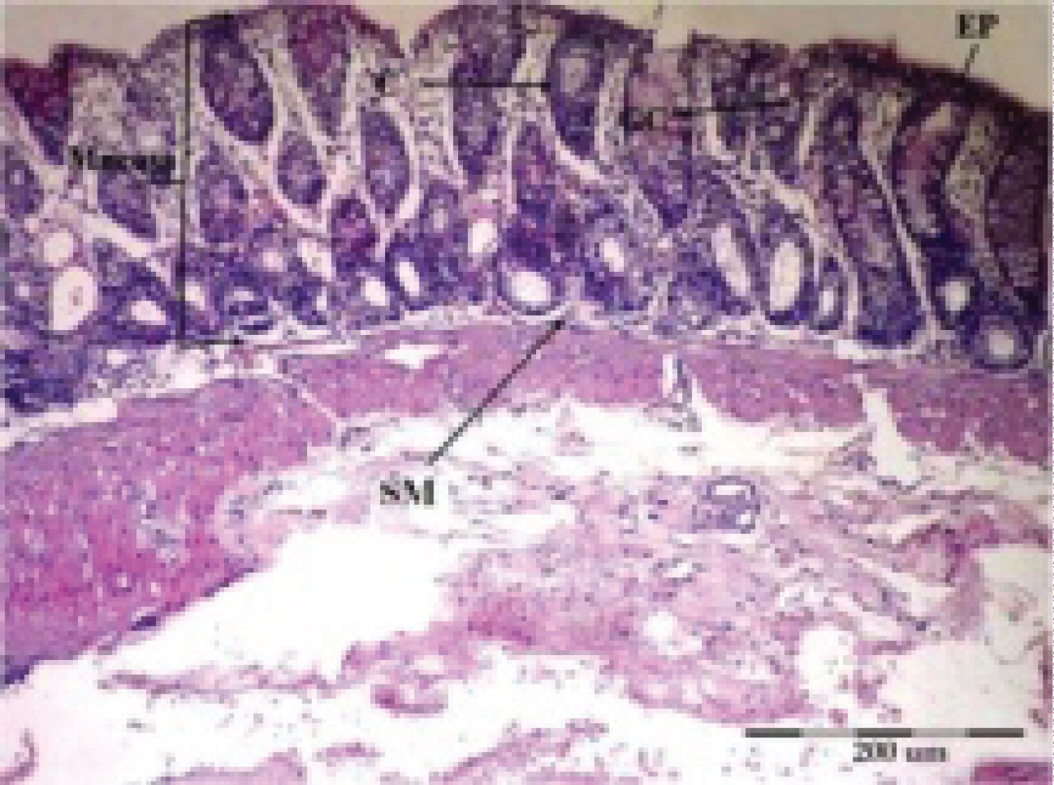 Figure 1: Muscularis externa, serosa layers are also observed.
View Figure 1
Figure 1: Muscularis externa, serosa layers are also observed.
View Figure 1
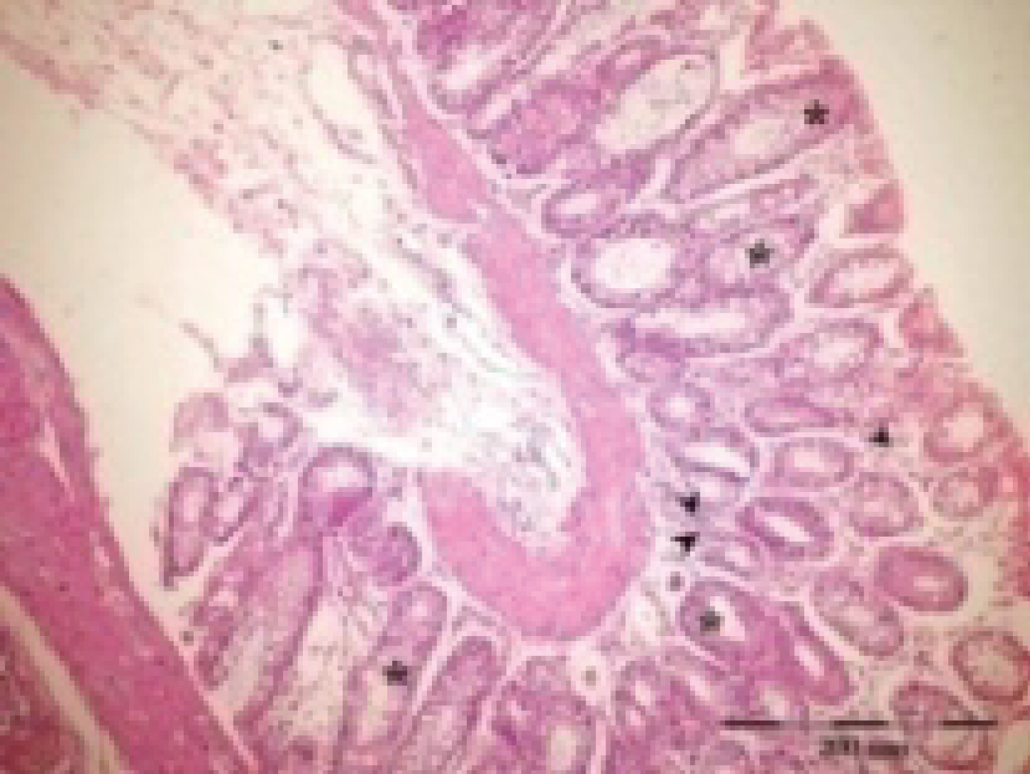 Figure 2: Crowded tubular glands (*) with irregular shapes and sizes invading the muscularis mucosa layer and abundant chronic inflammatory cells (▼) are also noticed.
View Figure 2
Figure 2: Crowded tubular glands (*) with irregular shapes and sizes invading the muscularis mucosa layer and abundant chronic inflammatory cells (▼) are also noticed.
View Figure 2
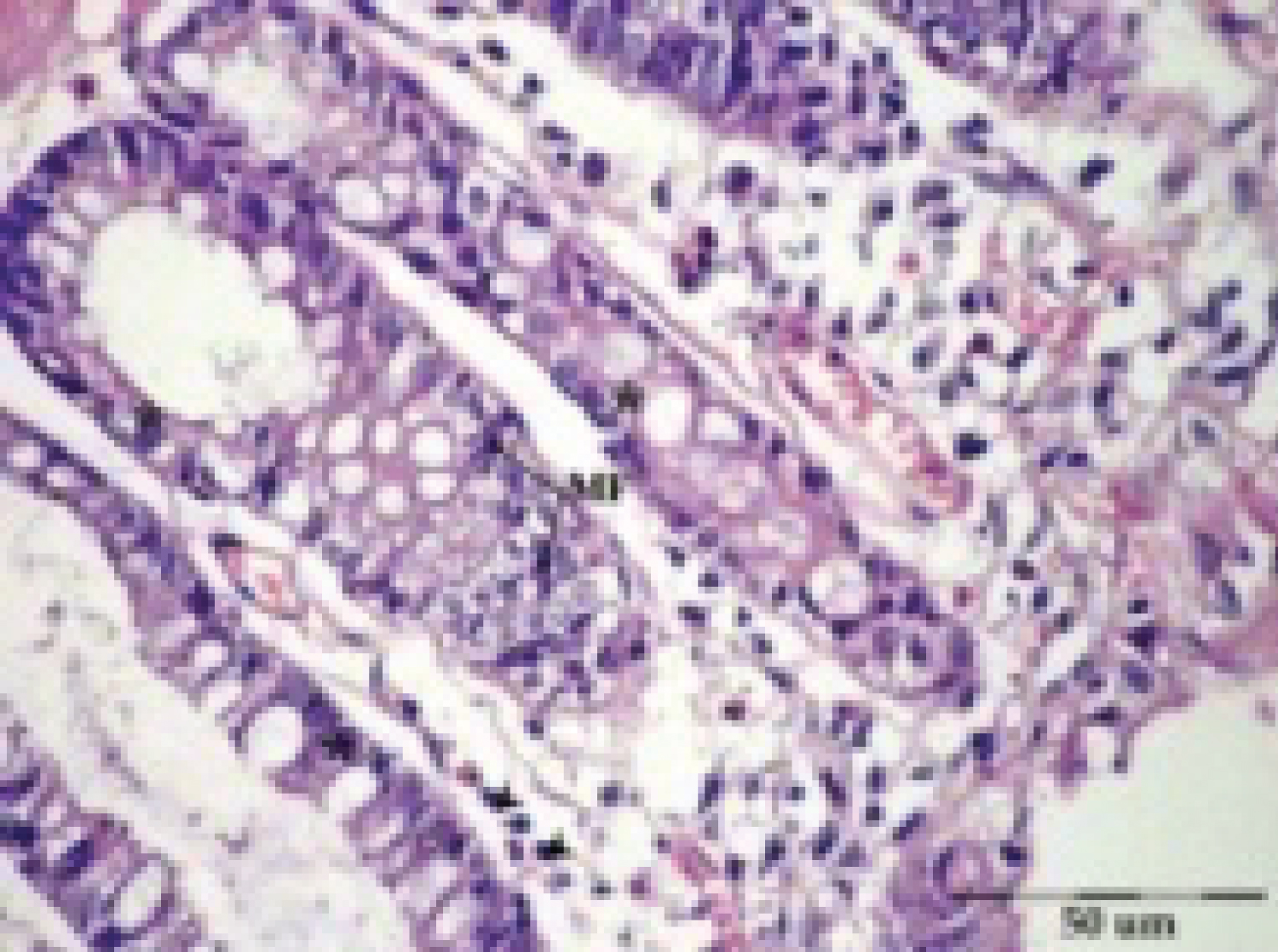 Figure 3: Architectural disturbance, Mitotic Figures (MF), and inflammatory cells (▼) are observed.
View Figure 3
Figure 3: Architectural disturbance, Mitotic Figures (MF), and inflammatory cells (▼) are observed.
View Figure 3
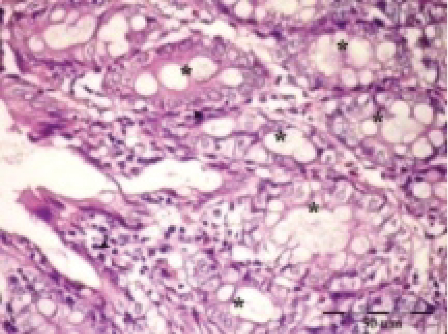 Figure 4: Some glands are seen lined by normal epithelial cells with round nuclei.
View Figure 4
Figure 4: Some glands are seen lined by normal epithelial cells with round nuclei.
View Figure 4
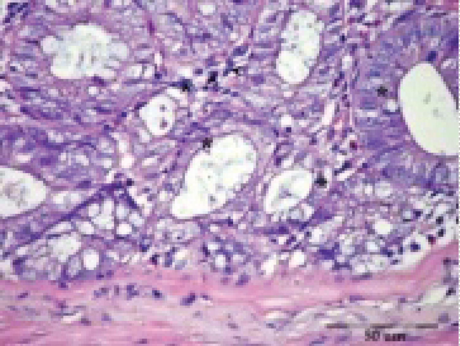 Figure 5: As well on colonic glands (*) showed dilated lumen and severe epithelial atypia with slim rod shaped stratified nuclei.
View Figure 5
Figure 5: As well on colonic glands (*) showed dilated lumen and severe epithelial atypia with slim rod shaped stratified nuclei.
View Figure 5
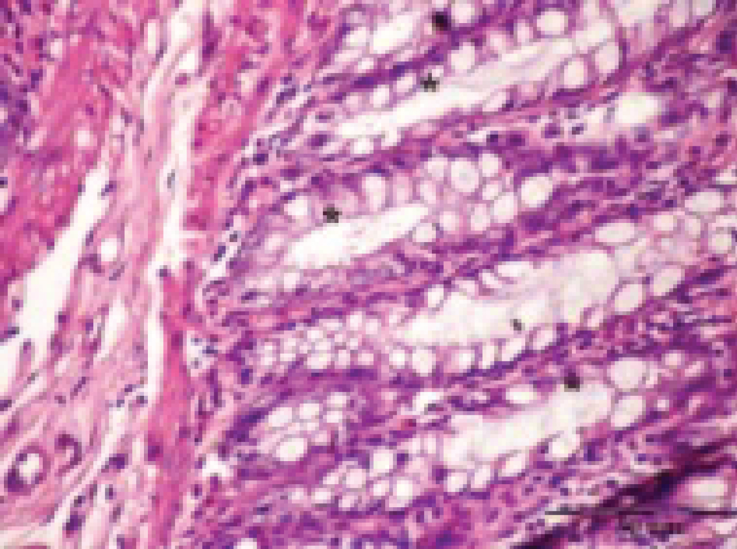 Figure 6: Colon sections of rats fed high fat diet and water ad libitum. (Group D) showed a regularly organized glands (*) lined mainly by epithelial and mucus secreting cells.
View Figure 6
Figure 6: Colon sections of rats fed high fat diet and water ad libitum. (Group D) showed a regularly organized glands (*) lined mainly by epithelial and mucus secreting cells.
View Figure 6
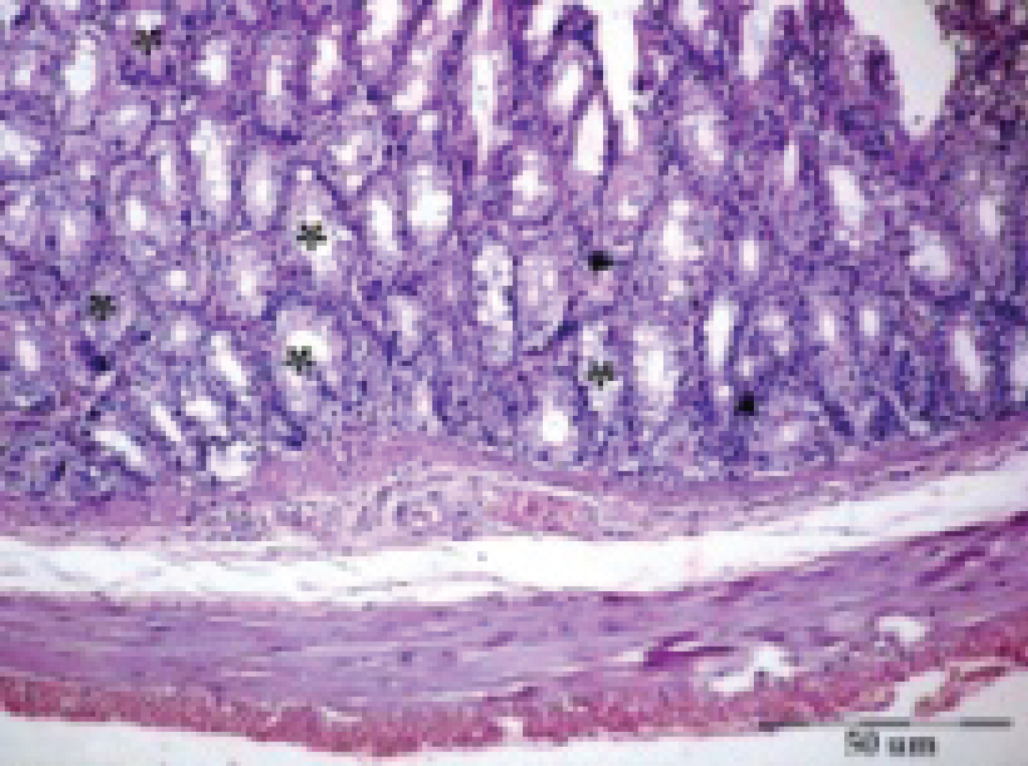 Figure 7: Group E showed the majority of colonic glands (*) with regular size and shape.
View Figure 7
Figure 7: Group E showed the majority of colonic glands (*) with regular size and shape.
View Figure 7
Normal rat colon section of group A, showing colon glands lined by columnar epithelium and abundant normal secretory cells. Positively stained nuclei located in the epithelial cells of the crypts (↑) (Figure 8). Colon sections of rats of group B received DMH -Standard diet and ad libitum access to water. Showing crypt multiplicity with parallel moderate increase in DNA (↑) (Figure 9). Colon section of group C of rats received DMH as in group B, fed high fat diet and water ad libitum. Showing aberrant crypts with stratified mucosa, occasional mitotic division and pyknotic nuclei (▼) which expressed marked increase of DNA (Figure 10). Colon section of group Dof rats fed high fat diet and water ad libitum. Showing predominance of closely packed glands (↑) with distended goblet cells and normal epithelial cells with small-sized nuclei stained dark purple for DNA (Figure 11). Colon sections of group E of animals received S.C. injections of DMH - Meloxicam - High fat diet and water ad libitum .The colon epithelium showed small non dividing nuclei stained for DNA reaction (↑) (Figure 12).
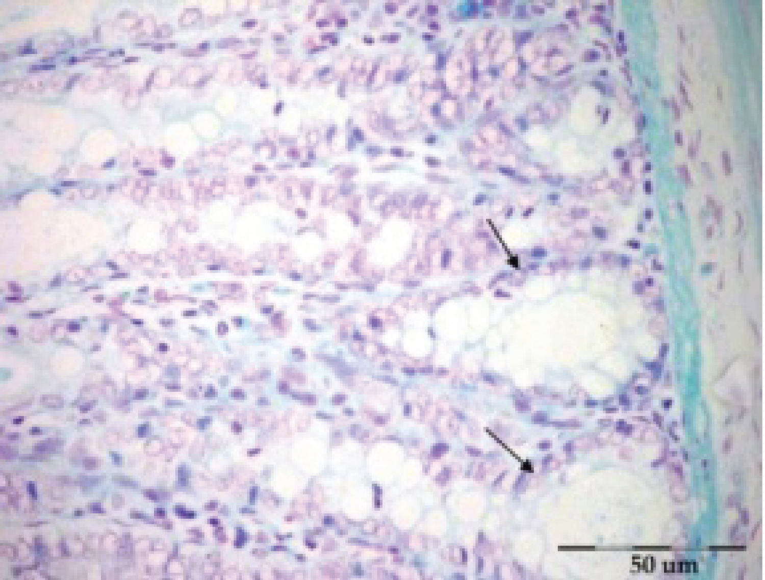 Figure 8: Positively stained nuclei located in the epithelial cells of the crypts (↑).
View Figure 8
Figure 8: Positively stained nuclei located in the epithelial cells of the crypts (↑).
View Figure 8
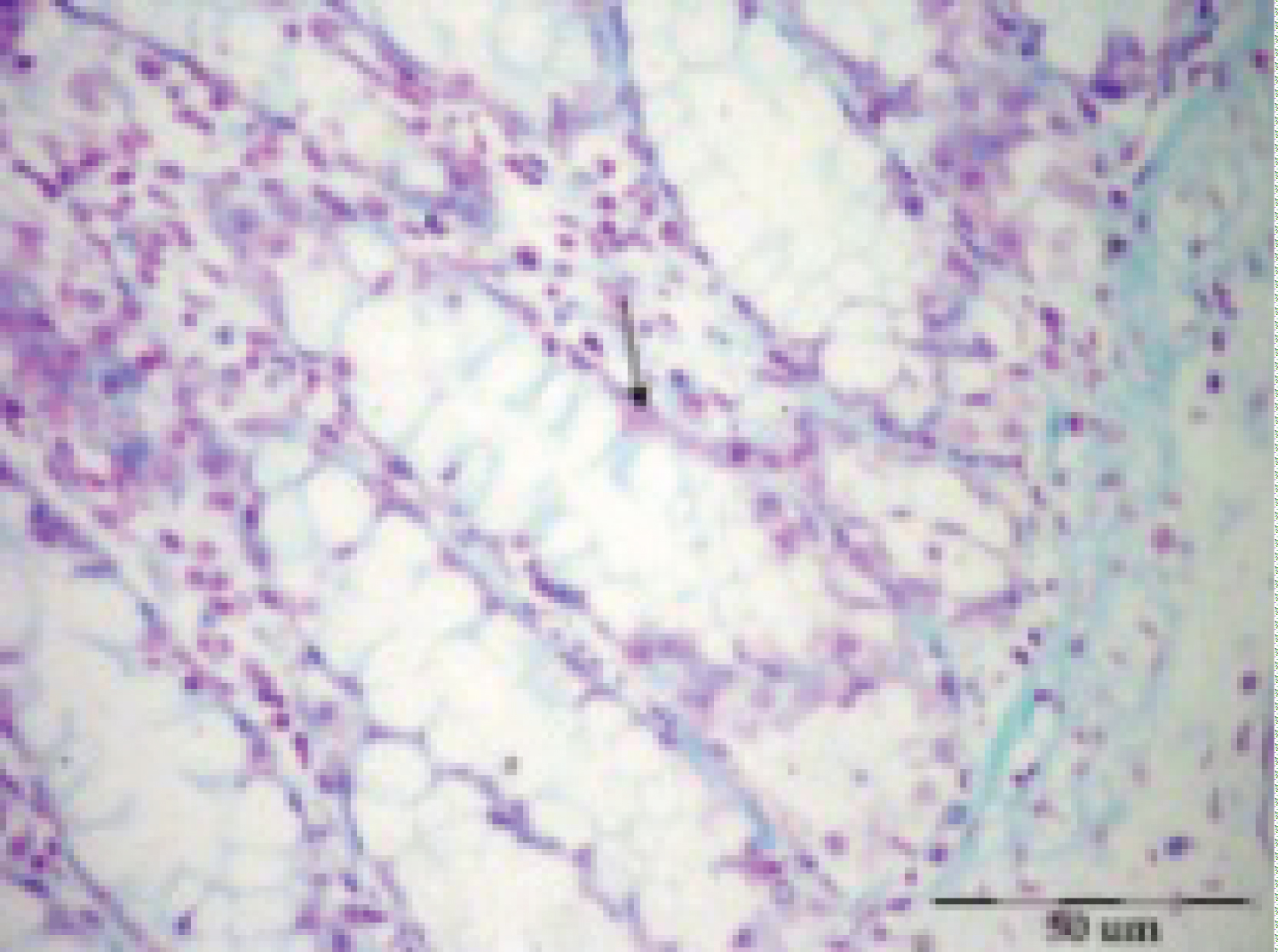 Figure 9: Showing crypt multiplicity with parallel moderate increase in DNA (↑).
View Figure 9
Figure 9: Showing crypt multiplicity with parallel moderate increase in DNA (↑).
View Figure 9
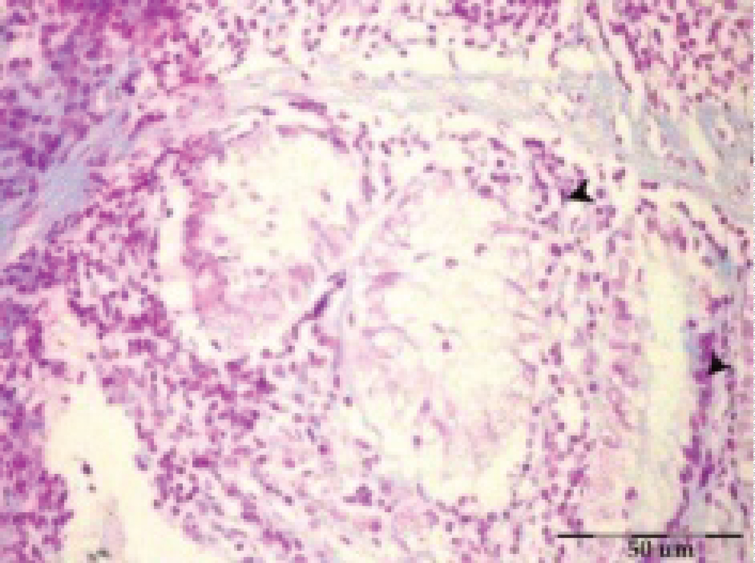 Figure 10: Showing aberrant crypts with stratified mucosa, occasional mitotic division and pyknotic nuclei (▼) which expressed marked increase of DNA.
View Figure 10
Figure 10: Showing aberrant crypts with stratified mucosa, occasional mitotic division and pyknotic nuclei (▼) which expressed marked increase of DNA.
View Figure 10
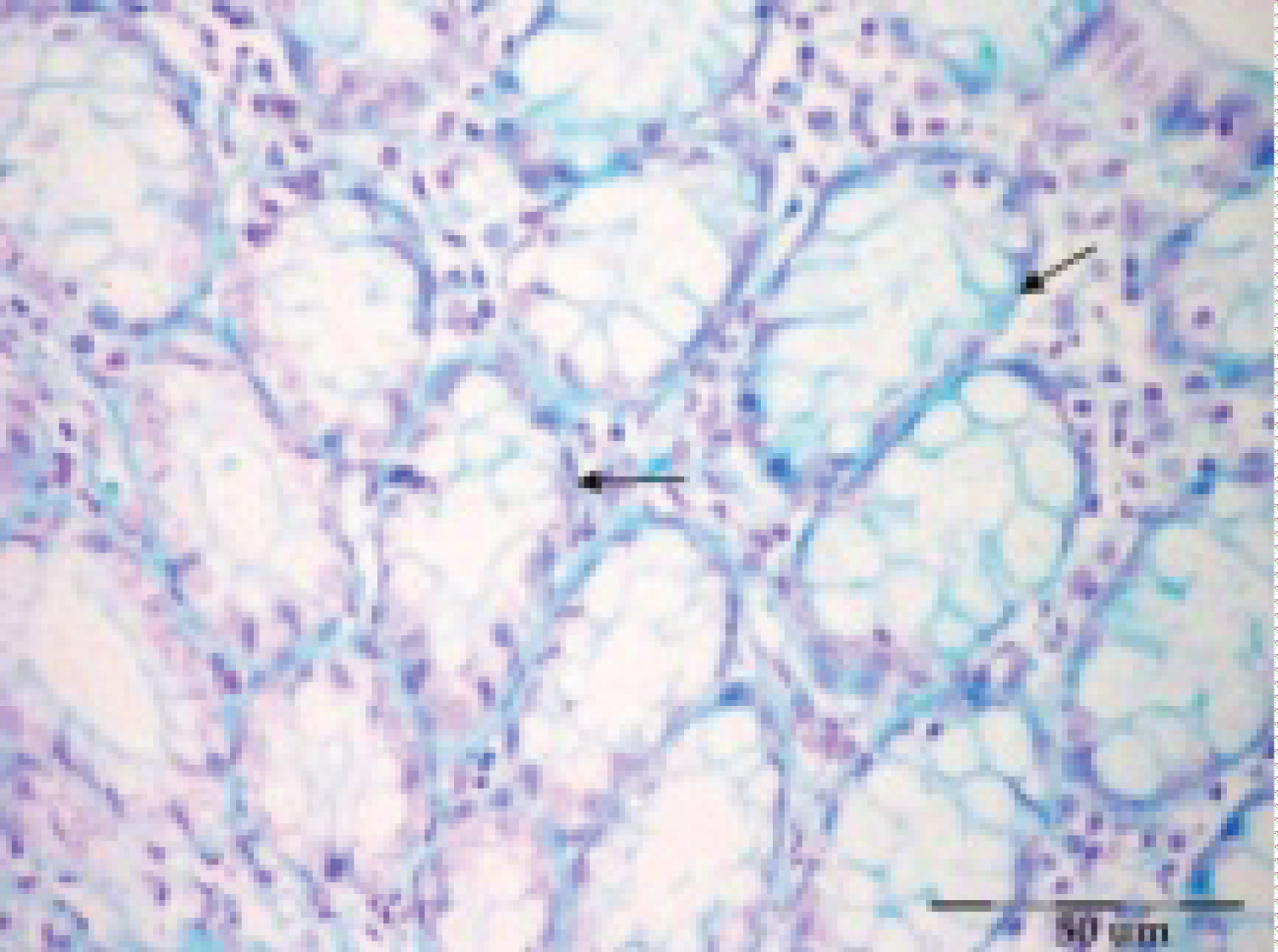 Figure 11: Showing predominance of closely packed glands (↑) with distended goblet cells and normal epithelial cells with small-sized nuclei stained dark purple for DNA.
View Figure 11
Figure 11: Showing predominance of closely packed glands (↑) with distended goblet cells and normal epithelial cells with small-sized nuclei stained dark purple for DNA.
View Figure 11
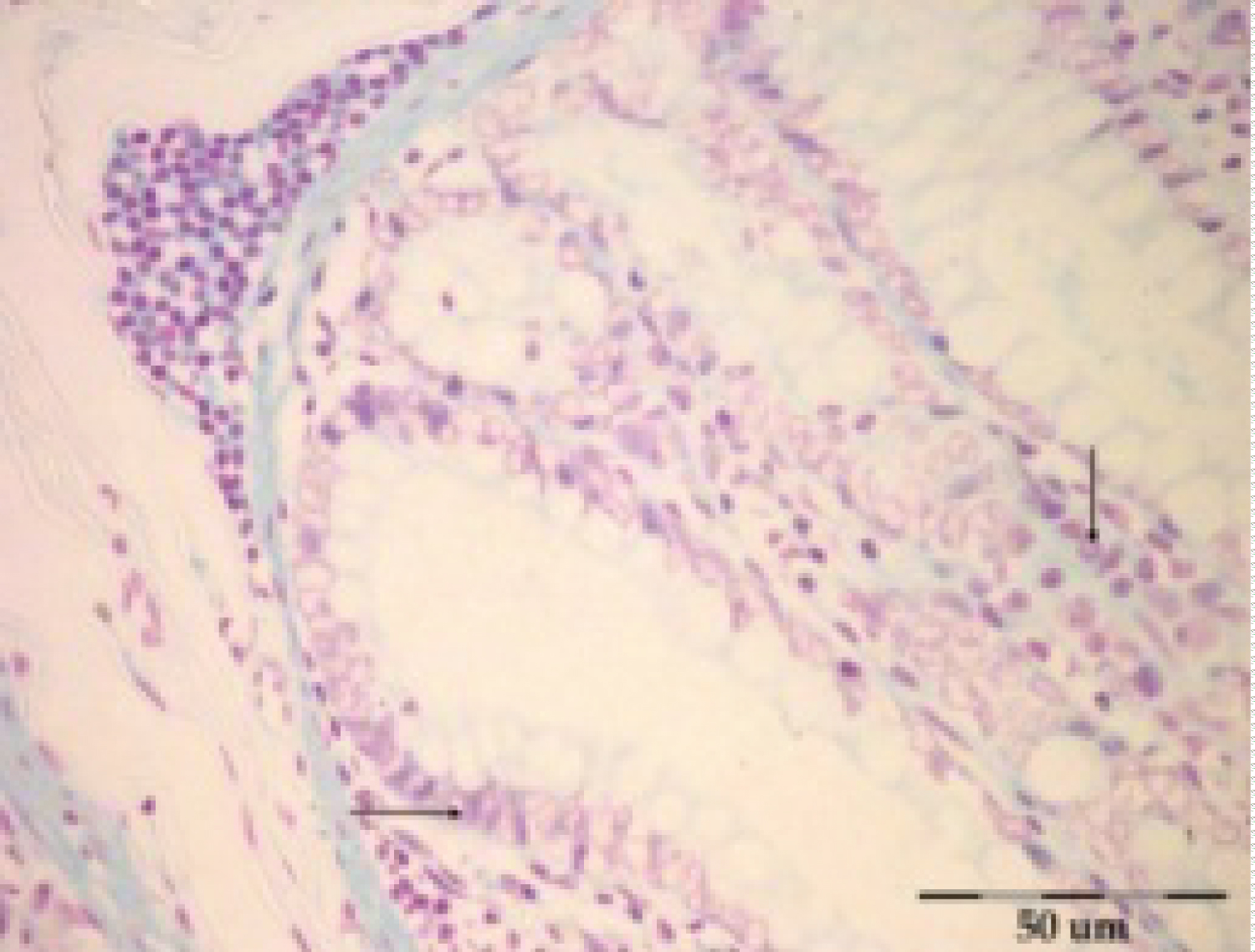 Figure 12: The colon epithelium showed small non dividing nuclei stained for DNA reaction (↑).
View Figure 12
Figure 12: The colon epithelium showed small non dividing nuclei stained for DNA reaction (↑).
View Figure 12
Colon sections of control group of rats received S.C. injections of saline solution. The goblet cells in most of crypts appeared few in number with moderate positive blue staining mucin (←) (Figure 13). Colon sections of group B received S.C. injections of 1,2 DMH, fed standard diet and water ad libitum.Increased number of goblet cells with increased blue staining mucin (↑) (Figure 14). Colon sections of group C of rats received 1,2 DMH as in group B -High fat diet. The colon crypts showed numerous proliferated crypts devoid of goblet cells. Mucin is almost completely depleted in most of ACs in rat colon (←) (Figure 15). Colon sections of group D of rats fed high fat diet and water ad libitum. Goblet cells with different sizes condensed in colon crypts lumen stained with different degree of blue coloration and other empty goblet cells after discharging their contents are observed (↑) (Figure 16). Colon sections of group E of rats received S.C.injections of 1,2 DMH-Meloxicam-High fat diet. Showed colon crypts with few goblet cells which contained less mucin (↑) (Figure 17).
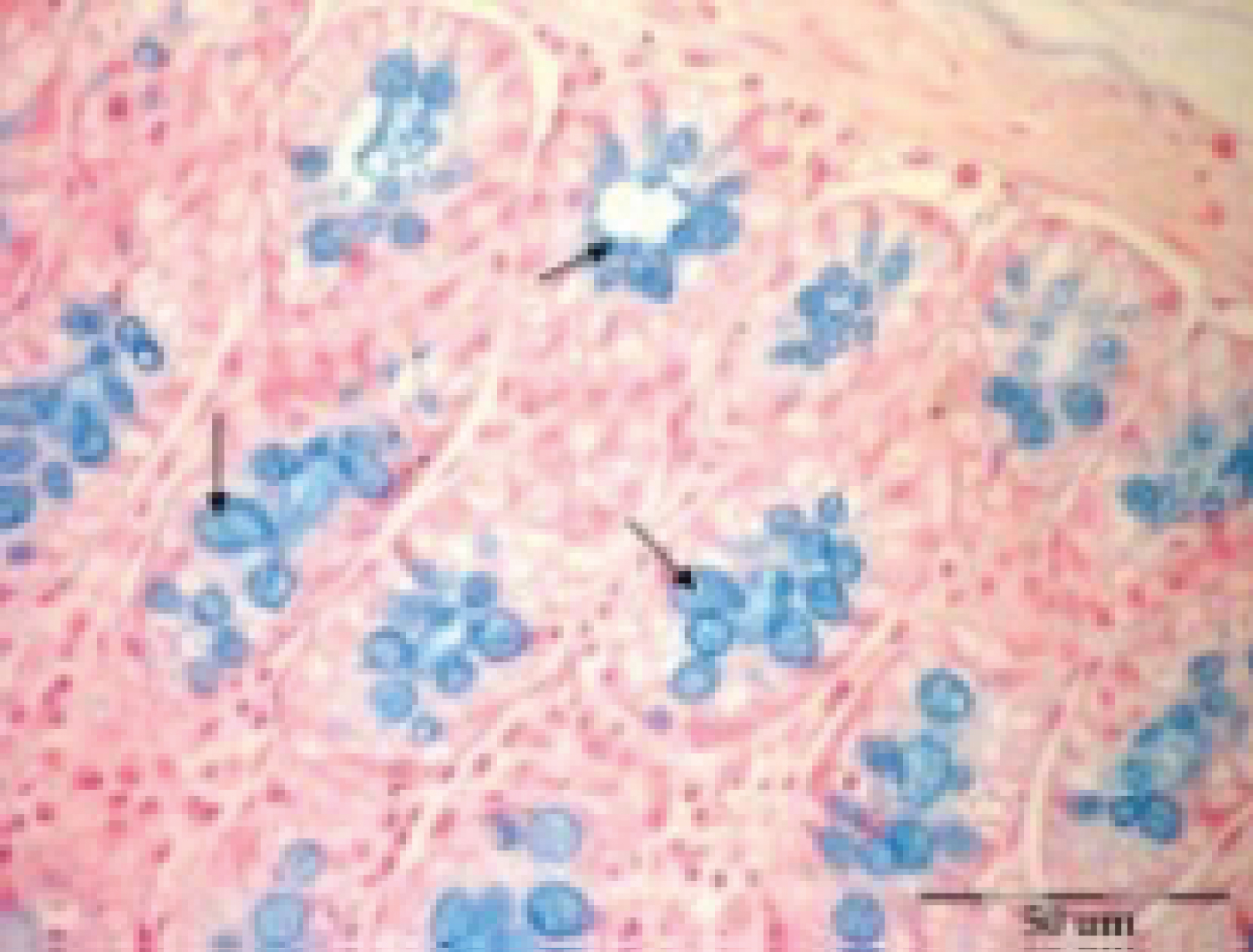 Figure 13: The goblet cells in most of crypts appeared few in number with moderate positive blue staining mucin (←).
View Figure 13
Figure 13: The goblet cells in most of crypts appeared few in number with moderate positive blue staining mucin (←).
View Figure 13
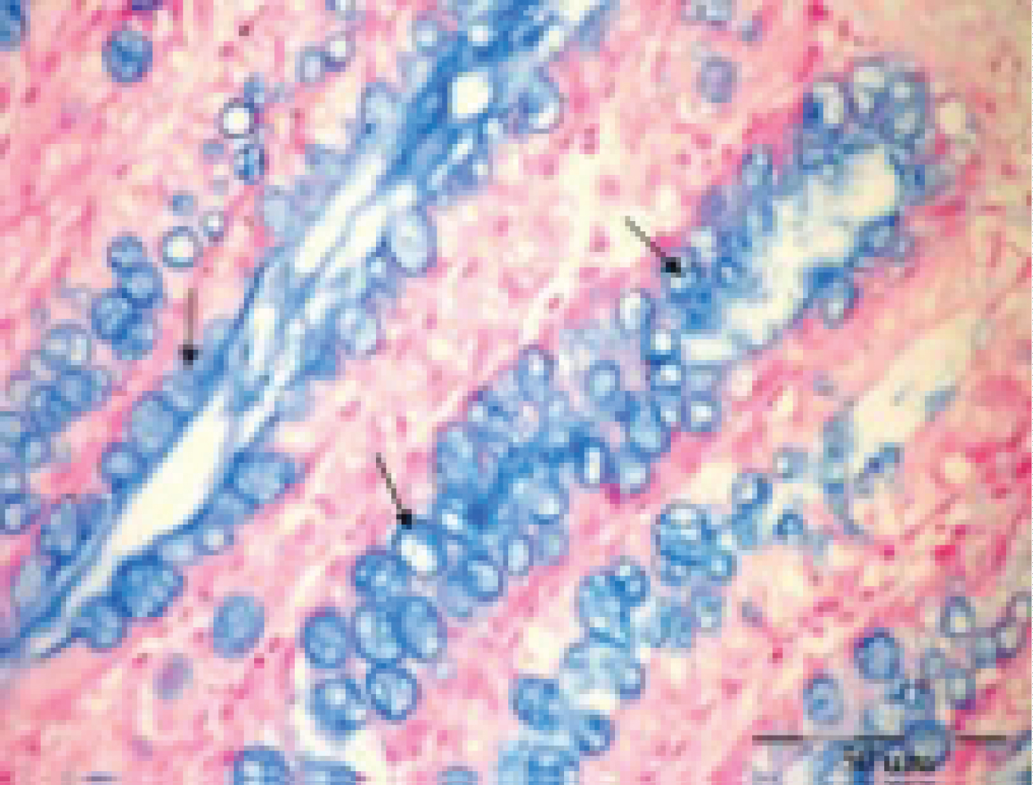 Figure 14: Increased number of goblet cells with increased blue staining mucin (↑).
View Figure 14
Figure 14: Increased number of goblet cells with increased blue staining mucin (↑).
View Figure 14
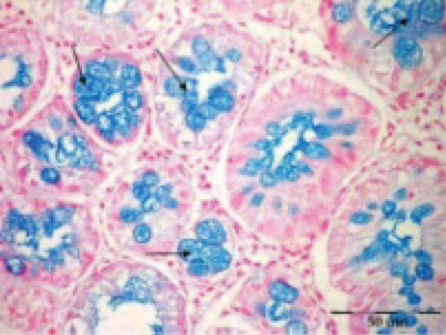 Figure 15: Mucin is almost completely depleted in most of ACs in rat colon (←).
View Figure 15
Figure 15: Mucin is almost completely depleted in most of ACs in rat colon (←).
View Figure 15
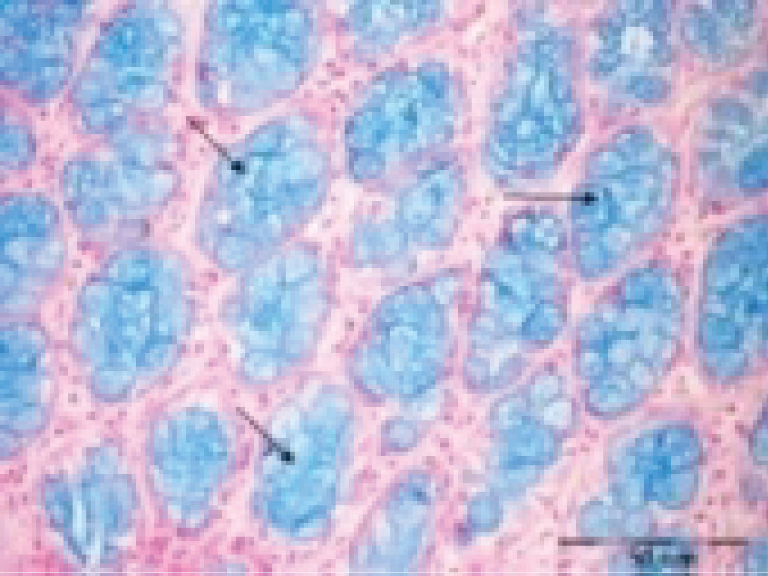 Figure 16: Goblet cells with different sizes condensed in colon crypts lumen stained with different degree of blue coloration and other empty goblet cells after discharging their contents are observed (↑).
View Figure 16
Figure 16: Goblet cells with different sizes condensed in colon crypts lumen stained with different degree of blue coloration and other empty goblet cells after discharging their contents are observed (↑).
View Figure 16
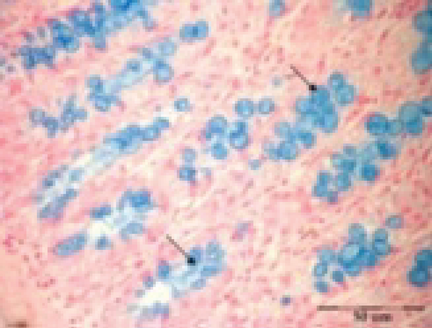 Figure 17: Colon sections of group E of rats received S.C. injections of 1,2 DMH-Meloxicam-High fat diet. Showed colon crypts with few goblet cells which contained less mucin (↑).
View Figure 17
Figure 17: Colon sections of group E of rats received S.C. injections of 1,2 DMH-Meloxicam-High fat diet. Showed colon crypts with few goblet cells which contained less mucin (↑).
View Figure 17
whole colon of Group B of rats received S.C. injections of 1,2 DMH, fed standard diet showing a small sized focus consisting of thickened crypts (↑), with increased pericryptal area; greater staining intensity due to thickened epithelium (Figure 18). ACF of whole mount colon of group C of animals received 1,2 DMH as in group B and fed high fat diet. Showing darkly staining proliferated crypt foci with thick epithelial lining (↑) (Figure 19). ACF in methylene blue-stained whole mount colon of group D of rats fed high fat diet and water ad libitum. Showing normal, moderate sized closely packed crypts (Figure 20). Whole colon of group E of animals received DMH- Meloxicam -High fat diet. Showing normal crypts with some thickened lining crypts (↑) (Figure 21).
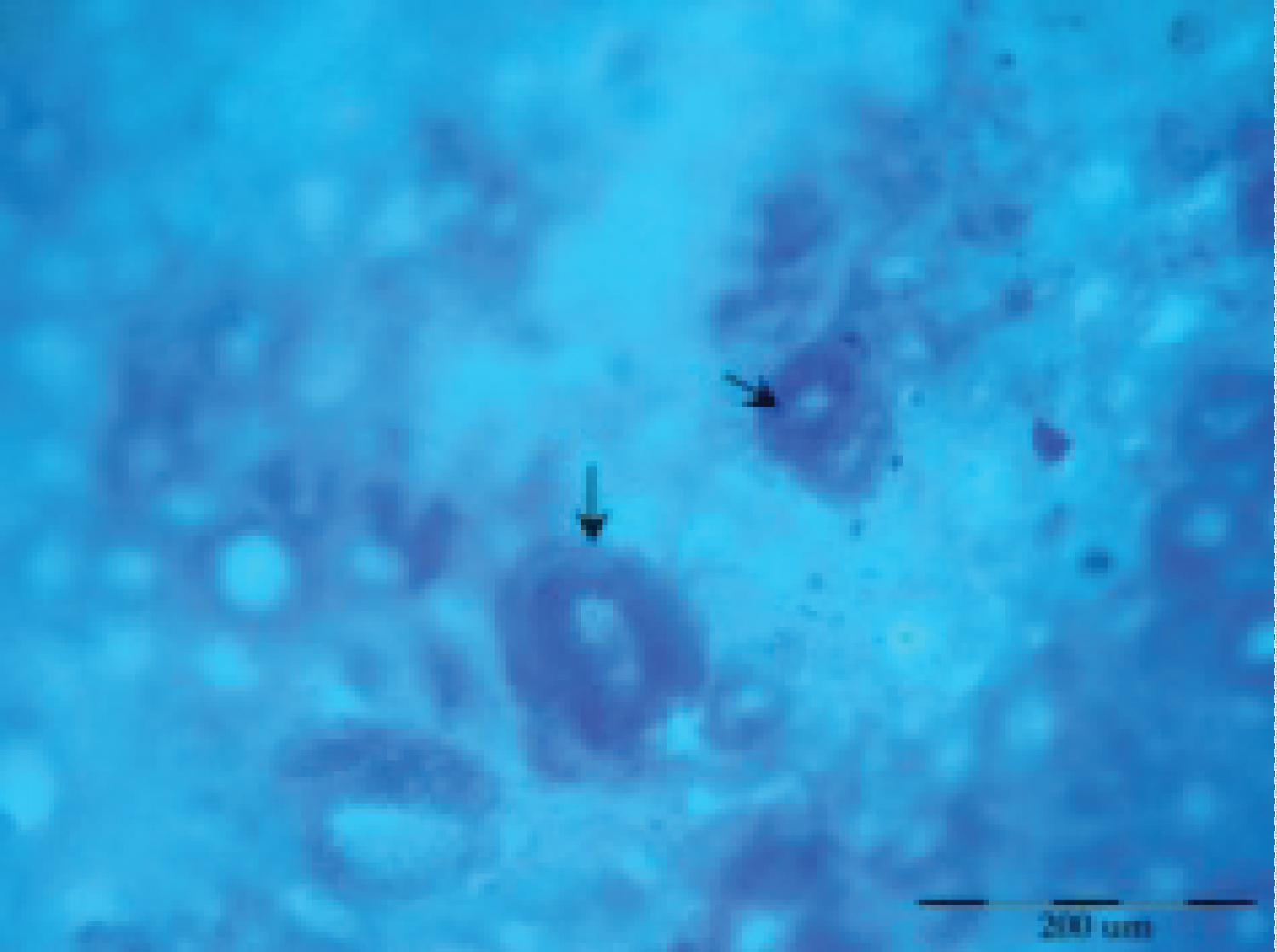 Figure 18: whole colon of Group B of rats received S.C. injections of 1,2 DMH, fed standard diet showing a small sized focus consisting of thickened crypts (↑), with increased pericryptal area; greater staining intensity due to thickened epithelium.
View Figure 18
Figure 18: whole colon of Group B of rats received S.C. injections of 1,2 DMH, fed standard diet showing a small sized focus consisting of thickened crypts (↑), with increased pericryptal area; greater staining intensity due to thickened epithelium.
View Figure 18
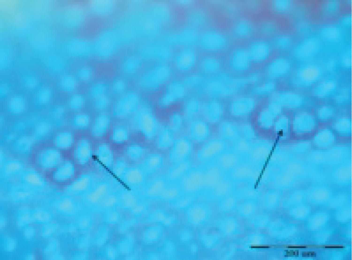 Figure 19: Showing darkly staining proliferated crypt foci with thick epithelial lining (↑).
View Figure 19
Figure 19: Showing darkly staining proliferated crypt foci with thick epithelial lining (↑).
View Figure 19
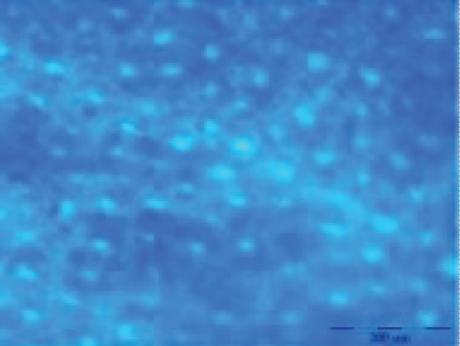 Figure 20: Showing normal, moderate sized closely packed crypts.
View Figure 20
Figure 20: Showing normal, moderate sized closely packed crypts.
View Figure 20
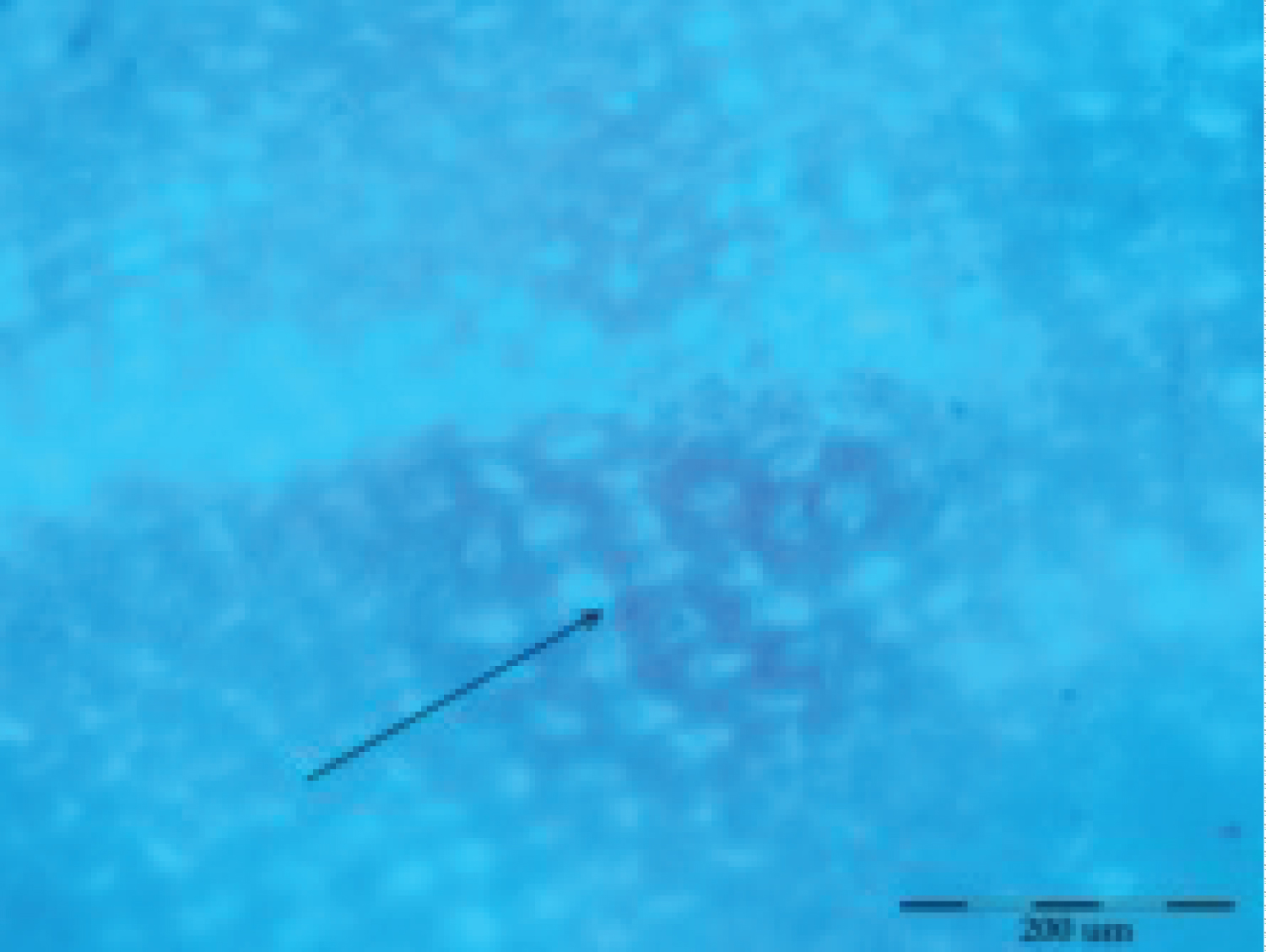 Figure 21: Whole colon of group E of animals received DMH- Meloxicam -High fat diet. Showing normal crypts with some thickened lining crypts (↑).
View Figure 21
Figure 21: Whole colon of group E of animals received DMH- Meloxicam -High fat diet. Showing normal crypts with some thickened lining crypts (↑).
View Figure 21
 Figure 22: Serum levels of CEA in groups A, B, C, D & E.
*: Significance was compared with group A.
View Figure 22
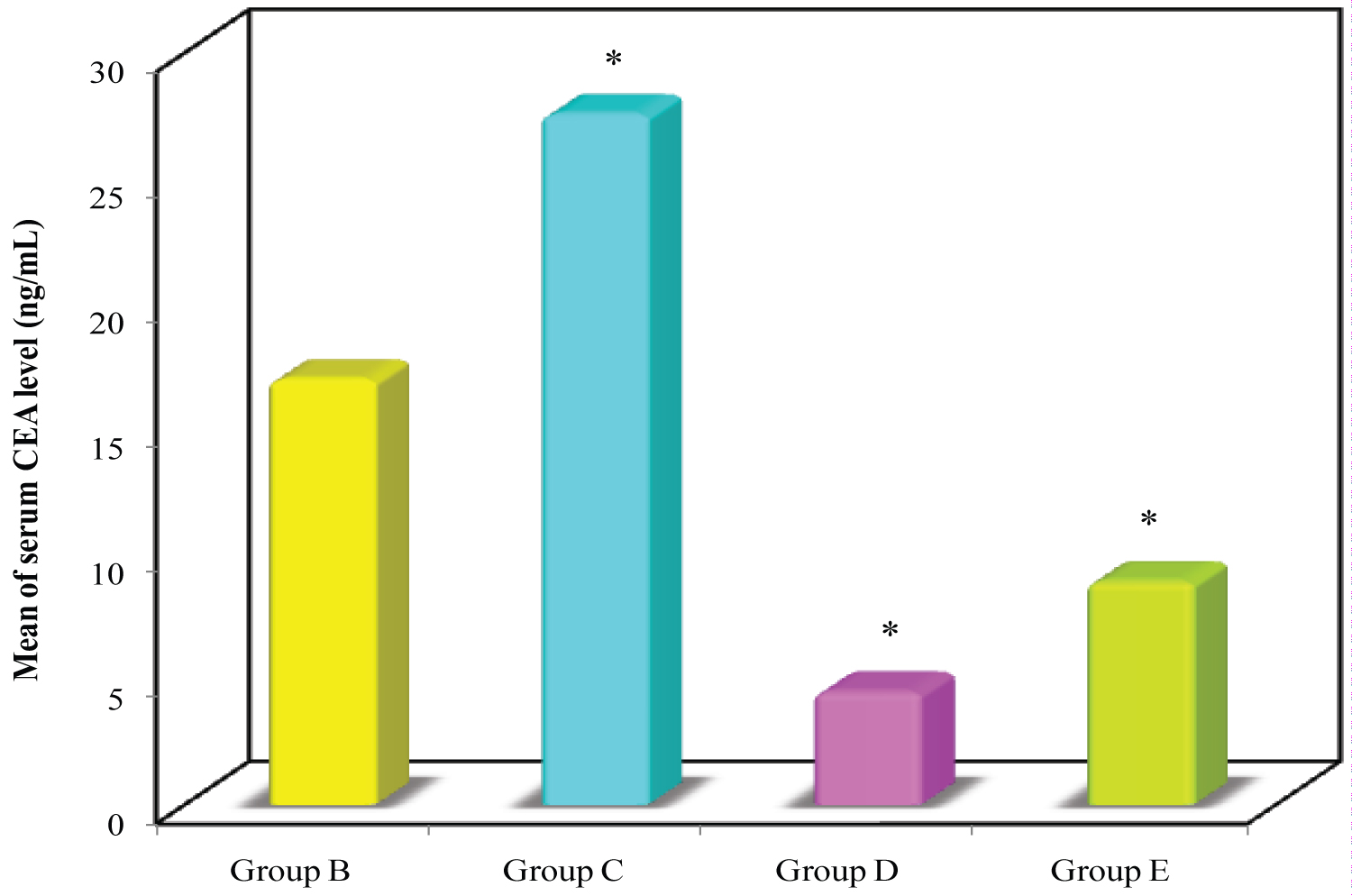 Figure 23: Serum levels of CEA in groups B, C, D & E.
*: Significance was compared with group B.
View Figure 23
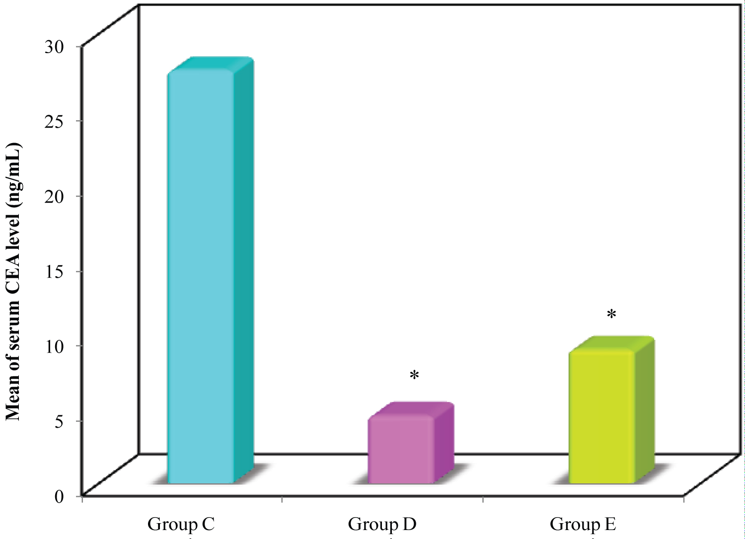 Figure 24: Serum levels of CEA in groups C, D, & E.
*: Significance was compared with group C.
View Figure 24
The Mean ± SD concentrations of serum CEA were 3.37 ± 0.26 ng/ml in the normal control group, 16.96 ± 2.89 ng/ml in rats with colorectal carcinoma, 27.54 ± 4.56 ng/ml in rats with colorectal carcinoma which were fed high fat diet, 4.50 ± 0.34 ng/ml in rats fed only on high fat diet and 8.90 ± 1.61 ng/ml in rats with colorectal carcinoma + high fat diet that were treated with meloxicam as an anti-inflammatory drug. The statistical analysis of their data showed that the serum levels of CEA in groups B and C were significantly greater than CEA levels in group A. The serum level of CEA in group E was significantly higher than group A. In group C, the serum level of CEA was significantly greater than group B. In groups D and E, the serum levels of CEA were significantly lower than in group B. In groups D and E, the serum levels of CEA were significantly lower than group C (Table 1).
Table 1: The serum levels of CEA (ng/ml) in the different studied groups. View Table 1
Cancer chemopreventive agents were considered as non-invasive and non-toxic that delayed, inhibit, or reverse carcinogenesis. Although many anticancer drugs have been developed, numbers of mortality and morbidity among cancer patients are high; therefore, it is necessary to implement the golden principle of “prevention is better than cure” [21].
Tumor formation in the large bowel is an intricate, multistep process influenced by an interplay between intrinsic and extrinsic factors, including age, gender, diet (intake of fat, fiber, alcohol and red meat), co-morbidities (inflammatory bowel diseases, obesity, diabetes mellitus) and lifestyle (physical activity, cigarette smoking) [22].
Aberrant crypt foci initially identified topographically on the colonic mucosa of rodents exposed to colorectal carcinogens have long been regarded as preneoplastic lesions [23].
1,2 DMH is a common colon carcinogen often used in developing CRC in various rodents. Perse and colleagues in 2011 recorded that 1,2 DMH was highly specific for colonic epithelium, inducing colorectal tumors in experimental animals. They stated that was the most widely used model of chemically induced colon carcinogenesis [24,25].
The aberrant crypt foci were considered as an early biomarker lesion for colorectal cancer. In the present study, induced ACF by the administration of 1,2 DMH to rat model fed balanced diet showed proliferation and crowding of the tubular glands which appeared irregular in shapes and sizes, touching the muscularis mucosa layer. The lining cells showed hyperchromatic nuclei and chronic inflammatory cells in between the glands. In a related study carried out by Naim and colleagues in 2009, ACF were considered as an early neoplastic cell lesion that was characterized by unstable colonic epithelia which encompassed many dysplastic crypts of the ACF that were enlarged and elevated when compared with the adjacent normal crypts. This was in accordance with the findings of the present study [26,27].
Moreover, rats which received 1,2 DMH and fed high fat diet showed overcrowding of the colonic tubules. The glands showed dysplastic changes evidenced by the absence of goblet cells, stratified absorptive cells with elongated hyperchromatic nuclei, lacking distinct nucleoli and infiltrated by chronic inflammatory cells. In accordance with these findings, previous work by Reddy in their study in 2000 [28] concluded that diet is one of the major factors accounting for the variability of cancer incidence and mortality at these sites. In addition, studies on different experimental animal models have supported the idea that high fat diet augmented the incidence of colon carcinogenesis, whereas low fat and high fiber present in fruits and vegetables diet, decreased the risk of colon cancer [28,29].
In the present work, examination of sections of colon of rats fed high fat diet, the structure of colonic mucosa was nearly similar to the control group. Regularly arranged tubular glands in which the secreting cells outnumbered the absorptive cells.
In contrast, Nakagama and their research team reported that increased quantity of fat presented, could have direct action on the colonocytes, causing significant increase in the colonocytes proliferation index, thus promoting colorectal cancer [30].
As regards to rats treated with meloxicam (a preferential COX 2 inhibitor) used in this study as a chemopreventive agent. Slight changes in colonic glands remained in the form of somewhat irregular size and shape lined by both types of cells. The mucous secreting cells exceeded the tall columnar cells. These results were evaluated in relation to the study done by Brown and colleagues in which several NSAIDs agents such as indomethacine, sulindic sulphone, celecoxib, and meloxicam were investigated. They found that the chemopreventive efficacy of these anti-inflammatory drugs are independent of cyclooxygenase inhibitor profile [31].
Chemical carcinogens were found to cause a genetic error by modification of the molecular structure of DNA that led to a mutation during DNA synthesis. DNA adducts formation resulted in either the activation of a proto-oncogene or the inactivation of a tumor suppressor gene which was considered a tumor initiating event [32].
The relevance of DNA damaged and repaired to the generation of cancer became evident when it was recognized that all agents that caused cancer also caused a change in the DNA sequence and thus were mutagens. All the effects of carcinogenic chemicals on tumor production can be accounted for, by the DNA damage and by the errors introduced into DNA during the cells efforts to repair this damage [33,34].
Sengottuvelan and colleagues indicated that 1, 2 DMH-induced DNA damage and oxidative stress in Wistar male rat colon carcinogenesis were suppressed/prevented effectively by resveratrol supplementation which ameliorated DNA damage [35].
The role of DNA content as a prognostic factor in colorectal cancer was highly controversial. Buhmeida in his study in 2009 showed that DNA content was not associated with clinical outcome. Others have reported that, some of these discrepant observations might be explained by differences in the technical aspects of recording the DNA contents or by differences of interpretation of the DNA histogram [36-38].
Cancer cell nuclei has indicated that measurements of nuclear morphometry and investigation of the distribution of histone protein/DNA complexes within the nucleus can be used to characterize the disease state and predict its progression [39].
In the present work Feulgen staining technique has been used for demonstrating the different degrees of DNA concentration in all the studied groups. The degree of the magenta color of Feulgen reaction revealed moderate DNA concentration in normal group associated with the normal columnar epithelial cells of the colon crypts.
Rats which were injected with 1,2 DMH received therapeutic dose of meloxicam and fed standard diet. There was occasional nuclear stratification and loss of polarity, irregular foci of increased multinucleated cells, with hyperchromatic nuclei due to increased concentration of DNA. However, it was shown by the current study that combinations of high-fat diet and 1,2 DMH has a higher incidence of colon aberrant foci with increased multinucleated cells and matched higher level in DNA. These observations were confirmed by Nairooz and colleagues stated that studies of rats fed high fat diet which differed significantly from the standard diet group showed nuclei typically irregular with positive DNA staining. The same finding was previously shown by Calle and colleagues in 2004 who confirmed that a high fat diet increased the risk of colon cancer [40,41].
Mucins are a family of high molecular weight, heavily glycosylated proteins produced by epithelial tissues. Their key characteristic are their ability to form gels; therefore they are a main component in most gel-like secretions, serving functions from lubrication to cell signaling to forming chemical barriers [42,43].
Secretory mucins were released from the apical surface of goblet cells by, baseline secretion or simple exocytosis and compound exocytosis. A wide array of bioactive factors, including hormones, neuropeptides, and inflammatory mediators, can induce compound exocytosis [44].
In the present study, the group of rats received 1,2 DMH and standard diet showed colorectal mucosa with abundant goblet cells predominant in the luminal epithelium in different phases of secretory activity with acid mucin stained by alcian blue. Corfield and their research team reported both the qualitative and quantitative changes occurred in the mucins in malignant transformation of colon. These changes included reduction in the total mucins output, reduction in sulphation (of neutral mucin), but an increase in sialylated mucin (of acidic mucin) [45].
Combination of high-fat diet and 1,2 DMH exhibited numerous alcian blue positive mucin stained goblet cells of different phases along the crypts. However, the rats that recieved HFD, showed goblet cells with different sizes condensed in the colon crypts lumens and stained by different degrees of blue color with other empty goblet cells forming mucin depleted foci.
However, the group treated with meloxicam drug, showed predominance of closely packed distended goblet cells, most of them appeared empty in the altered crypts, attained light blue coloration and contained less mucin due to expulsion of mucus. The present results were confirmed by the study of Nairooz and their colleagues [40] who reported that goblet cells appeared distended with blue acidic mucin secretion.
Methylene blue can positively stain metaplastic absorptive epithelium, such as intestinal-type metaplasia in the stomach, it does not stain non-absorptive epithelium, such as ectopic gastric metaplasia in a background of positive staining duodenal mucosa.
In the gastrointestinal epithelium the dysplastic epithelium areas and cancers absorb methylene blue in a different way than the normal mucosa. Thus, after staining with methylene blue these abnormalities appear as areas of absent or light staining or as a heterogeneous staining pattern against a background of uniformly blue-stained mucosa [46].
Intra-arterial methylene blue injection is recommended as a routine technique in the histopathologic study of colon cancer. Methylene blue-assisted Lymph Node (LN) dissection as a routine technique in the histopathologic examination of colorectal specimens [47].
The gene for CEA was one of the most widely expressed genes in cancer cells. It was expressed in 95% of colorectal, gastric, and 50% of breast cancers. In the current study, serum CEA levels were significantly higher in experimental animals of group B&C compared with group A of control rats. This means that serum CEA can be used to detect colorectal preneoplastic changes. These findings were proved by Abd-Elmoneim, et al. [48] who reported elevation of CEA levels in rats recieved DMH-induced colon cancer versus their normal- counterparts.
Although treatment with meloxicam was effective in reducing serum levels of CEA, its level was still slightly elevated in group E of animals given 1,2 DMH, meloxicam and fed high fat diet compared with the control group. This can be attributed to the presence of some residual tumor cells after treatment [49].
The significant increase in serum CEA levels in group C compared with group B can be attributed to the effect of the promoting action of fat after colorectal cancer initiation by 1,2 DMH. Furthermore, the significantly decreased CEA levels in group D compared with group B can be interpreted as evidence of the ability of 1,2 DMH to initiate colorectal carcinoma compared with fat which can only promote cancer after its initiation. The significant decline in serum CEA levels in group E compared with group B may be due to the therapeutic effect of meloxicam which killed most of colorectal cancer cells. Also, there was a significant reduction in serum CEA levels in group D compared with group C, which can be attributed to the combined action of both 1,2 DMH and fat in group C compared with the single promoting action of fat in group D. The efficiency and therapeutic effect of meloxicam has been defined by the significant decline in the serum CEA levels in group E of rats which were given meloxicam, when compared with group B & C [50].
The results of the current study confirmed the efficacy of meloxicam a Nonsteroidal Anti-Inflammatory Drug (NSAID). The growth of aberrant crypt foci in colon was delayed or uncompleted after meloxicam use and this was shown to be a good evidence of its effectiveness in colon cancer protection.
Immunoassay (ECLIA) of serum CEA levels were determined in all studied groups using a ready-for-use electrochem-iluminescence immunoassay Kit (ECLIA-Roche) according to manufacturer's protocol. The correlations between the changes in the serum CEA levels and the pathological outcomes were analysed. The statistical analysis of the data showed an elevated CEA level which indicated colon preneoplastic progression. The serum levels of CEA in group B and C were significantly greater than CEA level of group A. whereas the serum level of CEA in group E of meloxicam treated rats was similar to almost that of group A. In group C, the serum level of CEA was significantly greater than group B. The elevated level normalized after meloxicam treatment in group E.
Authors declare that they have no competing interests; financials or others.