Thyroid nodules are prevalent in the general population, especially in women and the elderly. Often, the diagnosis of nodular lesions is incidental, representing a very common finding, and usually these lesions are not clinically important since few of them carry a malignant neoplasm. Despite the low incidence of malignancy, it is imperative to exclude the presence of thyroid carcinoma.
This report aims is to identify the prevalence of thyroid nodules in the population of Baixada Santista (a geographic region of the state of São Paulo, Brazil), relate them to the population's epidemiologic characteristics (as well as age and sex), and execute macroscopic and histopathologic analyses. Thyroid glands were collected from 100 autopsies performed at the Baixada Santista Division of Postmortem Inspection at Guilherme Álvaro Hospital, in Santos, São Paulo, Brazil. The glands were fixed in formalin, then thick cuts of 1 mm each were made. When found, all nodules were sent for microscopic analysis.
We found that 39% of the cohort had undiagnosed thyroid nodules, with a greater prevalence in the female population (45; 24%) compared with males (34; 48%), with an average age of 68 and 62-years-old, respectively. Twelve (82%) of the nodules found presented a histologic diagnosis of papillary carcinoma.
Thyroid nodule, Autopsy, Thyroid gland, Thyroid neoplasms
Thyroid nodules are highly prevalent in humans, but this finding rarely has clinical importance [1-3]. The detection of these nodules is influenced by the epidemiological characteristics of the population and the sensitivity of the diagnostic methods. According to epidemiology, these findings are more common in women [3] and the elderly. The incidence increases with age in both sexes; in individuals with a history of ionizing radiation in the cervical region [4,5]; and in residents of regions with iodine deficiency [6]. In relation to diagnostic methods, several studies have demonstrated that thyroid nodules are identified in approximately 5% of the population through palpation [1] 19%-35% by ultrasonography [1] and 49%-57% by autopsy [7]. Although benign nodules are much more prevalent, in the presence of a nodular lesion it is essential to exclude thyroid cancer, which occurs in 5%-10% of cases and is mainly papillary carcinoma, which corresponds to the majority of malignant neoplasms [8].
This study aims to calculate the prevalence of thyroid nodules, to evaluate them for macroscopic and histopathological characteristics, and to correlate them with age and sex in the population of Baixada Santista (a coastal region of the state of São Paulo, Brazil).
A cross-sectional study with macroscopic and microscopic analysis of thyroid glands removed from 100 corpses was performed in the Division of Postmortem Inspection (SVO) of the Guilherme Álvaro Hospital, in Santos, São Paulo, Brazil. The study was carried out between March 6, 2017 and February 26, 2018. Patients' epidemiological information was obtained through the referral guide for performing a necropsy and was completed by the service of origin. The thyroid glands were collected near the laryngotracheal complex (Figure 1) by the attending physicians of the department, and were fixed in formalin. The dissection was performed (Figure 2) allowing separation of the entire gland from the laryngotracheal complex. Serial cross-sections of approximately 1 mm were performed to detect nodules (Figure 3). The nodular lesions were measured and characterized in aspect, borders, and staining, plus the presence of calcification, and were subsequently submitted to paraffinization. The resulting slides were stained with hematoxylin and eosin, allowing microscopic analysis. The inclusion criteria were: (i) Deaths submitted to autopsy at the Guilherme Álvaro Hospital in the Division of Postmortem Inspection; and (ii) Patients aged over 18-years-old. Exclusion criteria were: (i) Patients younger than 18-years-old; (ii) Those who had previous thyroid pathology diagnosed (whether benign or malignant) whether or not related to death; and (iii) Those with a poorly filled-out autopsy documentation.
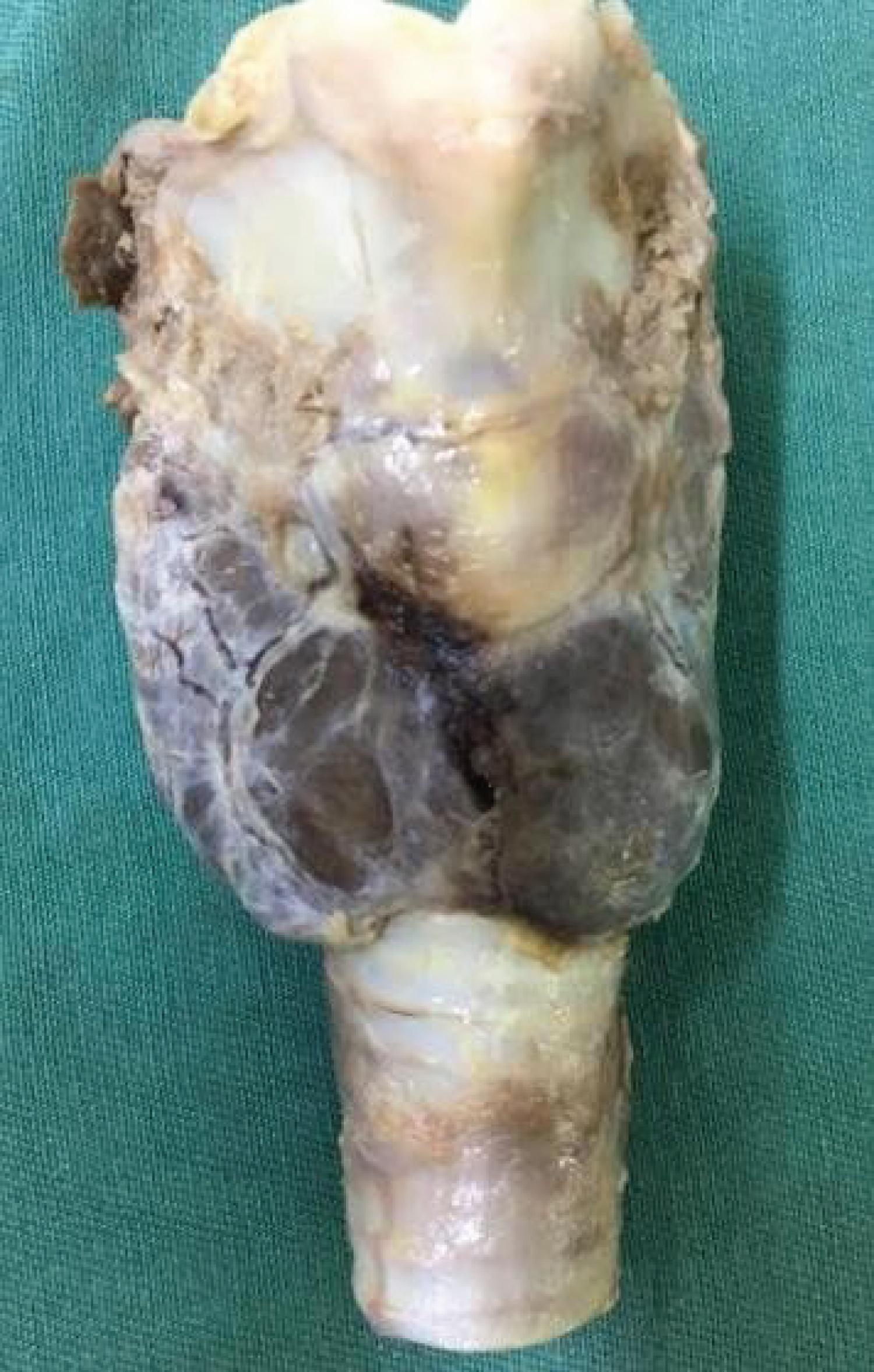 Figure 1: Laringotracheal complex.
View Figure 1
Figure 1: Laringotracheal complex.
View Figure 1
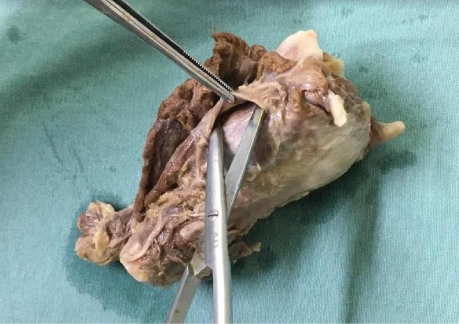 Figure 2: Dissection of the thyroid.
View Figure 2
Figure 2: Dissection of the thyroid.
View Figure 2
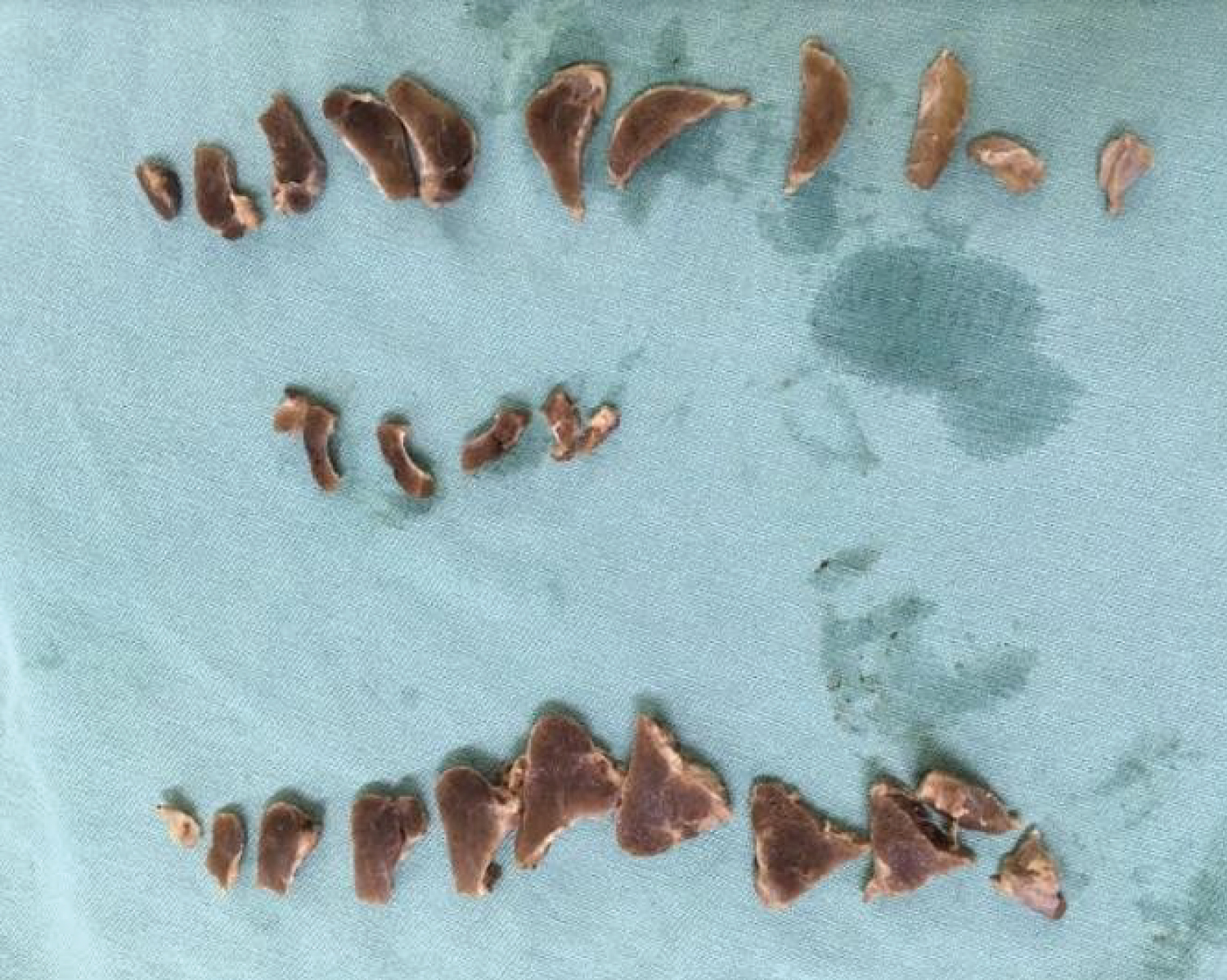 Figure 3: Cross-section of the gland.
View Figure 3
Figure 3: Cross-section of the gland.
View Figure 3
The research began after the approval of the Ethics and Research Committee of the Guilherme Álvaro Hospital (June 25, 2017; letter #16).
Thyroid samples were collected from 100 corpses: 42 females and 58 males. Of these thyroids, 39 (39%) presented nodules. The average age of the studied population was 63.27 years (range 19-100), and the average age of those with thyroid nodules was 68.62 years (range 27-100). Of the 58 thyroids extracted from the males, 20 (34.48%) had thyroid nodules; of the 42 thyroids from the females, 19 (45.24%) had this finding. Of the 39 thyroids with nodule findings, 30 presented a single lesion, 6 had two nodules, and 3 were multinodular.
Regarding the microscopic analysis, 12.82% of the nodules (5 of 39) were histologically diagnosed with papillary carcinoma, which means that 5% of the general population studied had undiagnosed papillary carcinoma when alive. Of these, 80% were of the classical variant and 20% of the follicular variant. The finding of papillary carcinoma was more common in the females (60%) than in the males (40%). The average age of the population with a histological diagnosis of papillary carcinoma was 61.6 years (range 49-78).
Regarding the macroscopic characteristics, nodules diagnosed histologically as papillary carcinoma had an average size of 1.0 cm; 40% had poorly delimited edges; 80% had a whitish appearance; and 40% had calcifications. These data contrast with the characteristics of nodules without carcinoma (mean size 0.67 cm; 5.88% were poorly delimited, 17.65% were whitish, and 8.82% calcified), as shown in Table 1.
Table 1: Macroscopic characteristics of the nodules. View Table 1
Several important papers regarding this subject provide new information very similar to this current report; however, a small discrepancy was noted. The main similarities and differences are detailed below.
According to the systematic review, "Epidemiology of thyroid nodules," [1] thyroid nodules were found in 50% of the autopsies of patients without a prior diagnosis of thyroid disease, and they were more frequent in women and the elderly. Of these, 4% were carcinomas. These findings correlate with the findings in our report.
The study, "Occult Thyroid Carcinoma: A study of 100 necropsies at the University Hospital Antonio Patricio de Alcalá," [9] found a prevalence of 35% of thyroid glands with nodules. Of these, 11.43% (4) presented thyroid carcinoma. These findings also match with our report.
In contrast, the cross-sectional study, "Gross and microscopic findings in clinically normal thyroid glands," [10] found a prevalence of 49.5% of thyroid glands with nodules in 1,000 dissections; again these were more frequent in women and the elderly. Also, among all the thyroid glands, 12.2% presented a single nodule and 37.3% were multinodular. These data do not match with our report, which showed that 30% had single nodules and 9% had multiple nodules (i.e. 39% of the thyroid glands were found with nodules).
Also, in the paper "Findings of thyroid nodules in autopsies in Western Mexico," [11] 300 autopsies were performed. Of these, 123 (41% of the population) presented thyroid nodules and, from these, 12 (4%) were malignant nodules. The nodules were more frequent in males (56%) than in females (44%)-a statistic that differs from most of worldwide literature, including the findings of our study.
There is a high prevalence of thyroid nodules in the studied population, but a few number includes in this report died from other causes, without a diagnosis of thyroid cancer, which allow us to infer that in most of the cases, the finding of the thyroid nodule would result in unnecessary distress to the patients. The introduction of new diagnostic techniques and increased medical surveillance can lead to massive increases in the detection of small and nonlethal papillary lesions, and the vast majority of these patients underwent to total thyroidectomy and radioiodine therapy, being exposed to its complications. These findings suggests that the watchful and waiting approach of these nodules can be considered as an option for patients with the diagnostic of small nodule of thyroid [12].
None.
None.
GMMJ and LSA conceived the experiment(s), GMMJ, LSA, PEOB and LRI conducted the experiment(s), LCB and RLPS analysed the results and statistics. LCB, PEOB and LRI prepared the manuscript. RLPS reviewed the manuscript.