

International Journal of Sports and Exercise Medicine
Eccentric Loading Increases Peak Torque Angle of the Ankle Plantar Flexors in Healthy Volunteers
Matthew Wellisch1*, Peter Hamer1, Luke Hopper2, Max Bulsara1 and James Debenham1
1School of Physiotherapy, University of Notre Dame, Australia
2Western Australian Academy of Performing Arts, Edith Cowan University, Australia
*Corresponding author: Matthew Wellisch, School of Physiotherapy, University of Notre Dame, 19 Mouat Street (PO Box 1225) Fremantle, Western Australia 6959, Australia, Tel: +61 404 887 640, E-mail: matthewwellisch@hotmail.com
Int J Sports Exerc Med, IJSEM-1-009, (Volume 1, Issue 2), Original Article; ISSN: 2469-5718
Received: May 01, 2015 | Accepted: May 21, 2015 | Published: May 24, 2015
Citation: Wellisch M, Hamer P, Hopper L, Bulsara M, Debenham J (2015) Eccentric Loading Increases Peak Torque Angle of the Ankle Plantar Flexors in Healthy Volunteers. Int J Sports Exerc Med 1:009. 10.23937/2469-5718/1510009
Copyright: © 2015 Wellisch M, et al. This is an open-access article distributed under the terms of the Creative Commons Attribution License, which permits unrestricted use, distribution, and reproduction in any medium, provided the original author and source are credited.
Abstract
Eccentric loading of the ankle plantar Flexor's (PF) has demonstrated clinical efficacy in the conservative treatment of Achilles tendinopathy, however, its mechanism of therapeutic benefit remains unclear. The purpose of this study was to examine the effects of PF eccentric loading on PF angle to peak torque (AtPT), peak torque (PT) and lower limb vertical stiffness. Thirty healthy volunteers were randomised to an eccentric (n=15) or concentric (n=13) exercise group. A 10-week loading programme of the ankle plantar flexors was completed. AtPT, PT and vertical stiffness were compared within and between groups before and after the interventions. AtPT increased in the eccentric group by 3.2° dorsiflexion (p=0.001) and decreased by 0.7° dorsiflexion (p=0.528) for the concentric group with significant post-intervention group differences (p< 0.001). PT levels were unchanged following the interventions for both groups (p>0.2); however, post-intervention the eccentric group showed a greater PT than the concentric group (p>0.05). Between group comparison showed no significant difference in vertical stiffness (p>0.5). However, the concentric group demonstrated a vertical stiffness increase of 765kNm-1 (p ≥ 0.05). This study demonstrates that a clinically-derived eccentric loading programme can produce an adaptive shift in AtPT of the ankle plantar flexors in a healthy population. These results support the theory that in part, eccentric loading derives its therapeutic benefit from mechanisms that influence plantar flexor motor performance.
Keywords
Achilles tendinopathy, Eccentric exercise, Plantar flexors, Muscle performance
Introduction
Achilles tendinopathy (AT) is a common overuse condition characterised by impaired physical function due to pain [1]. The incidence of AT in registered patients presenting to general practitioners is 1.85 per 1000, with average presentation age of forty-three [2]. Although the natural history of AT is unclear, early conservative treatment is thought to be optimal, with eccentric exercise demonstrated to be one of the more efficacious conservative treatments currently available [3-5]. Furthermore, eccentric loading protocols offer superior outcomes to concentric protocols in terms of pain and function [6-8] and the eccentric loading protocol outlined by Alfredson et al. [9] is widely used in the literature and clinically. Whilst the clinical efficacy of eccentric loading as an intervention for AT has been established the underpinning mechanisms behind its benefit are unclear. A number of theories have been proposed to explain the mechanism of efficacy for eccentric loading; one theory suggests mechanisms influencing pathology [10,11], whilst another suggests mechanisms influencing tendon structure [12-14]. However, it appears that these explanations alone are insufficient to account for the positive outcomes observed following eccentric loading, leaving a third biomechanical theory as an alternative explanation. This theory suggests mechanisms that positively influence motor performance [15].
Eccentric loading has been shown to improve many measures of motor performance, including peak torque (PT) [8,9], angle to peak torque (AtPT) [16,17] and vertical stiffness [18,19]. Interestingly, motor performance deficits in PT and vertical stiffness have been observed in individuals with AT [9,20,21]. Even though the mechanism of efficacy is not yet clearly understood, it is known that eccentric contractions serve as a mechanical stimulus that results in muscle fibre damage and a subsequent adaptive shift in the muscles length-tension relation to longer muscle lengths [22].
Whilst, eccentric loading has been demonstrated to increase PT, AtPT and vertical stiffness in other regions, this has not been demonstrated at the ankle; furthermore, it has not been demonstrated whether these changes are a unique feature of eccentric loading over concentric loading. Therefore the aim of this study was to investigate alterations in motor performance of the plantar flexors to a therapeutic eccentric loading protocol as compared to an equivalent concentric loading protocol. The logical foundation of this biomechanical investigation is to collect data from a healthy population to provide a baseline for the measures of interest. We hypothesised that eccentric loading would result in an increase in PT, AtPT and vertical stiffness of the ankle plantar flexors compared to a comparable concentric exercise. Given the relevance of eccentric loading in the management of AT, a better understanding of the mechanisms underpinning its efficacy should be sought. With a greater understanding of this, prescriptions of this therapeutic intervention can be progressed.
Methods
Participants
Thirty healthy participants were recruited from a local university community in Fremantle, Western Australia. Participants were excluded if they were pregnant, had received treatment for another lower quadrant disorder in the past 12 months, had a medical condition precluding physical activity, a history of AT or undergone any Achilles surgery. Participants were randomly allocated by a blinded independent research assistant to either the eccentric or concentric group using a random number generator in Microsoft Excel (Redmond, WA). Random allocation was performed prior to testing of the first participant and the results stored in sealed opaque envelopes. Two researchers were responsible for the collection of data. The first researcher was blinded to group allocation and was responsible for administering isokinetic dynamometer and force plate testing protocols pre and post-exercise. The second researcher was responsible for exercise protocol familiarisation of participants from both groups. Participants were blinded to the study's hypotheses. Participants were tested immediately prior to commencing their exercise protocol and post-exercise testing occurred within 48 hours of their final session. All testing and exercise protocols were completed on the left leg only.
Measures
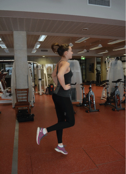
Vertical stiffness: A calibrated force platform (BP600900, AMTI, Watertown, USA) sampling at 200Hz and 'hardware zeroed' between trials to eliminate offset by thermal drift was used to capture the data to be used for vertical stiffness calculations. Participants performed barefoot sub-maximal single-limb hopping [23] at 2Hz according to the reliable and validated methods described by Joseph et al. [24] and Dalleau et al. [25]. Following 2 familiarisation trails interspersed by 60 s rests, participants hopped ten times on the force plate in time with a digital metronome (http://www.metronomeonline.com) (Figure 1).
.
Figure 1: Participant performing the hopping task on the force plate, hands on hips.
View Figure 1
Force plate data was filtered using a 2nd order low pass Butterworth filter with cut-off frequency of 50Hz during post-processing using custom-written software (LabVIEW, National Instruments, Version 8.2.1, U.S.). According to Dalleau et al. [25] vertical stiffness was calculated as:
Where M is the total body mass, Tc is the ground contact time and Tf is flight time. The first and last hops of the 10-hop trials were excluded from analysis [24].
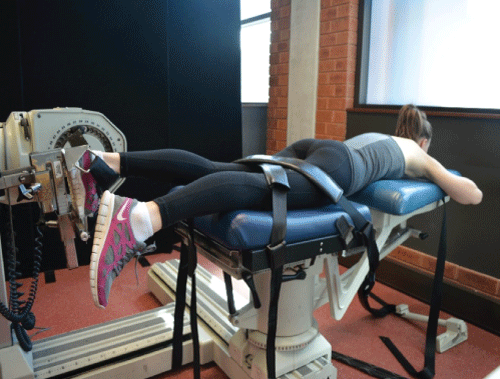
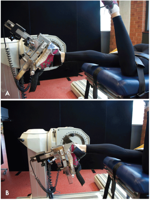
Isokinetic dynamometry: Eccentric plantar flexor PT and AtPT was measured using a regularly calibrated isokinetic dynamometer (KinCom; Chattanooga Group, Inc., Hixson, TN) with established reliability [26-28]. Eccentric contraction measures were recorded due to their importance in stretch-shortening cycle (SSC) function [29,30]. Participants were shod and tested in prone, with their lower limbs secured with straps stabilising the pelvis and thigh and their hands crossed, positioned under their forehead [28] (Figure 2). Each participant's foot and ankle were firmly attached to the footplate with straps (Figure 3). The knee angle was set at 10� to replicate a submaximal hopping position. The mechanical axis of the dynamometer was aligned with the midpoint of the lateral malleolus as the best-fit axis of the talocrural joint [27] and a contraction speed of 90�s-1 was used [9,31]. During testing, participants were instructed on a maximal contraction through the full range of motion and given consistent verbal encouragement throughout. Test range was set according to their measured anatomical range of motion, with limits set at end range plantar flexion minus 5� and end range dorsiflexion. Correction for gravity and limb weight was used and starting force was set at 90N. All KinCom parameters were recorded to ensure exact replication of positioning and setup during post-exercise testing. Following at least two familiarisation trials, five test contractions were recorded for analysis, with a 60s rest between trials. Individual contractions were discarded if a clearly disenable singular peak was not visible in the KinCom's graphical output of PT against angle in range.
.
Figure 2: Dynamometer test position in prone, hands on rest, thigh and pelvis secured, knee in 10 degrees of flexion.
View Figure 2
.
Figure 3: Dynamometer start position (A) and finish position (B). Note, right leg was fully extended during testing.
View Figure 3
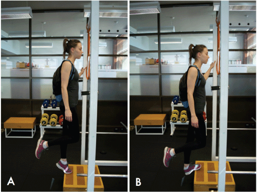
Eccentric and concentric loading: The eccentric loading protocol used for this study reflected the protocol outlined by Alfredson et al. [9]. An equivalent concentric loading protocol was outlined for the control group. Each group performed eccentric or concentric contractions on the test limb only, with participants instructed to perform the exercise on the left leg only, twice daily for ten weeks. Three sets of fifteen repetitions were performed in each session with 60 second rest between sets. Exercises were performed on the edge of a step, with the eccentric group starting heel raised with all body weight on the forefoot, before slowly lowering (Figure 4). The right leg was used to return the heel above the forefoot to the starting position. In contrast, the concentric group were required to raise their left heel above the forefoot from a heel lowered starting position with right leg being used to return the heel from the raised finish back to the starting position. Participants were instructed to maintain an upright body and straight leg whilst exercising. Each week, a weight of 1kg was progressively added to a backpack worn by the participants whilst exercising. Participants were given an exercise diary to record each exercise session and followed up regularly to encourage compliance. Participants continued loading until the day before post-exercise testing which occurred within ±5 days of their 10-week exercise finish date.
.
Figure 4: Eccentric exercise start position (A) and finish position (B). Start and finish position are reversed for the concentric exercise.
View Figure 4
IPAQ: The International Physical Activity Questionnaire (IPAQ) short form [32,33], which has established reliability and validity for measuring habitual physical activity, was administered during post-exercise testing.
Data analysis: Statistical Package for the Social Sciences (SPSS), Version 22.0 was used to conduct all statistical analysis with confidence intervals set at 95% and statistical significance level of p less than .05. A linear mixed model was used to analyse the data including the independent variable exercise group (concentric or eccentric), dependent variables PT, AtPT and vertical stiffness and covariates of age, gender, weight, exercise compliance and activity level. Factor and covariate analysis was performed using a fixed main effects model with type III sum of squares to obtain parameter estimates. Pairwise comparisons for difference in means utilising a Bonferroni adjustment for repeated measures were made for each dependent variable. Comparisons included an unadjusted within group analysis to examine difference in means for each dependent variable and comparisons within-group adjusted for age, gender, weight, exercise compliance and activity level. The covariates adjusted for in the model may all influence the dependent variable, therefore, to truly account for any intervention effect these influences were accounted for in the model. In addition, between groups analysis post-exercise was conducted to examine for differences in means. Between groups analysis was adjusted for the same covariates as within group but additionally controlled for pre-exercise measure values. Significant results were reported as the mean ± standard error.
Sample size was estimated using data from a previous study in which a similar hopping exercise was performed by 96 participants [25]. Prior data indicates that the difference in the change in vertical stiffness of matched pairs is normally distributed with a standard deviation of 9. If the true difference in the mean response of matched pairs is 7, we need 15 pairs of subjects to be able to reject the null hypothesis that this response difference is zero with probability (power) of 80 percent. The Type I error probability associated with this test of this null hypothesis is 5 percent. In addition, for angle to peak torque sample size was estimated using the data from a previous study in which a plantar flexor eccentric exercise was performed by 9 participants [34]. This data indicates if the true difference in the mean response of matched pairs is 1.5�, we will need to study 10 pairs of subjects.
Results
Of the initial 30 participants 2 were lost to follow-up, one participant incurred an ankle injury in their left leg as a result of external influences and the other discontinued citing personal reasons. Following post-exercise testing a concentric group participant was found to be involved in elite level sprint and rowing training during the study, including high intensity eccentric and plyometric training. However, in accordance with intention-to-treat analysis their data was included in the concentric group. As a result, a total of 28 participants completed the study; 15 eccentric and 13 concentric. Age, gender, weight, height, exercise compliance and IPAQ scores for both groups are displayed in Table 1. There was no significant difference in age, height, weight or compliance between groups. Missing values for PT and AtPT of 3 participants (2 eccentric, 1 concentric group) were recorded during post-exercise testing as they exceeded the force limits of the dynamometer and were therefore unable to be measured. Furthermore, two PT and AtPT values are missing for participant 3 in the concentric group as they exceeded the force limits on 2 of their 5 trials during post-exercise testing.
![]()
Table 1: Age, gender, weight, height, exercise compliance group means (standard deviation) and IPAQ details
View Table 1
Peak torque
There was no significant difference in PT from baseline to the post-exercise testing for the concentric exercise group (p=0.814) or the eccentric exercise group (p=0.216) (Table 2). The comparison adjusted for covariates revealed age, gender and activity level for the concentric group and age and activity level for the eccentric group were significant. However, the pre-post exercise analysis for both groups was still not significant. The post-exercise between group comparison revealed PT to be significantly greater in the eccentric group (p=0.043) (Table 3).
![]()
Table 2: Unadjusted exercise group means (standard errors) pre and post exercise
View Table 2
![]()
Table 3: Between group comparisons post-exercise, concentric minus eccentric, adjusted for age, gender, weight, compliance, IPAQ value and pre-exercise values
View Table 3
Angle to peak torque
Analysis revealed a significant increase in AtPT of the ankle plantar flexors to greater dorsiflexion within the eccentric group from pre-exercise 25.59 (± 0.63)� dorsiflexion to post exercise 28.69 (± 0.68)� dorsiflexion (p=0.001) (Table 2). Adjusting for covariates revealed no significance in age, gender, weight, exercise compliance or activity level. Furthermore, AtPT in the eccentric group was significantly greater than that of the concentric group (p=0.001) (Table 3). Concentric exercise did not have a significant effect on the AtPT of the plantar flexors (p=0.498).
Vertical stiffness
There were no significant changes in vertical stiffness from baseline to post-exercise for either the concentric group (p=0.187) or the eccentric group (p=0.511). Adjusting for covariates revealed a significant increase in the concentric group (p=0.005) with age, weight and activity level demonstrating significance. However, no significant difference in vertical stiffness was found between the concentric and eccentric groups post-exercise (p=0.468) (Table 3).
Discussion
The purpose of this study was to examine temporal changes in PT, AtPT and vertical stiffness in response to either a traditional eccentric loading protocol or a comparable concentric loading protocol control group. We found a significant shift in AtPT of 3.22 (± 0.93)� following the eccentric loading protocol (Table 2) and that AtPT of the eccentric group was significantly greater than the concentric group post-exercise (p=0.001) (Table 3). Additionally, PT was found to be significantly greater in the eccentric group post-exercise (p=0.043) (Table 3). Whilst we observed no difference in vertical stiffness between groups (p=0.468) (Table 3), a significant increase of 765kNm-� in vertical stiffness within the concentric group was observed (p=0.005).
In support of our hypothesis, the primary finding of this study demonstrates an actual change in the motor performance measure of AtPT at the ankle plantar flexors following eccentric loading in a healthy population. As a result, this adds to the plausibility that the mechanism of efficacy underpinning eccentric loading is related to its influence on this aspect of motor performance. The alteration in AtPT observed in this study is most likely due to sarcomerogenesis: a protective adaption of an increase of sarcomeres in series in response to eccentric induced muscle fibre damage resulting in a shift in AtPT towards longer muscle lengths [16,22]. The adaptive shift in AtPT towards longer muscle lengths is believed to be protective because at longer muscle lengths sarcomeres are less likely to be stretched onto the vulnerable descending limb of their length-tension relation [22]. The results of this study demonstrate an improved ability in the ankle plantar flexor musculotendinous units of participants to attenuate load at longer muscle lengths. We suggest therefore that patients with AT undergoing similar eccentric loading interventions are likely to experience the same phenomenon. This suggests the therapeutic benefit of eccentric loading may be derived from an increase in AtPT resulting in an improved ability of the plantarflexor musculotendinous unit to attenuate load at vulnerable longer muscles lengths. Further, biological plausibility exists that a 3.2� shift is clinically significant. Brughelli et al. [17] demonstrated an adaptive shift in AtPT of 6.5� in the quadriceps and 4.0� in the hamstrings of professional soccer players following an eccentric loading protocol. Given the differences in functional anatomy between quadriceps, hamstrings and plantar flexors, primarily excursion, it seems reasonable that the shift demonstrated in this study is less and to this extent.
This study builds on the findings of Jones et al. [34], who were the first to demonstrated an acute shift in AtPT at the ankle plantar flexors following a single bout of eccentric loading. Their study of 9 healthy individuals walking backwards on a treadmill measured an acute change of 3.9 (± 1.5)� that resolved back to baseline within 2 days. The findings of this study suggest in a healthy population following an initial disruption of sarcomeres as demonstrated by Jones et al. [34], an adaptive shift is evident in the ankle plantarflexors following a prolonged eccentric loading protocol.
Our findings support earlier studies demonstrating an adaptive shift in AtPT in human muscle following eccentric exercise. Brockett et al. [16], in their study of healthy volunteers were the first to demonstrate this, observing an adaptive shift in AtPT in the hamstrings at 10 days after a single bout of eccentric exercise. Furthermore, 6 participants were given a repeat dose of the exercise 8 days after the first. In response to the second bout of exercise, there was much less damage and soreness compared with the changes after the first dose. They suggest that this mechanism represents a protective adaptation reflective of sarcomerogenesis. Our findings are also consistent with those of Brughelli et al. [17] who studied professional male soccer players performing 4 weeks of eccentric exercises. They observed a significant increase in AtPT during knee flexion and extension in comparison to a concentric group.
Importantly, the results support those of Crill et al. [15], who were the first authors to study mechanical changes of the ankle plantarflexors following eccentric exercise in AT patients. Their study comprised 25 AT patient's undergoing 8 weeks of eccentric loading according to the Alfredson et al. [9] protocol. They demonstrated an increase in medial gastrocnemius fascicle length which they proposed is likely due to sarcomerogenesis, suggesting eccentric loading is an effective treatment of AT due to this biomechanical alteration.
Supporting our hypothesis, this study revealed significantly greater PT in the eccentric group compared to the concentric group following the exercise period. However, no significant changes were observed within group suggesting the between group difference is unlikely to be of clinical significance. It is well accepted strength gains resulting from resistance training require approximately 2-6 sets of ≤6 repetitions at ≥85% of 1 repetition maximum [35]. Given a young, healthy, active population the exercise prescriptions in this study likely did not meet these requirements for most participants and consequently an increase in PT within each group was not observed.
To date, research into the effects of eccentric loading on PT has focused on acute outcomes. Initially, eccentric loading results in an immediate reduction in PT, resolving towards baseline within a week [16,36]. However, the within group findings of this study are consistent with those of Brughelli et al. [17] who observed no significant changes in PT of quadriceps or hamstrings after a 4-week eccentric training period. Alfredson et al. [9] in their study of 15 recreational athletes with AT, observed a significantly lower PT in the injured leg compared to the uninjured leg and also demonstrated a significant increase in PT following a 12 week eccentric loading protocol. These findings, combined with the results of this study suggest that once pathology is introduced the system no longer operates at an optimal level in regards to strength.
Contrary to our hypothesis we observed no significant change in vertical stiffness following the eccentric loading protocol and whilst the increase in stiffness with concentric loading was significant, its magnitude is unlikely to be clinically meaningful. This is inconsistent with existing literature, e.g. Elmer et al. [19]. The most likely explanation for stiffness not changing in response to eccentric loading is that whilst the stiffness contribution of the ankle plantar flexors to lower limb stiffness is important [37], it is only one component of the motor system responsible for producing lower limb stiffness with other significant contributions from the knee and hip extensors. Recognising that lower limb stiffness is derived from multiple regions [38], it is possible that the mechanical changes induced at the ankle are insufficient to produce changes at a regional level. Elmer et al. [19] found a significant increase in vertical stiffness in healthy volunteers following a 7 week (3 sessions per week) progressive eccentric cycling protocol. The eccentric cycling protocol outlined by these authors was designed to affect the knee extensors (quadriceps), as opposed to our protocol aimed at the ankle plantar flexors.
There are several limitations to our study including the use of a healthy population. Although it is useful to initially investigate a healthy population to gather baseline measures; there is limited scope to draw comparisons with a pathological population. Additionally, our average participant age for the eccentric group was 23 years which according to the study by de Jonge et al. [2] is not representative of an AT population with average age of 43 years. The dynamometer we employed had a force limit of 2000N, meaning that individuals could not participate in the study if they exceeded this force limit on pre-exercise testing. Additionally, missing values were recorded for PT and AtPT of 4 participants during post-exercise testing. Only eccentric PT and AtPT of participants and not concentric was measured, however, changes in the eccentric contraction are of primary interest due to their importance in optimal SSC function [30]. Average exercise compliance level of 73% for the eccentric group and 68% for the concentric group and differences of ± 5 days in number of days exercised due to post-exercise scheduling; although this does not provide a true measure of full adherence to the 10 week protocol as outlined by Alfredson et al. [9], it does reflect exercise levels observed in clinical practice, providing additional external validity. Participants were responsible for the weekly addition of approximately 1kg to their backpacks. Therefore load was not strictly standardised to any individual anthropometric or performance (e.g. body mass or PT) metric. However, we believe this to be acceptable, reflective of normal clinical practice where small variations in load prescription are common [9]. Finally, participants were tested within 48 hours of their last exercise session. Although this is within the demonstrated recovery time of acute alterations in AtPT due to muscle damage, the prolonged timeframe of the exercise protocol suggests an adaptive shift is being witnessed.
This study is important as it provides further evidence supporting the mechanical theory underpinning the mechanism by which eccentric loading imparts its positive clinical effect, and this may assist in further research informing prescriptions of such exercises. It has been well established that eccentric loading is a mainstay in the treatment and prevention of hamstring injuries [39]. This understanding has been in part driven by the earlier work of Brockett et al. [16] who observed a shift in AtPT in the hamstrings following eccentric loading. This comparable study to our own provides further support that eccentric loading is a beneficial clinical tool to be used in the treatment of ankle plantarflexor injuries and that alteration in AtPT may provide a satisfactory mechanistic explanation.
This study raises two areas of interesting speculation, firstly, regarding pathogenesis. The primary pathogenetic mechanism in AT is thought to be excessive loading in conjunction with a combination of intrinsic contributing risk factors [40,41] and extrinsic [41]. This study also adds plausibility to the theory that AT is in part driven by altered motor performance, resulting in a subsequent inability to attenuate load within its physiological limits, leading to fatigue loading, failed healing and the development of tendinopathy.
Interestingly, the significant change in AtPT at an average compliance level of 73% for the eccentric group in this study raises speculative questions regarding dosage levels. Additionally, it has been demonstrated that a lower dosage of eccentric loading may provide similar clinical outcomes for subjects with AT [42].
Conclusion
This is the first randomised controlled study to demonstrate an adaptive shift in AtPT at the ankle plantar flexors following a prolonged eccentric loading protocol. The results of this study provide further evidence that eccentric loading may derive its therapeutic benefit from mechanisms influencing motor performance.
Ethics Approval
All procedures of the study have been approved by the Human Research Ethics Committee of the University of Notre Dame Australia (013153F), in conformity with the National Statement on Ethical Conduct in Human Research (2007). All participants provided written, informed consent before proceeding with testing.
Acknowledgement
There was no external funding for this study.
References
-
Sorosky B, Press J, Plastaras C, Rittenberg J (2004) The practical management of Achilles tendinopathy. Clin J Sport Med 14: 40-44.
-
de Jonge S, van den Berg C, de Vos RJ, van der Heide HJ, Weir A, et al. (2011) Incidence of midportion Achilles tendinopathy in the general population. Br J Sports Med 45: 1026-1028.
-
Magnussen RA, Dunn WR, Thomson AB (2009) Nonoperative treatment of midportion Achilles tendinopathy: a systematic review. Clin J Sport Med 19: 54-64.
-
Sussmilch-Leitch SP, Collins NJ, Bialocerkowski AE, Warden SJ, Crossley KM (2012) Physical therapies for Achilles tendinopathy: systematic review and meta-analysis. J Foot Ankle Res 5: 15.
-
Woitzik E, Jacob C, Wong JC, C�t� P, Shearer HM, et al. (2015) The effectiveness of exercise on recovery and clinical outcomes of soft tissue injuries of the leg, ankle, and foot: A systematic review by the Ontario Protocol for Traffic Injury Management (OPTIMa) Collaboration. Manual therapy.
-
Mafi N, Lorentzon R, Alfredson H (2001) Superior short-term results with eccentric calf muscle training compared to concentric training in a randomized prospective multicenter study on patients with chronic Achilles tendinosis. Knee Surg Sports Traumatol Arthrosc 9: 42-47.
-
Silbernagel KG, Thome� R, Thome� P, Karlsson J (2001) Eccentric overload training for patients with chronic Achilles tendon pain--a randomised controlled study with reliability testing of the evaluation methods. Scand J Med Sci Sports 11: 197-206.
-
Yu J, Park D, Lee G (2013) Effect of eccentric strengthening on pain, muscle strength, endurance, and functional fitness factors in male patients with achilles tendinopathy. Am J Phys Med Rehabil 92: 68-76.
-
Alfredson H, Pietil� T, Jonsson P, Lorentzon R (1998) Heavy-load eccentric calf muscle training for the treatment of chronic Achilles tendinosis. Am J Sports Med 26: 360-366.
-
Knobloch K, Kraemer R, Jagodzinski M, Zeichen J, Meller R, et al. (2007) Eccentric training decreases paratendon capillary blood flow and preserves paratendon oxygen saturation in chronic Achilles tendinopathy. J Orthop Sports Phys Ther 37: 269-276.
-
Ram R, Meeuwisse W, Patel C, Wiseman DA, Wiley JP (2013) The limited effectiveness of a home-based eccentric training for treatment of Achilles Tendinopathy. Clin Invest Med 36: 197-206.
-
Fyfe I, Stanish WD (1992) The use of eccentric training and stretching in the treatment and prevention of tendon injuries. Clin Sports Med 11: 601-624.
-
Rees JD, Lichtwark GA, Wolman RL, Wilson AM (2008) The mechanism for efficacy of eccentric loading in Achilles tendon injury; an in vivo study in humans. Rheumatology (Oxford) 47: 1493-1497.
-
Grigg NL, Wearing SC, Smeathers JE (2012) Achilles tendinopathy has an aberrant strain response to eccentric exercise. Med Sci Sports Exerc 44: 12-17.
-
Crill MT, Berlet G, Hyer C (2014) Plantar flexor muscle architecture changes as a result of eccentric exercise in patients with achilles tendinosis. Foot Ankle Spec 7: 460-465.
-
Brockett CL, Morgan DL, Proske U (2001) Human hamstring muscles adapt to eccentric exercise by changing optimum length. Med Sci Sports Exerc 33: 783-790.
-
Brughelli M, Mendiguchia J, Nosaka K, Idoate F, Arcos AL, et al. (2010) Effects of eccentric exercise on optimum length of the knee flexors and extensors during the preseason in professional soccer players. Phys Ther Sport 11: 50-55.
-
Pousson M, Van Hoecke J, Goubel F (1990) Changes in elastic characteristics of human muscle induced by eccentric exercise. J Biomech 23: 343-348.
-
Elmer S, Hahn S, McAllister P, Leong C, Martin J (2012) Improvements in multi-joint leg function following chronic eccentric exercise. Scand J Med Sci Sports 22: 653-661.
-
Maquirriain J1 (2012) Leg stiffness changes in athletes with Achilles tendinopathy. Int J Sports Med 33: 567-571.
-
Debenham JR, Travers MJ, Gibson W, Campbell A, Allison GT (2014) Achilles tendinopathy alters stretch shortening cycle behaviour during a sub-maximal hopping task. J Sci Med Sport
-
Proske U, Allen TJ (2005) Damage to skeletal muscle from eccentric exercise. Exerc Sport Sci Rev 33: 98-104.
-
Farley CT, Blickhan R, Saito J, Taylor CR (1991) Hopping frequency in humans: a test of how springs set stride frequency in bouncing gaits. J Appl Physiol (1985) 71: 2127-2132.
-
Joseph CW, Bradshaw EJ, Kemp J, Clark RA (2013) The interday reliability of ankle, knee, leg, and vertical musculoskeletal stiffness during hopping and overground running. J J Appl Biomech 29: 386-394.
-
Dalleau G, Belli A, Viale F, Lacour JR, Bourdin M (2004) A simple method for field measurements of leg stiffness in hopping. Int J Sports Med 25: 170-176.
-
Karnofel H, Wilkinson K, Lentell G (1989) Reliability of isokinetic muscle testing at the ankle. J Orthop Sports Phys Ther 11: 150-154.
-
Chester R, Costa ML, Shepstone L, Donell ST (2003) Reliability of isokinetic dynamometry in assessing plantarflexion torque following Achilles tendon rupture. Foot Ankle Int 24: 909-915.
-
M�ller M, Lind K, Styf J, Karlsson J (2005) The reliability of isokinetic testing of the ankle joint and a heel-raise test for endurance. Knee Surg Sports Traumatol Arthrosc 13: 60-71.
-
Komi PV (2000) Stretch-shortening cycle: a powerful model to study normal and fatigued muscle. J Biomech 33: 1197-1206.
-
Cormie P, McGuigan MR, Newton RU (2010) Changes in the eccentric phase contribute to improved stretch-shorten cycle performance after training. Med Sci Sports Exerc 42: 1731-1744.
-
Sapega AA (1990) Muscle performance evaluation in orthopaedic practice. J Bone Joint Surg Am 72: 1562-1574.
-
Craig CL, Marshall AL, Sj�str�m M, Bauman AE, Booth ML, et al. (2003) International physical activity questionnaire: 12-country reliability and validity. Med Sci Sports Exerc 35: 1381-1395.
-
Kim Y, Park I, Kang M (2013) Convergent validity of the international physical activity questionnaire (IPAQ): meta-analysis. Public Health Nutr 16: 440-452.
-
Jones C, Allen T, Talbot J, Morgan DL, Proske U (1997) Changes in the mechanical properties of human and amphibian muscle after eccentric exercise. Eur J Appl Physiol Occup Physiol 76: 21-31.
-
Rhea MR, Alvar BA, Burkett LN, Ball SD (2003) A meta-analysis to determine the dose response for strength development. Med Sci Sports Exerc 35: 456-464.
-
Nosaka K, Newton MJ, Sacco P (2005) Attenuation of protective effect against eccentric exercise-induced muscle damage. Can J Appl Physiol 30: 529-542.
-
Farley CT, Morgenroth DC (1999) Leg stiffness primarily depends on ankle stiffness during human hopping. J Biomech 32: 267-273.
-
Hobara H, Inoue K, Omuro K, Muraoka T, Kanosue K (2011) Determinant of leg stiffness during hopping is frequency-dependent. Eur J Appl Physiol 111: 2195-2201.
-
Thorborg K (2012) Why hamstring eccentrics are hamstring essentials. Br J Sports Med 46: 463-465.
-
Maffulli N, Kader D (2002) Tendinopathy of tendo achillis. J Bone Joint Surg Br 84: 1-8.
-
Maffulli N, Sharma P, Luscombe KL (2004) Achilles tendinopathy: aetiology and management. J R Soc Med 97: 472-476.
-
Stevens M, Tan CW (2014) Effectiveness of the Alfredson�s Protocol 2 Compared with a Lower Repetition Volume Protocol for Mid-portion Achilles Tendinopathy: a Randomized Controlled Trial. J Orthop Sports Phys Ther 44: 59-67.
