A 57-year-old male presented with a four week history of shortness of breath on exertion, fatigue and melaena with no underlying medical or family history. Recent blood tests with his primary care physician demonstrated an iron deficiency anaemia with a haemoglobin of 7 g/dL and MCV of 68 fl.
Oesophagogastroduodenoscopy (OGD) was performed in the first instance which did not show any evidence of recent bleeding or cause of the melaena. Of note, a CEA level was normal at 1.2 ng/ml. A colonoscopy was performed due to ongoing "dark" rectal bleeding. A friable ulcerated ileocaecal valve (ICV) with puckering of the surrounding mucosa and spontaneous bleeding was found (Figure 1A and Figure 1B). It was not possible to intubate the terminal ileum due to the presence of the lesion. Biopsies were taken and the histology was consistent with a moderately differentiated adenocarcinoma (Figure 1C). A computed tomography scan did not show any evidence of distant metastasis. The patient was referred for a right hemicolectomy and was subsequently staged as PT 3 N2 B (Figure 1D). His haemoglobin recovered postoperatively and he was referred for adjuvant chemotherapy.
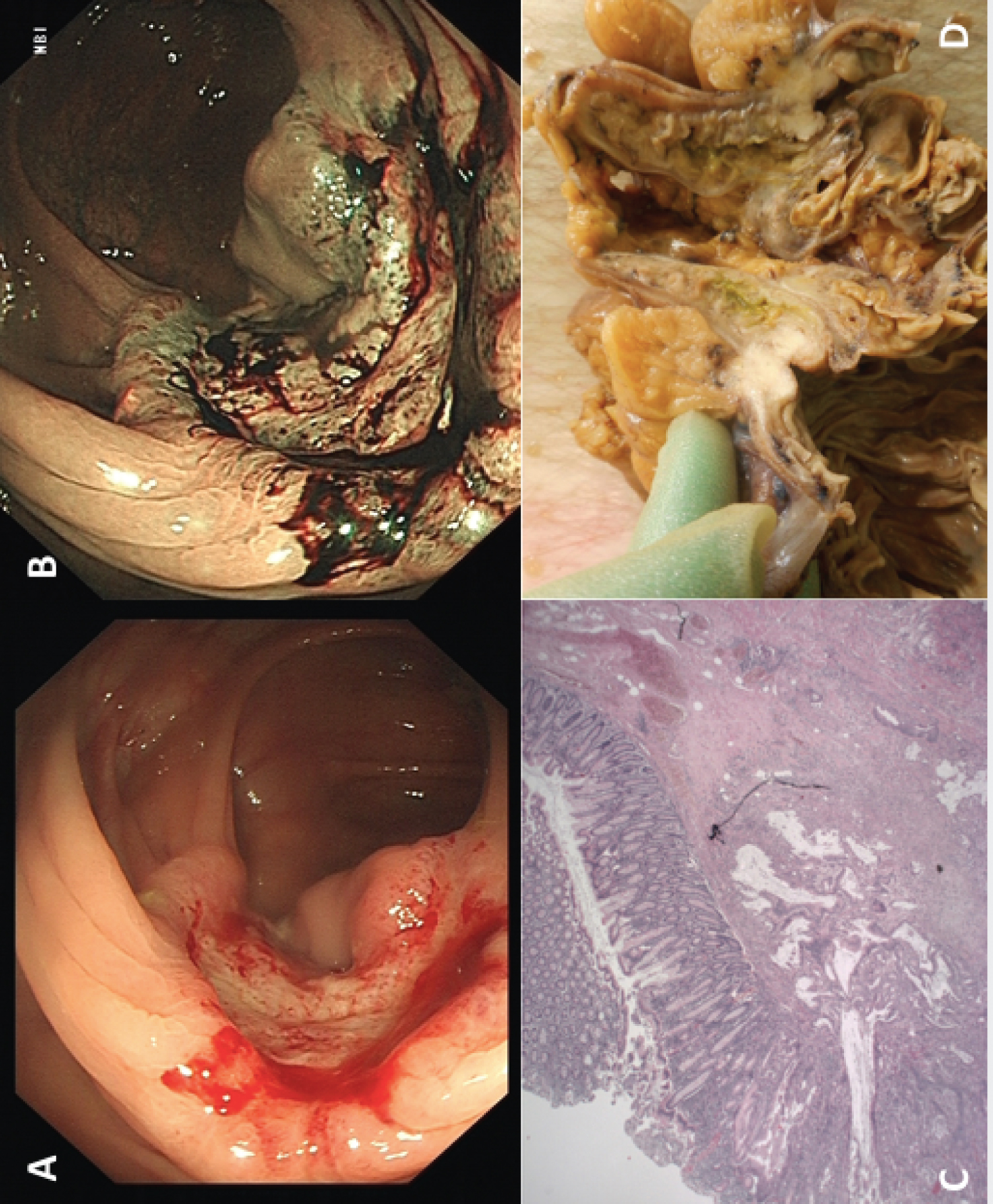 Figure 1: (A) White light image of the ulcerated ileocaecal valve lesion; (B) Narrow band imaging of the ulcerated ileocaecal valve lesion; (C) Resected gross specimen; (D) Histology demonstrating adenocarcinoma.
View Figure 1
Figure 1: (A) White light image of the ulcerated ileocaecal valve lesion; (B) Narrow band imaging of the ulcerated ileocaecal valve lesion; (C) Resected gross specimen; (D) Histology demonstrating adenocarcinoma.
View Figure 1
Adenocarcinoma of the ileocaecal valve is rare and commonly appears as non-specific ICV thickening radiologically [1,2]. Right sided colonic lesions including of the ICV should be considered as a cause of melaena when OGD is normal.
The authors of this image of the month have no conflict of interest to disclose.
No grants or financial supports were obtained.
Neasa Mc Gettigan: Conception, drafting of the article and final approval, Edric Leung-drafting of the article; Subhasish Sengupta: Critical revision of the article for important intellectual content; final approval of the article.