

Journal of Otolaryngology and Rhinology
Calcitonin-Secreting Moderately Differentiated Neuroendocrine Carcinoma of Larynx with Solitary Papillary Thyroid Carcinoma: A Case Report with Immunohistochemical Study
Hanadi Fatani1*, Faraj Alotaiby1 and Saleh F Aldhahri2
1Department of Oral and Maxillofacial Pathology, Collage of Dentistry, Qassim University, Alumblyda, Saudi Arabia
2Department of Otolaryngology, College of Medicine, King Saud University, Riyadh, Saudi Arabia
*Corresponding author:
Dr. Hanadi Fatani, MD, Consultant Anatomical Pathology, Head & Neck Subspecialty, King Fahad Medical City, Pathology and Clinical Laboratory Administration, Anatomical Pathology Department, P.O. Box 59046 Riyadh 11525, Saudi Arabia, Tel: +011-288-9999 Ext. 11540, E-mail: emsaffana@yahoo.com
J Otolaryngol Rhinol, JOR-2-023, (Volume 2, Issue 4), Case Report
Received: May 04, 2016; Accepted: August 10, 2016; Published: August 12, 2016
Citation: Fatani H, Alotaiby F, Aldhahri SF (2016) Calcitonin-Secreting Moderately Differentiated Neuroendocrine Carcinoma of Larynx with Solitary Papillary Thyroid Carcinoma: A Case Report with Immunohistochemical Study. J Otolaryngol Rhinol 2:023.
Copyright: © 2016 Fatani H, et al. This is an open-access article distributed under the terms of the Creative Commons Attribution License, which permits unrestricted use, distribution, and reproduction in any medium, provided the original author and source are credited.
Abstract
Introduction:Primary neuroendocrine tumors (NET) of the larynx with elevated serum calcitonin are rare in occurrence. Till date only one case has reported associated metastatic NET to thyroid.
Case report:A 51-year-old female presented with 2 months' history of sore throat, change in voice, odynophagia, along with neck and thyroid nodule. CT/PET scan showed thickening of epiglottis with enhancement after intravenous contrast. Laser resection of epiglottis was done and histopathology revealed moderately differentiated neuroendocrine carcinoma. A week later patient underwent total thyroidectomy with bilateral modified radical neck dissection. Adjuvant external beam radiotherapy to the larynx and neck was given post-operatively.
Results:Patient had high serum calcitonin level. Thyroid section showed a micro papillary thyroid carcinoma (PTC) which was calcitonin negative. Epiglottic and lymph node sections were positive for calcitonin and negative for TTF-1.
Conclusions:This is the first case of primary calcitonin-secreting laryngeal NET metastasizing to neck lymph nodes, and showing rare association with solitary PTC.
Keywords
Neuroendocrine carcinoma of larynx, Micro papillary thyroid carcinoma, Thyroidectomy, Epiglottis, Serum calcitonin
Introduction
Neuroendocrine tumors (NET) of the larynx, comprising only 0.6% of the laryngeal neoplasms are characterized by the presence of neurosecretory granules and marker hormones, most commonly calcitonin [1,2]. According to World Health Organization (WHO), laryngeal NET are classified into well-differentiated neuroendocrine carcinoma (typical carcinoid), moderately differentiated neuroendocrine carcinoma (MDNC) also known as large cell carcinoma or atypical carcinoid, poorly differentiated neuroendocrine carcinoma (small cell carcinoma), and paraganglioma [3]. MDNC is reportedly the most frequent among all NET of larynx [4]. There is a high propensity for MDNC to microscopically mimic medullary carcinoma of thyroid (MTC), particularly when presenting as metastasis to thyroid. Both of these carcinomas stain positive for calcitonin and carcinoembryonic antigen (CEA) [5]. It has been suggested that calcitonin-secreting NET of larynx represent ectopic thyroid medullary carcinoma due to laryngo tracheal rests of thyroid tissue [6]. Till date several cases of MDNC have been published suggesting elevated/normal calcitonin levels, out of which only one case reported metastasis to thyroid [2].
However the biological and histological variability of MDNC is extensive and it may even be associated with differentiated thyroid cancer including papillary and follicular cancer [7]. In this report we present an atypical carcinoid of the epiglottis whose clinicopathological features were first mistaken as an adenocarcinoma and then as MTC with metastasis to cervical lymph node. However, immunocytochemical studies and histopathology of thyroid proved it to be solitary papillary carcinoma of thyroid. To the best of our knowledge, this is the first case of calcitonin secreting MDNC of larynx presenting along with solitary papillary thyroid carcinoma.
Case Report
Clinical presentation and physical examination
A 51-year-old female presented with 2 months' history of sore throat, change in voice, mild odynophagia, left ear pain, dysphagia accompanied with weight loss. There was no noisy breathing, sense of fullness or pressure in the neck, hemoptysis or dyspnoea at rest or on exertion. There was no history of smoking, alcohol consumption, hypertension, radiation exposure and no family history of thyroid carcinoma or NET of larynx. On clinical examination of the neck, a non-tender, firm, right sided thyroid nodule was present along with a palpable right level II non tender firm cervical lymph node. No other lymphadenopathy was found in the neck. Oral cavity, oropharynx and bilateral ear examinations were normal. Physical examination was also otherwise normal as was routine hematological and biochemical profile.
Imaging (CT scan, PET scan and ultrasound)
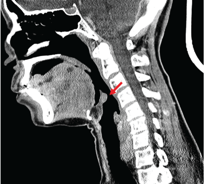
The fiber optic endoscopic examination revealed irregularity in the tip of epiglottis with normal vocal cord mobility, base of tongue and pharynx. There was no mucosal ulceration suggesting lesion to be sub-epithelial. Upon radiographic evaluation, chest X-ray was normal. Computed tomography (CT) revealed significant thickening of the epiglottis with enhancement after intravenous contrast along with right cervical level II enhancing lymph node measuring 1.5 cm and ipsilateral thyroid nodule. A small nodule was also noticed in the left lobe of thyroid gland (Figure 1).
.
Figure 1: Computed tomography (CT) scan report showing sagittal view of the epiglottis.
View Figure 1
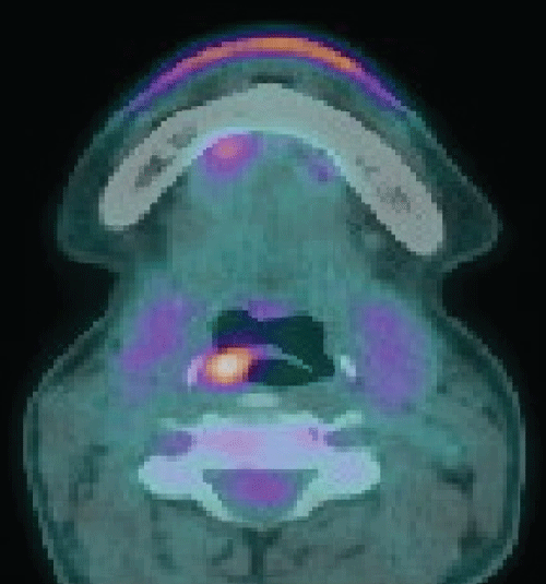
Ultrasound of the thyroid showed enlargement of homolateral nodes which appear slightly hyperechoic with increased internal vascularity suggesting inflammatory process. Further, the lesion was neither cystic nor calcified. PET scan showed the same finding as the CT with mild hypermetabolic right cervical lymph node. No retrosternal extension, lung or bone metastasis was observed (Figure 2).
.
Figure 2: Positron emission tomography-computed tomography (PET-CT) scans image demonstrating axial view of the epiglottis.
View Figure 2
Biopsy and thyroid function test
Biopsy of the epiglottis suggested poorly differentiated mucin-secreting adenocarcinoma. The thyroid function test was normal with serum TG: 0.04 (ref 1.40-78), TSH: 4.140 miu/L (ref 0.270-4.200), T4: 23.7 pmol/L (ref 12-22).
Epiglottic resection, gross and histopathology
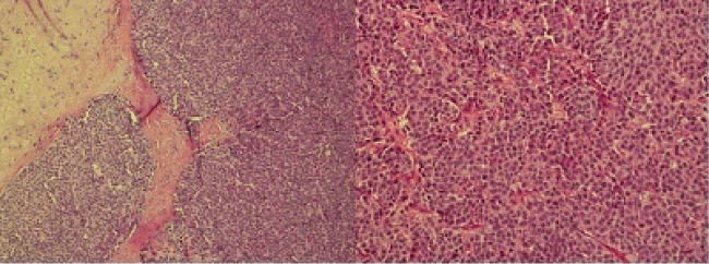
Laser resection of the epiglottis was done. Neck dissection was planned to be done after the final pathology report. Intra-operatively the tumor was confined to right epiglottis with no gross extension to the vallecular or the base of epiglottis, aryepiglottic fold, arytenoid and false vocal cord. Mapped frozen sections for medial, lateral and deep margins also came negative. Grossly the lesion specimen was a single piece of elongated grey tan tissue measuring 2.0 × 1.3 × 0.5 cm with round grey tan mass measuring 1.0 × 0.9 × 0.5 cm. Histopathologic examination exhibited the classical appearances of atypical carcinoids. Organoid pattern, cellular pleomorphism, and punctate necrosis were present (Figure 3). The neoplastic cells were larger and the nuclei were vesicular containing prominent nucleoli. There were no mucinous and amyloid changes. Mitoses numbered approximately five per 10 high power fields. This confirmed the diagnosis of moderately differentiated NET.
.
Figure 3: Photomicrographs of the lesion. A) 10x H & E section shows sheet of neuroendocrine cells with nesting arcitecture; B) 20x section shows epithelioid cells arranged in organoid pattern with "salt and pepper" chromatin.
View Figure 3
Thyroidectomy, neck dissection, gross and histopathology
A week later, the patient underwent total thyroidectomy with bilateral modified radical neck dissection (type III). Intra-operatively only the right level II cervical lymph nodes were noticed to be abnormal, and were removed. Histopathology of lymph node exhibited a moderate degree of pleomorphism, prominent nucleoli, necrosis and readily identifiable mitoses, suggesting it to be atypical carcinoid.
Grossly total thyroidectomy specimen weighed 26 grams, measuring: right lobe - 5 × 3 × 1.5 cm, left lobe - 5 × 2.5 × 1 cm, isthmus - 2 × 1.3 cm with pyramidal lobe 2 × 0.5 cm. The outer surface of each lobe was brown tan, homogenously covered with thin membrane and blood vessels.
Histologically, a complex, branched, randomly oriented papillae with fibrovascular cores associated with follicles were found. Bland-appearing spindle cells exhibiting fine chromatin, indistinct nucleoli and rare mitoses with minimal inflammation were observed, giving a picture of micro papillary thyroid carcinoma. Tumor was multifocal with no lymphovascular and perineural invasion or extra thyroidal extension. Additionally prominent stromal desmoplasia and pseudovascular pattern suggested Hashimoto's thyroiditis.
Immunohistochemistry
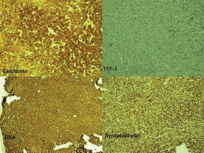
Epiglottis and lymph node sections were positive for CKpan, Synaptophysin, and Chromogranin A, while CD56, CK7, CK20, and P63 were negative. Ki67 was reactive in about 10% of the neoplastic cells. CEA antigen and calcitonin was positive and thyroid transcription factor-1 (TTF-1) was negative in epiglottic and lymph node section, while thyroid lesion was negative for calcitonin (Figure 4).
.
Figure 4: Immunohistochemistry imaging. The tumor shows immunreactivity to calcitonin, carcinoembryonic antigen (CEA) and synaptophysin and negative to thyroid transcription factor (TTF-1).
View Figure 4
Post-operative follow up
Following thyroidectomy and neck dissection, the patient underwent octreotide scan which was negative and the serum calcitonin level was found to be marginally high (12, Ref < 8). Post operatively the patient did not have any complications and was discharged home on thyroxin and pain medications. Patient received adjuvant external beam radiotherapy to the larynx and the neck. Since then the patient has been under clinical surveillance and now, for over 2 years there is no evidence of recurrent disease or high calcitonin level.
Discussion
MDNC, though uncommon, is the second most common primary laryngeal malignancy following only squamous cell carcinoma [3]. Histologically atypical carcinoids are recognized as occasional secretors of calcitonin [2]. Also hypercalcitoninemia have been reported in several cases of primary laryngeal NET with metastatic cervical lymph nodes [8]. Despite having histopathologic similarity of MDNC larynx with MTC till date only one case by laborer et al. has shown associated metastasis of laryngeal NET to thyroid [2]. This is the first case of laryngeal NET with cervical metastasis and hypercalcitoninemia, but associated with solitary PTC.
It is prudent to highlight that the serum calcitonin level in the present case was marginally high as compared to earlier case reports of Sweeney (1200 ng/L; ref < 200), Smet et al. (3790 pg/L; ref < 100), Insabato et al. (970 pg/ml; ref < 300) and Labryer et al. (157 pg/ml; ref < 8) [2,6,8,9]. Woodruff et al. 1985 published 9 cases of MDNC, out of which 6 had high calcitonin and 3 were normal [10]. El-naggar et al. (1991) in his case reported normal serum calcitonin level. But, immunocytochemistry for laryngeal NET and cervical lymph node was positive for calcitonin (Table 1) [11]. Thyroidectomy in each of these patients failed to disclose a primary thyroid neoplasm.
![]()
Table 1: Comparison of reported MDNC of larynx with or without elevated serum calcitonin.
View Table 1
In the current case, immunohistochemistry for epiglottic tumor and lymph node were positive for calcitonin and levels were marginally high. This may be attributed to the fact that, serum calcitonin was measured after removal of both the epiglottis and thyroid lesion. However no association with MTC has been found in this case as the thyroid lesion came out to be primary papillary thyroid carcinoma. The relation of high calcitonin with laryngeal MDNC and MTC is attributed to the microenvironment in which their precursor cells develop; however it is difficult to assign any reason for associated PTC [12].
PTC has a tendency to metastasize early to ipsilateral local lymph nodes, with ~20% of patients having lymph node metastases as the first presentation [13,14]. But the immunohistochemistry and histology pattern of lymph node in the current case suggested metastasis from laryngeal NET. Distinguishing laryngeal NET with MTC or any other thyroid lesion as primary or secondary is important, as treatment modality and survival in both are different. MTC is associated with 5 yr survival rate of ~80%, if confined to thyroid and 50% with nodal involvement, while micro PTC has excellent prognosis [15-17]. On the other hand, laryngeal atypical carcinoid has an aggressive clinical behavior which increases with high metastatic index, as in this case (Ki67 reactive in ~10% of the neoplastic cells), having 10 years survival of ~10-20% [7].
Conclusion
Despite the histological similarity between MDNC and MTC, presence of high calcitonin in laryngeal NET shall have cautious pointing towards the presence of metastasis to thyroid. To the best of knowledge, till date only one case associated with MDNC metastasis to thyroid was reported, while present case has associated PTC. More clinical and epidemiological studies on laryngeal NET are required for identifying a trend of associated primary or secondary of thyroid lesion.
Acknowledgement
We express our deep gratitude towards WorkSure® India for providing medical writing support in this case report.
References
-
Giordano G, Corcione L, Giordano D, D'Adda T, Gnetti L, et al. (2009) Primary moderately differentiated neuroendocrine carcinoma (atypical carcinoid) of the larynx: A case report with immunohistochemical and molecular study. Auris Nasus Larynx 36: 228-231.
-
LaBryer L, Sawh R, McLaurin C, Scofield RH (2015) Calcitonin-Secreting Neuroendocrine Carcinoma of Larynx with Metastasis to Thyroid. Case Rep Endocrinol 28.
-
Barnes L, Eveson JW, Reichart P, Sidransky D (2005) World Health Organization Classification of Tumours, Pathology and Genetics of Head and Neck Tumours, IARC Press.
-
Gillenwater A, Lewin J, Roberts D, El-Naggar A (2005) Moderately differentiated neuroendocrine carcinoma (atypical carcinoid) of the larynx: a clinically aggressive tumor. Laryngoscope 115: 1191-1195.
-
Hirsch MS, Faquin WC, Krane JF (2004) Thyroid transcription factor-1, but not p53, is helpful in distinguishing moderately differentiated neuroendocrine carcinoma of the larynx from medullary carcinoma of the thyroid. Mod Pathol 17: 631-636.
-
Sweeney EC, McDonnell L, O'Brien C (1981) Medullary carcinoma of the thyroid presenting as tumours of the pharynx and larynx. Histopathology 5: 263-275.
-
Ferlito A, Silver CE, Bradford CR, Rinaldo A (2009) Neuroendocrine neoplasms of the larynx: an overview. Head Neck 31: 1634-1646.
-
Smets G, Warson F, Dehou MF, Storme G, Sacre R, et al. (1990) Metastasizing neuroendocrine carcinoma of the larynx with calcitonin and somatostatin secretion and CEA production, resembling medullary thyroid carcinoma. Virchows Arch A Pathol Anat Histopathol 416: 539-543.
-
Insabato L, De Rosa G, Terracciano LM, Lupoli G, Montedoro D, et al. (1993) A calcitonin-producing neuroendocrine tumor of the larynx: a case report. Tumori 79: 227-230.
-
Woodruff JM, Huvos AG, Erlandson RA, Shah JP, Gerold FP (1985) Neuroendocrine carcinomas of the larynx. A study of two types, one of which mimics thyroid medullary carcinoma. Am J Surg Pathol 9: 771-790.
-
El-Naggar AK, Batsakis JG, Vassilopoulou-Sellin R, Ordonez NG, Luna MA (1991) Medullary (thyroid) carcinoma-like carcinoids of the larynx. J Laryngol Otol 105: 683-686.
-
Galante L, Gudmundsson TV, Matthews EW, Tse A, Williams ED, et al. (1968) Thymic and parathyroid origin of calcitonin in man. Lancet 2: 537-538.
-
Pacini F, Castagna MG, Brilli L, Pentheroudakis G (2009) Differentiated thyroid cancer: ESMO clinical recommendations for diagnosis, treatment and follow-up. Ann Oncol 20: 143-146.
-
Wada N, Duh QY, Sugino K, Iwasaki H, Kameyama K, et al. (2003) Lymph node metastasis from 259 papillary thyroid microcarcinomas: frequency, pattern of occurrence and recurrence, and optimal strategy for neck dissection. Ann Surg 237: 399-407.
-
Grunwald F Biersack HJ, Grounwald F (2005) Thyroid cancer. Berlin: Numbers from National Cancer Database in the US: 10.
-
Sippel RS, Kunnimalaiyaan M, Chen H (2008) Current management of medullary thyroid cancer. Oncologist 13: 539-547.
-
Kuo SF, Lin SF, Chao TC, Hsueh C, Lin KJ, et al. (2013) Prognosis of multifocal papillary thyroid carcinoma. Int J Endocrinol.
