Primary Central Nervous System Lymphoma (PCNSL) is a rare non-Hodgkin type neoplasm, which crosses the midline. We report an unusual case of a 71-year-old Caucasian female who was shown to have PCNSL by a tissue biopsy after the brain Magnetic Resonance Imaging (MRI) showed Central Nervous System (CNS) lesions crossing the corpus callosum. We propose that PCNSL should be considered in the differential diagnosis of midline crossing lesions. Awareness of this is imperative for treatment decisions for such patients.
Primary CNS Lymphoma (PCNSL), Midline crossing lesions, Corpus callosum
Primary Central Nervous System Lymphoma (PCNSL) is a rare aggressive neoplasm found within the brain, commonly in the corpus callosum, deep gray matter structures or the periventricular region [1]. PCNSL is a non-Hodgkin type tumor predominantly composed of diffuse large B-cells and accounts for less than 2% of malignant brain tumors [1,2]. According to WHO 2008 classification, PCNSL is considered a mature B-cell neoplasm [3]. It occurs in 0.47 per 100,000 people/year [2]. We report a 71-year-old Caucasian female who presents with subtle neurological symptoms, who was found to have extensive brain lesions on Magnetic Resonance Imaging (MRI) that crossed the midline. Based on this report it is imperative to consider a broad differential diagnosis and have the provisional diagnosis set before subjecting these patients to stereotactic neurosurgical biopsy.
We report a case of a 71-year-old Caucasian female who presented with two to three weeks of horizontal diplopia and coordination difficulty in the right upper extremity. She also reported weight loss and a low grade fever. Patient had a medical history of hypertension, hyperlipidemia, hypothyroidism, and anxiety and a family history of hypertension only. Patient reported she was a former smoker of 4-5 cigarettes a day for 2-3 years in her 20s, drinks alcohol socially, and has never participated in illicit drug use.
Her physical finding of note included right homonymous upper quadrantanopia, binocular diplopia evident upon looking to the left, normal other cranial nerves, right upper extremity strength of graded 5/5- proximally and 4+/5 distally, right upper extremity past pointing and subtle ataxia on the right, including finger to nose and the heel to shin test, as well as gait ataxia.
MRI of the brain was performed with and without contrast and showed multiple hyper-intense lesions involving the basal ganglion, thalamus, midbrain, pons, temporal lobe, occipital lobe, and enhancing corpus callosal lesions. There was no evidence of edema, necrosis, or ring enhancement, as shown in Figure 1. Lesions were found to cross the midline via the corpus callosum.
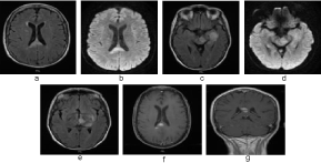 Figure 1: MRI of the brain a) Axial FLAIR showing corpus callosal lesion; b) Axial DWI of corpus callosal lesion; c) Axial FLAIR showing midbrain and temporal lobe lesion; d) Axial DWI of midbrain and temporal lobe lesion; e) Axial FLAIR showing thalamus and basal ganglion lesion; f) Axial T1 contrast showing enhancement of corpus callosal lesion; g) Coronal T1 contrast showing enhancement of corpus callosal lesion. View Figure 1
The differential diagnosis included neoplasms, demyelinating disorders and autoimmune and inflammatory conditions, infections, vascular causes, and trauma, which are discussed below along with representative MRI images.
Glioblastoma Multiforme (GBM) comprises 25% of all primary Central Nervous System (CNS) tumors and is the most aggressive type of brain tumor. It is a heterogonous necrotic mass with surrounding edema and ring enhancement as seen in Figure 2. They typical extend along white matter tracts including the corpus callosum [1].
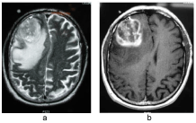 Figure 2: GBM in the brain a) Axial T2 showing heterogonous necrotic mass with surrounding edema with mass effect; b) Axial T1 contrast showing heterogeneous and ring enhancement [4]. View Figure 2
Lymphomas comprise 2% of all primary CNS tumors and occur in immune competent as well as immune compromised host, such as HIV patients and patients on immunosuppressant therapy. PCNSL occurs in 0.47 per 100,000 people/year [2]. Lymphomas can be multi-centric; with less enhancement and edema compared to GBM [1]. They also spread along white matter tracts and can cross the midline via the corpus callosum. Lymphomas, as well as GBMs, can present as a "butterfly" mass, as seen in Figure 3.
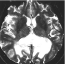 Figure 3: Axial T2 Butterfly mass of lymphoma [1]. View Figure 3
Gliomatosis Cerebri is a very slow growing, rare glial tumor, which affects the white matter and may progress to the other side of the brain through the corpus callosum. Gliomatosis Cerebri, as seen in Figure 4, does not usually have edema, necrosis and rarely enhance with contrast [6].
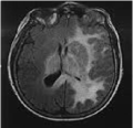 Figure 4: Axial FLAIR MRI showing a slowly progressive Gliomatosis Cerebri in the corpus callosum [5]. View Figure 4
Metastatic brain tumors can be found in the corpus callosum. Metastatic tumors can have single or multiple lesions with surrounding edema and heterogeneous ring enhancement, as seen in Figure 5.
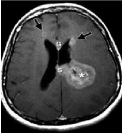 Figure 5: Axial T1 MRI with contrast showing heterogeneous enhancement in a brain metastatic tumor in the corpus callosum from lung cancer [7]. View Figure 5
Meningioma is a common primary brain tumor, but rare in the corpus callosum. It can sometimes become malignant. Meningioma can be seen in Figure 6 to have a dural tail, ring enhancement, and vasogenic edema extending into the parietal lobes creating a butterfly pattern.
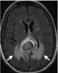 Figure 6: Axial FLAIR MRI showing meningioma with butterfly pattern of vasogenic edema [7]. View Figure 6
Neuromyelitis Optica (NMO) is a rare demyelinating disease that presents as optic neuritis or longitudinally extensive transverse myelitis in which NMO-IgG is antibody positive. NMO also has brain lesions along the lining of the ventricular wall and within the corpus callosum, as seen in Figure 7.
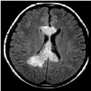 Figure 7: Axial FLAIR MRI showing corpus callosal NMO lesions [8]. View Figure 7
Multiple Sclerosis (MS) is a white matter disease affecting the corpus callosum. Hyperintense lesions are seen on Fluid-Attenuated Inversion Recovery (FLAIR) sequence of MRI in the periventricular, juxtacortical, corpus callosal, infratentorial regions. Some of these lesions may enhance, indicative of active lesions. A FLAIR sequence of MRI image of Multiple Sclerosis is shown in Figure 8.
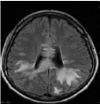 Figure 8: Axial FLAIR MRI showing corpus callosal Multiple Sclerosis lesions [8]. View Figure 8
Hereditary leukoencephalopathies are genetic, demyelinating diseases, which progressively affect white matter. Metachromatic Leukodystrophy is a type of hereditary leukoencephalopathy, causing dementia and peripheral neuropathy. It is attributed to the deficiency of Aryl sulfatase A with lesions starting in the periventricular region and spreading outwards. It can involve the splenium of corpus callosum as it continues to progress, as shown below in Figure 9.
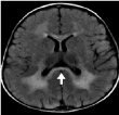 Figure 9: Axial FLAIR MRI showing lesions of metachromatic leukodystrophy in the splenium of the corpus callosum [7]. View Figure 9
Adrenoleukodystrophy is a type of hereditary leukoencephalopathy, which is X-linked. It is a disorder of peroxisomal fatty acid beta oxidation that results in the accumulation of very long chain fatty acids in the body, affecting various tissues. In the CNS, it most severely affects myelinated fibers. It starts dorsally in the brain and progresses anteriorly. Lesions seen in corpus callosum are as shown below in Figure 10.
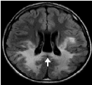 Figure 10: Axial FLAIR MRI showing Adrenoleukodystrophy with lesions in splenium of corpus callosum, starting dorsally and progressing anteriorly [7]. View Figure 10
Marchiafava-Bignami disease is a demyelinating disorder of the corpus callosum caused by vitamin B12 deficiency, that was thought originally to be due to drinking red wine [1,7]. The MRI of Marchiafava-Bignami disease shows lesions with edema in the early phases, T2 FLAIR hyperintensity, and a variable contrast enhancement, as seen in Figure 11, [7]. In chronic stages, the MRI will show lesions with necrosis, atrophy and minimal contrast enhancement [7].
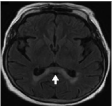 Figure 11: Axial FLAIR MRI showing demyelinating lesion in anterior and posterior corpus callosum [7]. View Figure 11
Acquired Demyelinating Encephalomyelitis (ADEM) can affect any part of the brain including the corpus callosum, as seen in Figure 12. Approximately 5% of ADEM cases are associated with vaccinations and others may be related to infections [9].
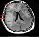 Figure 12: Axial FLAIR MRI showing an ADEM lesion in the anterior and posterior corpus callosum [9]. View Figure 12
Stroke in the splenium of corpus callosum may result from acute infarction due to posterior circulation especially involving the posterior pericallosal artery, as seen in Figure 13.
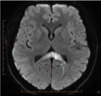 Figure 13: Axial Restricted diffusion confirmed on ADC MRI showing stroke lesion in the posterior corpus callosum [10]. View Figure 13
Posterior Reversible Encephalopathy Syndrome (PRES) may involve the corpus callosum, as seen in Figure 14, in addition to bilateral occipital brain regions. It may occur to due hypertension or could also be an autoimmune process.
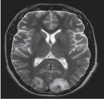 Figure 14: Axial T2 MRI showing PRES lesion in posterior corpus callosum [6]. View Figure 14
Neurosarcoidosis is a non-caseating granuloma, which affects the optic nerve, brain (including the corpus callosum), spinal cord, and peripheral nerves. The brain MRI is non-specific. Lesions may occur anywhere in the brain, including the leptomeninges, and may enhance without signs of edema [11].
Lupus Cerebritis is a connective tissue disorder, which may affect the central and peripheral nervous system. The brain MRI for Lupus Cerebritis shows enhancing demyelinating lesions that eventually lead to atrophy [12].
Mild Encephalopathy with Reversible Splenium Lesions (MERS) is a transient mild encephalopathy with an unknown etiology that has been described in children from Japan and East Asia [13]. Various infections have been thought to be associated with MERS. A brain MRI of MERS is shown in Figure 15.
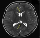 Figure 15: Axial T2 MRI showing a MERS lesion in the selenium of the corpus callosum [13]. View Figure 15
Other causes that should be considered in the differential include infections, vascular causes, such as infarct, arteriovenous malformation, and periventricular leukomalacia, trauma leading to hypoxic-ischemic encephalopathy.
As neoplasm was suspected, a brain biopsy was performed and histopathological and immunohistochemical stain slides were obtained, as shown in Figure 16.
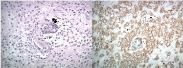 Figure 16: PCNSL in the biopsy of brain lesion is shown a) Atypical, large lymphoid cells clustered around a cerebral blood vessel; b) Large tumor cells with positive CD20 stain. View Figure 16
The patient was diagnosed with PCNSL with CD20 stain. Her whole body PET CT was negative for extra CNS Lymphoma. Bone marrow biopsy was not performed. So far she is treated with nine monthly infusions of High Dose Methotrexate with Leucovorin rescue, along with Rituxan. Her IELSG score was 3/5 [14,15]. Her expected 5 year survival is approximately 30%. Old CHOP therapy with alkylating agents was not used. No radiation has been given at this time, but it can be given if there is a recurrence. She has tolerated the chemotherapy well and is stable overall. Her major new manifestation has been some confusion and short-term memory loss, but her vison has improved. There has been no fatigue or systematic symptoms. A recent follow-up MRI has shown interval improvement with minimal enhancement, less than 1 cm, in the splenium of the corpus callosum near the biopsy site.
Our patient presented with minimum symptoms, considering the extent of disease on her MRI. After reviewing various MRIs and looking at different images of lesions that cross the midline, we went through the differential diagnosis and reached the conclusion that more than likely we were dealing with a neoplastic process.
We excluded HIV and other immunocompromised states. The patient was not in the age group for MS. Moreover, the pattern of lesions on the MRI was not consistent with a demyelinating conditions or vascular disorders. There was no history of trauma or any other signs of systemic illness to suggest other inflammatory conditions. Lymphoma was at the top of our differential because lesions were multi-focal without vasogenic edema, as you would expect to find in GBM, and there were no signs of necrosis. Metastasis was considered, but the pattern of enhancement was not ring enhancing type, as seen with these lesions.
Stereotactic brain biopsy confirmed the diagnosis of PCNSL. An early suspicion of lymphoma allowed for an early decision not to initiate steroids, which would have been given in case of metastasis or GBM. The initiation of steroids prior to biopsy would have most likely resulted in the disappearance or shrinking of the brain lesions, delaying a definitive diagnosis. A proper exhaustive differential, after reviewing the MRI images, was crucial in reaching the correct diagnosis of PCNSL and getting timely treatment for our patient.
PCNSL is a rare non-Hodgkin type neoplasm that must be considered the differential of corpus callosum lesions that cross the midline. Reviewing the images and considering PCNSL in the differential is important from both a diagnostic and timely therapeutic standpoint.
I am thankful to Dr. David Straus for performing the stereotactic brain biopsy, Dr. Harry Kellermire for providing the pathological slides and Pooja Pokharna for formatting. I am also thankful to Dr. Philip Baldwin for providing oncological update on the patient.