

Obstetrics and Gynaecology Cases - Reviews
Adenomyoma of Endocervical Type of the Cervix Uteri with Reactive Atypia and Goblet Cell Differentiation - A Case Report and Review of the Literature
Steffen Hauptmann1*, Katja Mohr2 and Regina Grosse2
1Medical Service Center for Gynecology, Cytology, and Histology, Martin Luther University of Halle-Wittenberg, Halle, Germany
2Department of Gynecology, Martin Luther University of Halle-Wittenberg, Halle, Germany
*Corresponding author: Steffen Hauptmann, MD, Medical Service Center for Gynecology, Cytology, and Histology, Martin Luther University of Halle-Wittenberg, Eisenbahnstr, 50-52, D-66424 Homburg (Saar), Germany, Tel: +49-6841-993252, Fax: +49-6841-993253, E-mail: steffen.hauptmann1@gmx.de
Obstet Gynecol Cases Rev, OGCR-2-060, (Volume 2, Issue 5), Case Report; ISSN: 2377-9004
Received: July 16, 2015 | Accepted: September 28, 2015 | Published: September 30, 2015
Citation: Hauptmann S, Mohr K, Grosse R (2015) Adenomyoma of Endocervical Type of the Cervix Uteri with Reactive Atypia and Goblet Cell Differentiation - A Case Report and Review of the Literature. Obstet Gynecol Cases Rev 2:060. 10.23937/2377-9004/1410060
Copyright: © 2015 Hauptmann S, et al. This is an open-access article distributed under the terms of the Creative Commons Attribution License, which permits unrestricted use, distribution, and reproduction in any medium, provided the original author and source are credited.
Abstract
In this report we present the rare case of a 53 years old woman with an adenomyoma of endocervical type of the cervix uteri with reactive atypia and focal goblet cell differentiation. The epithelium of the lesion was MUC5AC positive, making this marker invalid in excluding the most important differential diagnosis of this lesion, the minimal deviation adenocarcinoma. The most relevant literature is reviewed and the differential diagnoses are discussed.
Introduction
Cullen firstly described Adenomyomas in 1896 [1]. They are much more common in the corpus than in the cervix uteri and belong to the group of mixed Mullerian tumors of the female genital tract. They represent the second most common variety of this tumor group within the endometrium after carcinosarcoma [2]. Three general categories of adenomyoma are known: (i) adenomyoma of endocervical type, (ii) atypical polypoid adenomyoma and (iii) adenomyoma of usual type. Endocervical type adenomyoma has mucinous, sometimes microcystic dilated glands, usually without epithelial atypia. However, papillary infoldings, irregular shape with leaf-like architecture, and foci of tubal and endometrioid metaplasia may occur. The stroma is composed of smooth muscle cell proliferation. Atypical polypoid adenomyoma has angulated glands lined by endometrioid epithelial cells with atypia, frequently accompanied by squamous epithelial metaplasia. They are most frequently localized within the lower uterine segment. An adenomyoma of usual type consists of endometrioid glands, surrounded by endometrioid stroma and a smooth muscle cell proliferation in the periphery. Adenomyomas of any type occur either as polypoid tumors or as non-polypoid, but circumscribed intramural lesions [3]. In the cervix, adenomyomas, and particularly those harboring atypia, are rare and might cause problems in the differential diagnosis [4,5]. Moreover, endocervical adenomyomas containing goblet cells have never been described before. Therefore, we report on one currently observed case and have reviewed the relevant literature.
Case Report
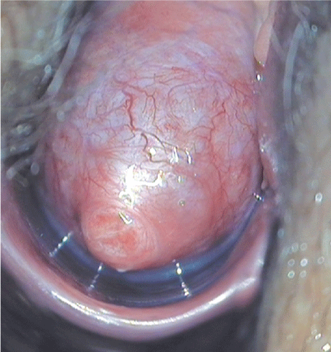
A 53 years old postmenopausal female presented with a feeling of pressure and heaviness in the bladder region as well as abnormal vaginal bleeding. During hysteroscopy a slight enlargement of the anterior part of the cervix was noticed. Curettage revealed only immature squamous metaplasia of the endocervical epithelium and atrophic endometrium. Therefore, the cervical lesion was colposcopically monitored for 1.5 years. Due to an accelerated growth up to a size of 2 cm within 12 months and the presence of abnormal glandular cells within a repeated Pap smear (Pap III) a hysterectomy was recommended. Upon admission, colposcopy revealed a 3.5 cm measuring bulging, resilient tumor involving the anterior part of the portion (Figure 1). The ultrasonic picture was that of a sharply demarcated, partly cystic lesion with a papillary internal architecture. The patient was treated by vaginal hysterectomy. Surgery and postoperative follow-up were without complications.
.
Figure 1: Colposcopic view. A bulging tumor of the anterior part of the cervix with smooth surface and prominent submucosal vascularization is visible.
View Figure 1
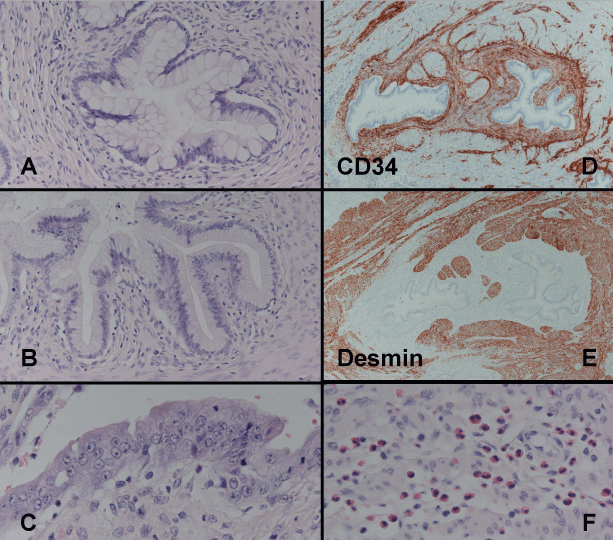
Histology revealed a well circumscribed but not encapsulated proliferation composed of thick bundles of smooth muscle cells without atypia and no mitotic activity. In between there were branched glandular complexes with papillary infoldings. The epithelium was mainly single layered, in some areas multilayered with slight atypia in the latter. Epithelial differentiation ranged from endocervical to intestinal with widespread occurrence of goblet cells (Figure 2). The goblet cells were positive for CK20 and MUC2. The endocervical epithelium was CK7 positive, but ER and PR were negative. It strongly expressed MUC5AC and focally to a lesser extent, also MUC6 in a diffuse cytoplasmic pattern as well as MUC4 and CEA along the luminal surfaces. The Ki-67 labeling was approximately 5% throughout the lesion with a focal increase up to 20% in atypical areas. The latter also showed a weak p53 positivity of approximately 30% of the atypical cells. Sequencing of exons 5 to 9 of the p53 revealed wild type sequence. An intense mixed inflammatory cell infiltration was found particularly in the vicinity of the multilayered slightly atypical epithelial regions. Both, synaptophysin and chromogranin stains were negative. The epithelial complexes were surrounded by a proliferation of CD34-positive fibroblastoid cells, which expressed ER and a few single cells were Ki-67 positive. The smooth muscle components of the lesion expressed ER, PR and desmin but were negative for Ki-67. Using PCR no HPV infection was detectable. The uterine wall contained a substantial number of adenomyotic foci.
.
Figure 2: Glandular proliferation containing numerous goblet cells, surrounded by a proliferation of fibroblast-like cells (A). In other areas the glands show endocervical differentiation (B). Some glands are lined by a multi-layered, atypical epithelium with a reduced amount of cytoplasmic mucin and enlarged nuclei with irregularly distributed chromatin and prominent nucleoli (C). The periglandular stroma contains a substantial amount of eosinophilc granulocytes (F). Immunohistochemistry reveals CD34-positive Periglandular fibroblasts (D) surrounded by desmin-positive smooth muscle proliferation (E).
View Figure 2
Discussion
Mixed mesenchymal-epithelial tumors occur almost exclusively in the female genital tract and reflect the biphasic differentiation potential of the Mullerian duct. These mixed mullerian tumors (MMT) usually arise within the endometrium where carcinosarcoma (CS) is the most frequent type of MMT. Tumors with only one malignant component are much less frequent, with adenosarcoma (AS) being more common than carcinofibroma [2,6]. In locations of the female genital tract devoid of endometrial tissue MMTs are uncommon. This is particularly true for the cervix uteri where only single cases of MMTs have been reported. Only one out of 200 CSs but one out of 10 AS arise within the cervix. In contrast to the endometrium, cervical MMTs are more frequent benign (adenofibromas and adenomyomas) than malignant. Adenomyomas (AM) are encountered in both the uterine cavity and the cervix where the former are much more frequent than the latter but they also occur in the broad ligament, the uterosacral ligament, the ovary, and the Bartolin gland [6-8]. According to our review of the literature (Table 1) only 36 cases of endocervical AMs have been reported so far, whereas several hundred of uterine AMs have been published [9-17].
![]()
Table 1: Cervical adenomyomas reported in the literature.
View Table 1
Endometrial AMs occur predominantly in postmenopausal, nulliparous woman as rounded lesion of the lower uterine segment with pushing, non-infiltrative border and are mainly of usual type. They occur either as well-circumscribed intramural nodules or as polypoid intracavitary mass. Their glands are lined by a single layered endometrioid epithelium, which may show squamous, tubal, or mucinous metaplasia. The glands are surrounded by endometrioid stroma, and a peripheral proliferation of smooth muscle cells showing the same variability of histoarchitecture and differentiation as described for leiomyomas.
Approx. 25% of endometrial AMs have severe epithelial atypia categorizing them as atypical polypoid adenomyoma (APA). These atypical tumors are always polypoid intracavitary lesions occurring in younger woman. Their epithelial proliferation is more complex, and they frequently contain squamous morules, sometimes with necrosis. Intramural AMs virtually never have epithelial atypia [6,7,15,16]. Therefore, an AM like intramural lesion with epithelial atypia always raises suspicion of endometrioid carcinoma. There are several reports of endometrioid adenocarcinoma within APA [18], and the risk of carcinoma elsewhere within the endometrium is approximately 10%. These data and the high risk to recur if treated by curettage alone are the reasons for performing a hysterectomy in these patients [15].
Endocervical AMs occur in multiparous woman with a mean age of 41 years (range 21-56 years) as whitish to yellow, solid, usually exophytic-polypoid, less common as circumscribed intramural nodules, which may contain grossly visible mucinous cysts. They may prolaps through the external cervical os inducing hemorrhage and severe reactive tissue change by chronic hypoxia. With a mean size of 5.8 cm (range 1.3-23 cm) endocervical AM are much larger than endometrial AMs. Histologically they usually have a lobulated architecture with a central located dilated irregular shaped gland, sometimes with papillary infoldings, surrounded by smaller glandular proliferations, resulting in a leaf-like architecture. The glandular epithelium has an endocervical phenotype with scant CEA and Ca125 positivity along the luminal margins, expression of CK7, as well as nuclear positivity for ER and PAX8. Some cases have endometrioid, tubal or squamous foci [9-19]. Recently a mesonephric differentiation has been reported in endocervical AM [18]. An intestinal differentiation within AMs of any type has, to our knowledge, never been described before. Goblet cell differentiation in general is a rare event within cervical lesions. When present in a significant amount, a Peutz-Jeghers syndrome should be considered. A thin rim of fibrous stroma and an intense peripheral smooth muscle proliferation are surrounding the glands of AM. The fibrous stroma may contain prominent adipose tissue, and symplasmic type multinucleate cells within the smooth muscle proliferation have been described [18]. In general, the mitotic rate in both glandular and stromal component is very low with no more than one mitotic figure in ten high-power fields [13,19], with the exception of one case with a focal increase of proliferation up to 20% within atypical areas as reported by Mikami et al. [12]. Epithelial atypia, however, is a rare event in AM of endocervical type and has been reported only once [12]. The slightly atypical epithelial cells found in this case had an open chromatin and were always associated with an inflammatory infiltrate around them. Moreover, the nuclear p53 reactivity was weak and below 50% and sequencing of exons 5 to 9 did not reveal mutations. Therefore, these probably are reactive atypia with stress and/or proliferation-induced p53 wild type upregulation. The absence of HPV, the p16 negativity and the low Ki-67 index of approximately 5% also exclude an adenocarcinoma in situ of usual type. The so called endometrioid type of endocervical AM is entirely composed of endometrioid glands and probably represents very deep located endometrial AMs. Cervical AM does rarely recur after polypectomy but never spread beyond the uterus [18,20].
The most important differential diagnosis of cervical AM is the minimal deviation adenocarcinoma (MDA) [4,5,13]. In contrast to the polypoid and circumscribed growth of AM, MDA is an ill-defined infiltrative mass, diffusely expanding the cervical wall. Nuclear atypia, although usually only of slight degree, periglandular stromal desmoplasia and loss of epithelial ER expression are features of MDA, whereas abundant stromal smooth muscle proliferation and ER positive epithelium without atypia are typical for cervical AM. However, an inflammatory stromal reaction by ruptured glands with mucin extravasation in AM might simulate a desmoplastic response, and may induce focal reactive epithelial atypia. MDA produces only neutral mucin of gastric type (MUC5AC-positive), and contains neuroendocrine cells, whereas AM produces a wide range of mucins, but in rare cases gastric mucins too as described previously as well as in this case [17,21]. Therefore, MUC5AC expression is not helpful in this differential diagnosis.
Lobular endocervical gland hyperplasia (LEGH) is a cervical glandular proliferation entering the differential diagnosis of AM too. LEGH is usually not polypoid, and is completely composed of gastric type epithelium. The cells are more cuboidal with pale eosinophilic cytoplasm, and are ER-negative. Moreover, there is usually no associated stromal smooth muscle component.
Another differential diagnosis of AM is adenosarcoma (AS), because the stromal component of AS not infrequently shows only a low degree of a typicality. However, the stroma of AS is more fibroblast like with periglandular stromal condensation, whereas in AM there is an ordered stromal architecture with periglandular fibroblastic differentiation surrounded by smooth muscle proliferation. Moreover, AS has a "phylloides like" or "leaf-like" structure with polypoid and papillary proliferations at the surface, a feature not seen in AM [22,23]. The pathogenesis of AMs is not clear. Some authors believe that these lesions are hamartomas (endocevicosis), comparable to intramural endometriosis. This idea is supported by the fact that in most cases of AM the uterus harbors foci of intramural endometrioisis as well [4]. Other authors think that hormonal dysbalances, particularly of estrogen or prolactin, play a role by inducing myofibroblastic differentiation and proliferation [12,15].
In summary, endocervical AMs are rare benign tumors, which may show gastric or intestinal differentiation. The two most significant histopathological differential diagnoses are minimal deviation adenocarcinoma of the cervix uteri and adenosarcomas.
References
-
Cullen TS (1896) Adenomyoma uteri diffusum benignum. Johns Hopkins Hosp Rep 6: 133-157.
-
McCluggage WG, Kubik-Huch RA (2003) Mixed epithelial and mesenchymal tumors. In: Pathology and genetics of tumours of the breast and female genital organs. World Health Organization classification of tumours. IARC Press, Lyon: 284-286.
-
Mazur MT (1981) Atypical polypoid adenomyomas of the endometrium. Am J Surg Pathol 5: 473-482.
-
Ramos P, Valenzuela P, Santana A, Ruiz A, Solano J (2003) Atypical polypoid adenomyoma of the uterine cervix: a diagnostic problem. J Obstet Gynaecol 23: 319-321.
-
Jakus S, Edmonds P, Dunton C, Holland G (2002) Atypical polypoid adenomyoma mimicking cervical adenocarcinoma. J Low Genit Tract Dis 6: 33-38.
-
McCluggage WG, Haller U, Kurman RJ (2003) Mixed epithelial and mesenchymal tumours. In: Pathology and genetics of tumours of the breast and female genital organs. World Health Organization classification of tumours. IARC Press, Lyon: 245-249.
-
Wight TC, Ferenzczy A (2002) Benign diseases of the cervix. In: Kurman RRJ (ed) Blaustein's Pathology of the female genital tract. 5th (edn) Springer: 225-252.
-
Zaloudek C, Hendrickson MR (2002) Mesenchymal tumors of the uterus. In: Kurman RRJ (ed) Blaustein's Pathology of the female genital tract. 5th (edn) Springer: 561-615.
-
Athas JM, Bluemke DA, Isacson C Sheth S (1996) Large cervical adenomyoma occurring in a first-trimester gravid uterus: radiologic-pathologic correlation. AJR Am J Roentgenol 167: 514-515.
-
Nasu K, Sugano T, Miyakawa I (1995) Adenomyomatous polyp of the uterus. Int J Gynecol Obstet 48: 319-321.
-
Kuwbara H, Ohno M, Moriwaki S (1999) Uterine adenomyoma of endocervical type. Pathol Internat 49: 1019-1021.
-
Mikami Y, Maehata K, Fujiwara K, Manabe T (2001) Endocervical adenomyoma. A case report with histochemical and immunohistochemical studies. APMIS 109: 546-550.
-
Gilks CB, Young RH, Clement PB, Hart WR, Scully RE (1996) Adenomyomas of the uterine cervix of endocervical type: a report of ten cases of a benign cervical tumor that may be confused with adenoma malignum. Mod Pathol 9: 220-224.
-
Uppal S, Heller DS, Cracchiolo B. (2003) Adenomyoma of the cervix: report of a case and review of the literature. J Low Genital Tract Dis 7: 218-220.
-
Longacre TA, Chung H, Rouse RV, Hendrickson MR (1996) Atypical polypoid adenomyofibromas (atypical polypoid adenomyomas) of the uterus: a clinicopathological study of 55 cases. Am J Surg Pathol 20: 1-20.
-
Fukunaga M, Endo Y, Ushigome S, Ishikawa E (1995) Atypical polypoid adenomyomas of the uterus. Histopathology 27: 35-42.
-
Ota S, Ushijima K, Nishio S, Fujiyoshi N, Takemoto S, et al. (2007) Polypoid endocervical adenomyoma: a case report with clinicopathologic analyses. J Obstet Gynecol Res 33: 363-367.
-
Casey S, McCluggage WG. (2015) Adenomyomas of the Uterine Cervix: Report of a Cohort Including Endocervical and Novel Variants. Histopathology 66: 420-429.
-
Matsuzaki S, Matsuzaki S, Tanaka Y, Fujita M, Yoshino K, et al. (2014) Large uterine cervical adenomyoma excised by vaginal approach: case report, images, and literature review. J Minim Invasive Gynecol 21: 954-958.
-
Sugiyama T, Ohta S, Nishida T, Okura N, Tanabe K (1998) Two cases of endometrial adenocarcinoma arising from atypical polypoid adenomyoma. Gynecol Oncol 71: 141-144.
-
Ishii K, Hosaka N, Toki T, Memose M, Hidaka E (1998) A new view of the so-called adenoma malignum of the uterine cervix. Virchows Arch 432: 315-322.
-
McCluggage WG (2010) Mullerian adenosarcoma of the female genital tract. Adv Anat Pathol 17: 122-129.
-
Soslow RA, Oliva E, Oliva E (2008) Mullerian adenosarcomas: an immunophenotypic analysis of 35 cases. Am J Surg Pathol 32: 1013-1021.
