A 49-years-old nulliparous woman came to our attention reporting pain and discomfort in the vagina as well as an important dyspareunia, which prevented a normal sex life by a few months. The gynecological examination showed a small paraurethral lesion easily bleeding and a small sub urethral ulcerated lesion. A biopsy specimen of vagina reported localization of Langerhans cell histiocytosis (LCH). LCH is a group of idiopathic disorders characterized by the presence of cells with characteristics similar to bone marrow-derived Langerhans cells juxtaposed against a backdrop of hematopoietic cells, including T-cells, macrophages, and eosinophils. Lesions on the mucosa are rarer, but they may occur on the oral mucosa or in the gastrointestinal, urogenital, and vaginal tracts. In this case, the axial CT examination shows extrapulmonary manifestation for vaginal involvement. Considering the symptoms reported by the patient local therapy with hyaluronic acid was recommended. The patient reported a significant improvement in symptoms, from a VAS score of 10 to a VAS score of 6 after 6 months and a VAS score of 2 after 1 year. Therefore, a local therapy with hyaluronic acid can be a valuable aid in treating symptoms of vaginal histiocytosis, especially vaginal dryness and dyspareunia.
Histiocytosis, Vagina, Dyspareunia, Hyaluronic acid
Langerhans cell histiocytosis (LCH) is a group of idiopathic disorders characterized by the presence of cells with characteristics similar to bone marrow-derived Langerhans cells juxtaposed against a backdrop of hematopoietic cells, including T-cells, macrophages, and eosinophils. It occurs in children, adolescents, and young adults, and it is currently thought to represent a disorder of immune regulation. It encompasses a continuum of disease with varying clinical presentation, severity of involvement, organ dysfunction, and prognosis. This continuum ranges from a benign localized process (e.g., eosinophilic granuloma), to a chronic progressive form (e.g., Hand-Schuller-Christian syndrome), to acute systemic illness (e.g., Letterer-Siwe disease). Although the etiology of LCH is unknown, it is generally accepted that it represents the manifestation of an undefined immunologic perturbation related to the manner in which macrophages process antigens and surface-associated antigen antibody complexes; LCH is classified according to its anatomic extent of disease. It tends to involve sites in which Langerhans cells are physiologically present or sites which have a resident cell population of the mononuclear phagocyte system. However, the process of LCH does not always begin with these cells but may also start with unique initiators that cause the phenotypic transformation of macrophages to pathologic Langerhans cells [1].
Lesions on the mucosa are rarer, but they may occur on the oral mucosa or in the gastrointestinal, urogenital, and vaginal tracts [2]. Langerhans cell hystiocitosis of the lower female genital tract was first reported by Andrews in 1939 [3]. Multiple, vulvar, vaginal, and/or cervical lesions are usually found concomitant with or secondary to mucocutaneous or systemic disease. Less commonly, the genital lesions portend subsequent systemic manifestation [4,5]. Rarely, LCH may present as solitary or multiple genital lesions without involvement of other organs [6].
We present the case of a woman with manifestations and symptoms of the disease only in the genitals and in which were then detected alterations in the lung.
A 49-years-old nulliparous woman came to our attention reporting pain and discomfort in the vagina as well as an important dyspareunia, which prevented a normal sex life by a few months. Medical and family history of the patient resulted both negative.
The only clinical findings of some interest was the presence of a pap test done four months earlier, according to screening, which reported a histological report of atypical squamous cells of undetermined significance (ASC-US) and HPV DNA test which resulted negative. A subsequent biopsy specimen of painful not infiltrated vaginal ulcera, localized in the middle third of the left sidewall reported fragments of vaginal mucosa with ulcerative acute inflammation with numerous eosinophils. It was therefore repeated a biopsy of the cervix and vagina using hematoxylin and eosin techniques, reporting fragments of mucosa with aggregates of epithelioid cells with irregular nuclear and intense ulcerative eosinophilic inflammation (Figure 1 and Figure 2). It was reported localization of Langerhans cell histiocytosis (S100 +, CD1a +, CD4 +, CD68 +/-, CD30-, CD56- , CD34- , MPO- share proliferating MIB1 20-30%).
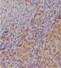 Figure 1: Ulceration Malpighian with a large inflammatory infiltrate in the cervical stroma; at high magnification, numerous eosinophils which tend to form microabscesses, lymphocytes and plasma cells. View Figure 1
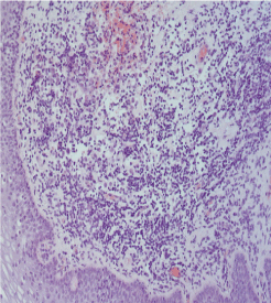 Figure 2: Focal infiltrate in the superficial dermis consists mainly of small lymphocytes and occasional histiocytes (H-E ×10). View Figure 2
The patient was then directed to the gynecology department and to that of respiratory diseases for a better diagnostic assessment.
According to the specialists, the patient has performed several investigations: Blood tests, gynecological ultrasound, abdominal ultrasound, abdomen RMN, CT, lung function tests, and bone marrow biopsy.
Blood tests showed a neutrophilic leukocytosis, hemoglobin (16 g/dl), hematocrit (46.3%), red blood cells (5.23 × 10⁶/uL) and platelets (427000/uL) at the upper limits of normal. Infectious tests were all negative.
Abdominal ultrasound did not show alterations, while chest x-ray showed diffuse parenchymal consolidation of lattice-nodular appearance in the hilarious regions bilaterally. A subsequent axial CT examination shows the presence of irregular cysts with centrolobular distribution involving upper lung zones with costophrenic sparing and micronodular pattern in the middle zone.
Dynamic contrast-enhanced MRI examination of lower abdomen on axial plane shows extrapulmonary manifestation for vaginal involvement (white arrow).
Lower abdominal MRI performed with and without contrast medium, showed three cystic formations of the right hepatic lobe, two to level of the dome with dimensioned about 3 mm and another one localized to the VII segment about 8 mm. Pancreas, spleen and kidneys were normal.
Bone marrow biopsy noted normal hematopoietic lines (with a bone marrow cellularity of 40-50%) and poor infiltrated B and T lymphoid.
Considering the systemic signs of the disease, a therapy with cortisone 25 mg daily was recommended. Regarding the gynecological examinations, visual examination showed a small paraurethral lesion easily bleeding and a small suburethral ulcerated lesion (Figure 3). The introduction of the speculum evoked intense pain and allowed to observe small bleeding lesions in vaginal walls bilaterally. The gynecological ultrasound was performed by transabdominal because the patient complained of intense pain at the introduction of the transducer in the vagina. Uterus and ovaries were normal. There were no hyperplastic lymph nodes in the groin. Considering the symptoms reported by the patient we recommended a therapy with hyalouronic acid 2% topically and orally. A questionnaire was provided to the patient (Table 1), with the request to fill it every week in order to assess the possible benefits of therapy. At the first administration the questionnaire the patient reported: vaginal bleeding: absent; dyspareunia: severe (VAS 10); vulvar itching: absent; vaginal itching: absent; rectal tenesmus: absent; tenesmus vaginal: absent; dysuria: mild (VAS 3). After one month the patient didn't report any improvement. After three months she reported a slight improvement in dyspareunia (VAS 8), while it was still impossible to introduce a normal speculum in vagina and to have sexual intercourse. After six months we repeated biopsies of vulva and vagina. The patient reported an improvement in dyspareunia with a VAS score of 6. We were able to introduce a normal speculum in vagina with only mild pain reported by the patient. After one year, the patient reported normal sexual intercourses and we were able to perform transvaginal ultrasound as well as to insert a speculum in vagina. The patient reported dyspareunia with a VAS score of 2.
 Figure 3: Vulvar appearance before the start of therapy with hyaluronic acid. View Figure 3
At small magnification, there is ulceration of the Malpighian with a large inflammatory infiltrate in the cervical stroma; high magnification, they notice numerous eosinophils which tend to form microabscesses, lymphocytes and plasma cells. A careful observation reveals a neoplastic proliferation confined in the stroma. The neoplastic cells are medium sized, oval with dendritic processes; coffee bean nuclei like, with finely dispersed chromatin, the nuclear membranes and inconspicuous nucleoli thin. The cytoplasm is moderately represented, vacuolated, weakly eosinophilic. The neoplastic cells express CD1a, S100, CD 68. The set of morphological and immunohistochemical findings are indicative of Langerhans cell histiocytosis. The staining with hematoxylin eosin (10x, 20x) (Table 1): Focal infiltrate in the superficial dermis consists mainly of small lymphocytes and occasional histiocytes (CD1a +, CD68 +, S100 +).
Table 1: Questionnaire administered to the patient. View Table 1
The current patient was treated with cortisone for her systemic disease, but she didn't refer any improvement of vaginal and vulvar symptoms, which constitute the most serious problem for her. The use of hyaluronic acid in its dual forms, soft capsules orally, and vaginal gel topically, has, albeit after a few months, alleviated genital symptoms of the patient, bringing her back to a state of physical and mental wellbeing.
In addition to systemic therapy for histiocytosis, carried out in this case with cortisone, it has proven very useful the local treatment of the first symptom complained by the patient and recognized as the more disabling at the time, namely vaginal dryness. Therefore, a local therapy with hyaluronic acid can be a valuable aid in treating symptoms of vaginal histiocytosis.