Necrotizing fasciitis is a severe, rare, potentially lethal soft tissue infection that develops in the scrotum and perineum, the abdominal wall or the extremities [1]. It progresses rapidly and septic shock may ensue [1,2]. When the anterior abdominal wall is involved it is referred to as Meleney's gangrene [3]. It is seen as a serious complication of surgery which evolves rapidly and could carry a high mortality rate of 30-50%, approaching up to 90% in diabetic patients [3-6]. It is a synergistic infection involving microaerophilic non-haemolytic streptococci and aerobic haemolytic staphylococci but may also involve various other aerobes and anaerobes [2,7,8]. It requires a high index of suspicion for early diagnosis and treatment. Unfortunately, the early symptoms and signs of this condition are non-specific and the diagnosis can be easily missed [9,10]. We report the development of cardiovascular collapse followed by development of Meleney's gangrene in a 38-year-old P3+ [2] woman after an uneventful abdominal myomectomy. We wish to share the difficulty encountered in making the diagnosis from the non-specific symptoms and management protocol adopted to ensure full recovery of the patient especially in a low resource setting.
Meleney, Gangrene, Necrotizing, Fasciitis, Cardiovascular, Collapse
Abdominal myomectomy is a commonly performed gynaecological procedure for varying indications especially in symptomatic women with future desire for fertility. It is a fairly safe procedure especially with haemostatic steps such as use of cervico-isthmic catheters to reduce blood loss during surgery [11]. With meticulous obliteration of dead spaces created by enucleation of leiomyomas, post-operative infective and febrile morbidities are grossly reduced.
Necrotizing fasciits of the anterior abdominal wall is a very rare condition especially following gynaecological procedures [12]. It is described classically as a synergistic infection involving microaerophilic non-haemolytic streptococci and aerobic haemolytic staphylococci but may also involve various other aerobes and anaerobes [2,7,8] beta haemolytic streptococcus acting in synergy with certain anaerobic organisms [1,2]. Predisposing factors for this condition include immune-compromise, diabetes mellitus and low socio-economic status among others [2].
We report the case of a 38-year-old P3+ [2] woman who had abdominal myomectomy on account of symptomatic uterine fibroids (menorrhagia and pain) of 3 years duration. She was not a known diabetic or hypertensive. The uterus was enlarged up to 26 weeks and her body mass index was 22.3 kg/m2. Her pre-operative haemogram and kidney function tests were within normal limits and fasting blood sugar was normal.
She had abdominal myomectomy following laid down unit protocols and thirty (30) pieces of leiomyomas of varying sizes were removed and submitted for histopathological assessment. They demonstrated varying degrees of degenerative changes. Layered closure of the anterior abdominal wall was done using delayed absorbable sutures (polygycolic acid). Of particular note was a thickened sub-cutaneous layer measuring approximately 6 cm in thickness. The intra-operative blood loss was 180 mls. She had a smooth immediate post-operative course with a post-operative packed cell volume of 30%. She was discharged on the 4th post-operative day but decided to wait till the 6th post-operative day to sort-out post-discharge formalities.
On the 6th post-operative day while in a seated position, she developed a sudden onset of tiredness and profuse sweating. While attempting to return to a supine position she fell and lost consciousness. Patient was noticed to be in cardiovascular collapse as the pulse rate was 118 beats per minute while the blood pressure was 50/30 mmHg and she was febrile to touch. Random blood sugar was 5.7 mmol/L. She was immediately resuscitated with intravenous infusion of normal saline and intranasal oxygen. Within 30 minutes of resuscitation, patient regained consciousness and the blood pressure normalized to 100/70 mmHg. Further examination of the abdomen revealed a well healed pfannensteil scar and a minimal area of blister adjoining the edge of the zinc-oxide-coated plaster measuring approximately 3 × 4 cm. Her temperature was 38.7 °C.
Abdominal ultrasound was done which excluded any intra-abdominal collection but there was minimal stasis of peristaltic motions on visualization of bowel loops. Repeat packed cell volume was 30% and E/U/Cr was essentially normal. Blood samples were obtained for blood culture and serial abdominal girth measurement did not reveal any progressive abdominal distension and there was no area of undue tenderness.
She was recommenced on 'nil per oris' due to a reduced perception of bowel sounds and a presumed diagnosis of suspected paralytic ileus with? gram negative septicaemia. The wound care was continued with methylated spirit and gentian violet while special attention was given to the blistered area which was presumed to be a reaction to the zinc oxide coated plaster. Intravenous antibiotics were recommenced and patient monitoring was continued.
She appeared to be making good clinical progress until she attempted to sit out of bed on the 7th post-operative day when she experienced another episode of fainting attack with deterioration of her vital signs. Blood pressure was 60/30 mmHg, pulse rate was 117 beats per minute and the temperature was 37.9 °C. Resuscitation produced an instant normalization of her vital signs and patient was nursed in supine position. Her blood pressure became 100/70 mmHg while her pulse rate was 101 beats per minute. Some induration was noticed on the anterior abdominal wall superior to the pfannensteil incision which was intact and healing well. A presumed diagnosis of anterior abdominal wall cellulitis was made and the antibiotic regimen was continued.
On the 8th post-operative day a discolouration was noticed around the blister site which was now ruptured and discharging sero-sanguinous fluid. A swab sample was obtained for microscopy culture and sensitivity while intravenous gentamycin was added to her antibiotic regimen.
On the 9th post-operative day the discolouration noted around the former blister site was noticed to be spreading upwards and laterally on both sides and the patient developed superficial tenderness in both flanks and her temperature became 39 °C. The microscopy culture and sensitivity returned on the 10th post-operative day and returned growth of Pseudomonas aeruginosa and Proteus mirabilis which were maximally sensitive to augmentin, cefixime, cefuroxime and ceftriaxone. Her antibiotic was changed to include intravenous ceftriaxone 1 g 12 hourly. An assessment of necrotizing fasciitis (Meleney's gangrene) was made and the need for wound debridement was explained to the patient.
A consult was sent to the general surgical team and the patient was scheduled for wound debridement under general anaesthesia. Intraoperative findings revealed area of extensive gangrene involving a portion of skin above the pfannesnteil incision and extending to the subcutaneous tissue immediately beneath the skin and extending laterally to both lumbar regions and stopping just short of the umbilicus superiorly (Figure 1). All gangrenous tissue was excised and additional chemical debridement was done using 2% hydrogen peroxide solution while the subcutaneous layer was packed with EUSOL soaked gauze on either side. The patient continued on daily wound dressing, vital signs normalized and the wound granulated progressively (Figure 2 and Figure 3) till a secondary closure was done on the 60th post-operative day (Figure 4).
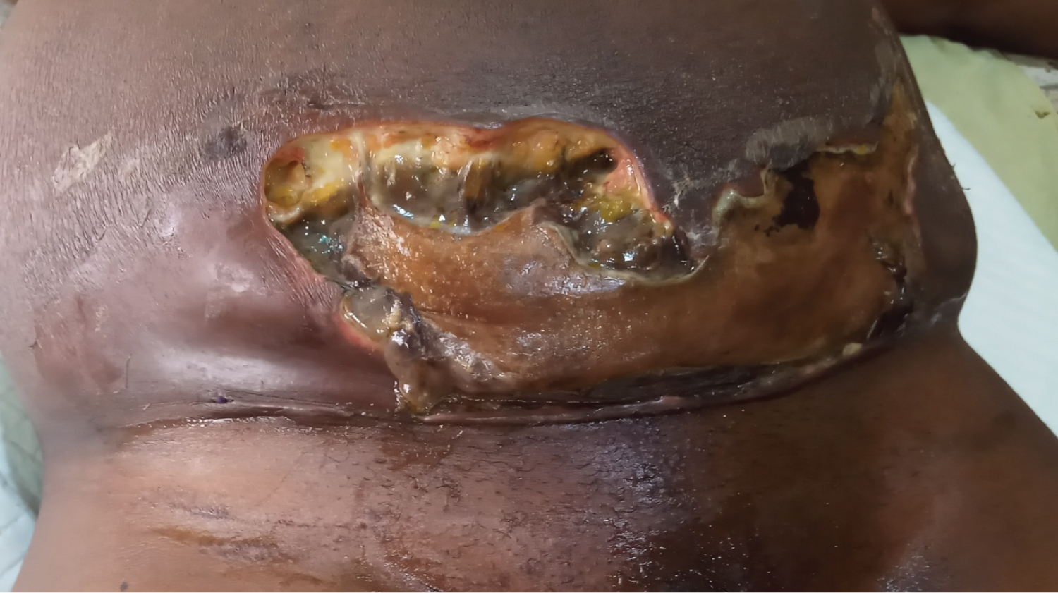 Figure 1: Wound just before debridement.
View Figure 1
Figure 1: Wound just before debridement.
View Figure 1
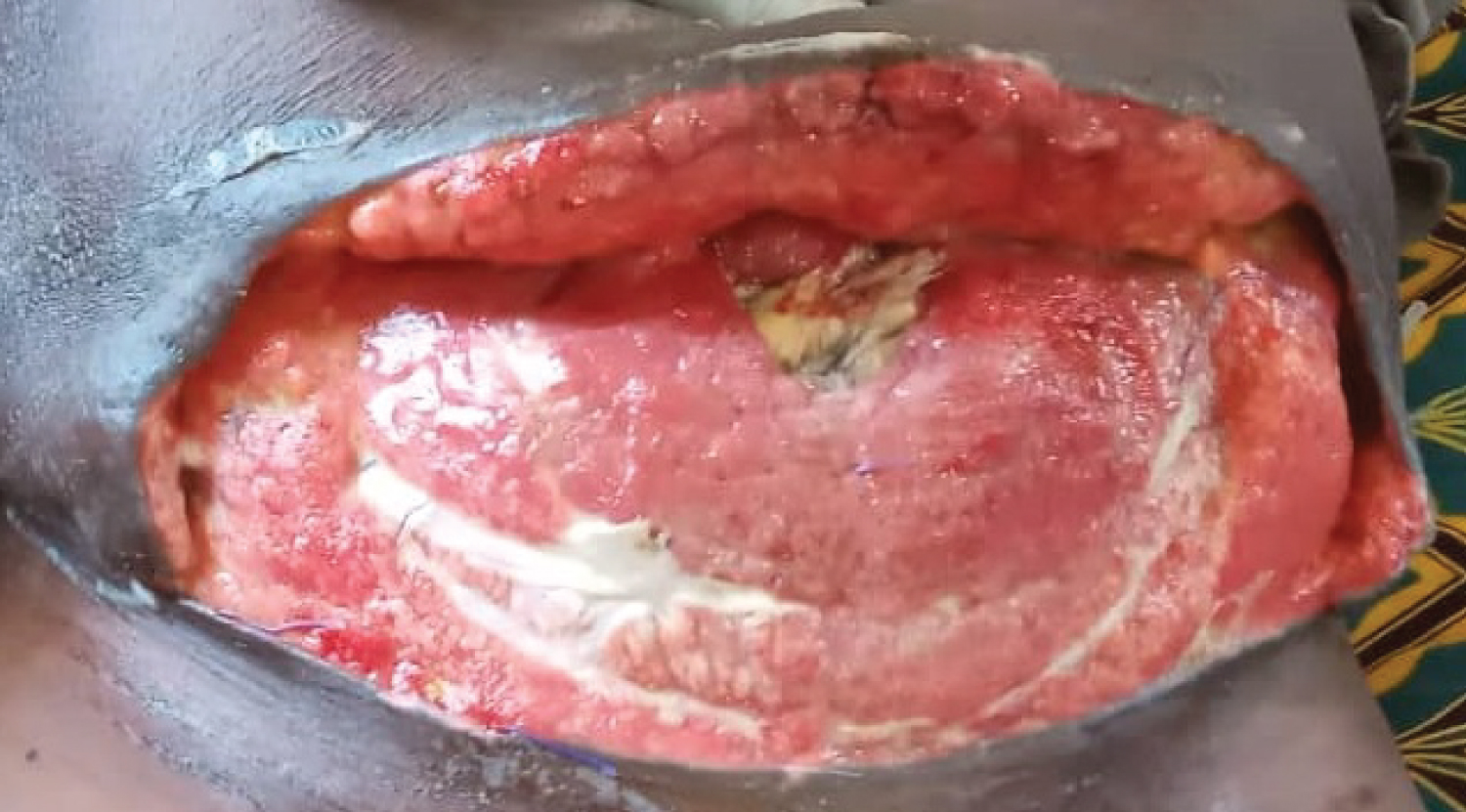 Figure 2: Wound 8 Days after debridement and Wound dressing.
View Figure 2
Figure 2: Wound 8 Days after debridement and Wound dressing.
View Figure 2
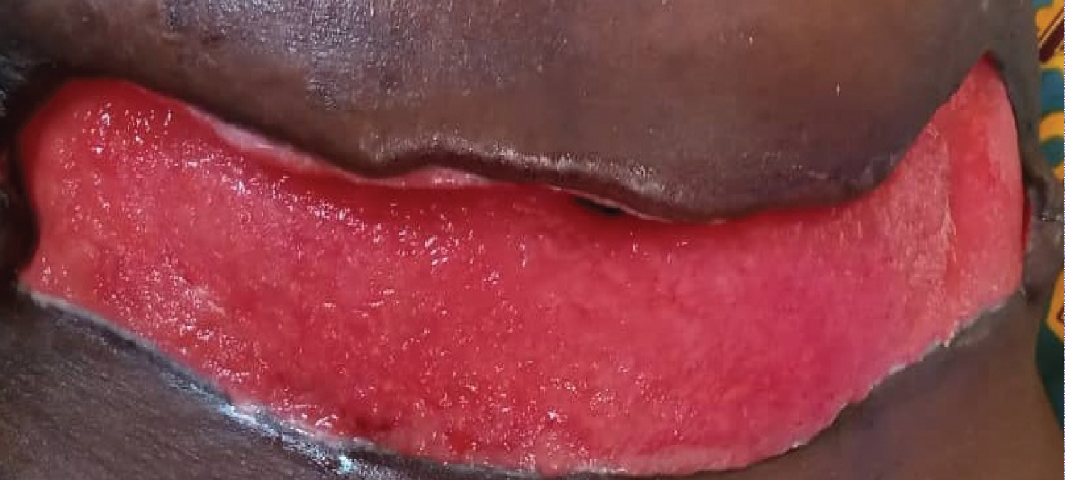 Figure 3: Wound 24 Days after debridement and Wound dressing.
View Figure 3
Figure 3: Wound 24 Days after debridement and Wound dressing.
View Figure 3
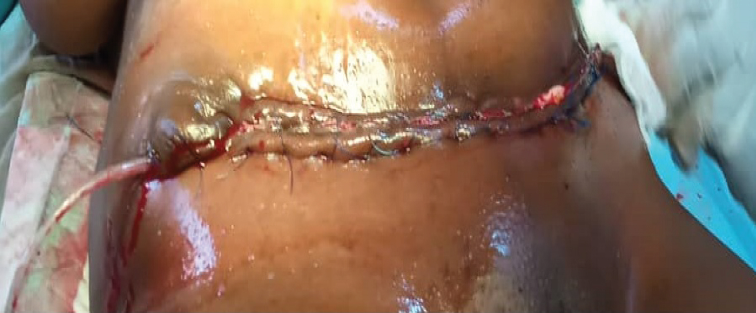 Figure 4: Secondary wound closure 45 Days after debridement.
View Figure 4
Figure 4: Secondary wound closure 45 Days after debridement.
View Figure 4
She was eventually discharged from the ward on the 70th post-operative day (Figure 5) and followed up in the gynaecological clinic. Her visits to the gynaecological clinics have been uneventful.
 Figure 5: Wound at discharge.
View Figure 5
Figure 5: Wound at discharge.
View Figure 5
Necrotizing fasciitis is a rare condition especially the anterior abdominal wall variety which is commonly referred to as Meleney's gangrene [1,3]. Meleney's gangrene is known to be a rapidly progressing necrotizing fasciitis caused by bacteria acting in a synergistic manner to effect maximum tissue destruction within a very short period of time [3]. It complicates anterior abdominal wall surgeries but in very rare instances minor injuries such as abrasions or scratches to the skin of the abdomen might be the portal of entry [5,6,8].
The organisms responsible for this condition belong to a vast array of aerobes and anaerobes acting synergistically to result in an infectious morbidity which has a mortality of 30-50% and even up to 90% especially in individuals with compromised immunity and diabetics [3-6].
The patient above is not a known diabetic and there is no clinical history suggestive of immune-compromise. She however had a thickened abdominal fatty layer which caught attention during surgery. Despite a normal body mass index, this truncal obesity was the only identifiable risk factor in this patient [13].
The immediate post-operative period in this patient was uneventful and the first sign of danger was a sudden onset of fainting attack post-discharge which was suspected to be from gram negative endotoxic shock. This cardiovascular collapse could be the earliest warning sign in some patients [14], and if missed could result into delay in treatment and mortality. The skin changes on the patient's abdominal wall were not specific except for a bullous reaction superior to the incision line which was thought to be due to zinc oxide toxicity and it was associated with non-specific induration of surrounding skin which was thought to be a straight forward cellulitis. However a progressive worsening of the bullous reaction ranging from rupture to discolouration of surrounding skin and apparent gangrene of the skin and subcutaneous tissues developed rapidly. This leads credence to the fact that a high index of suspicion is needed for the diagnosis of necrotizing fasciitis as the early symptoms and skin changes are non-specific and delay in definitive management could be life-threatening [15].
Parenteral antibiotics and extensive surgical debridement is the documented management of choice [16,17] and in this case the persistent fever observed in the patient crashed within a few hours of debridement and recommencement of parenteral antibiotics. Surgical debridement was combined with chemical debridement using 2% hydrogen peroxide. This was followed up with daily dressing using gauze packs soaked in eusol which was later changed to pure honey dressing.
The patient was provided with psychological support and placed on high protein and high-calory diet in addition to blood transfusion to optimize her haematocrit above 30%. These steps resulted in progressive improvement of her wound as evidenced by progressive granulation and reduction of wound size.
The wound was dressed daily for a total of 44 days and the patient had secondary wound closure on post-operative day 45. The wound eventually healed completely around the 60th post-operative day and the patient was eventually discharged. The progression of the disease is as shown in the attached images.
Early diagnosis, institution of parenteral antibiotics and extensive debridement with subsequent wound dressing are proven measures for optimal care and reduction of mortality in necrotizing fasciitis [18,19].
Meleney's gangrene is a fatal complication of surgery but early diagnosis, parenteral antibiotics and extensive wound debridement are important steps in the reduction of mortality. Sudden onset of shock in a post-operative patient that was hitherto stable after exclusion of haemorrhagic complication of surgery should strongly raise a suspicion for an evolving necrotizing fasciitis.
There was no financial support from any source for the conduct of this study. This research did not receive any specific grant from funding agencies in the public, commercial, or not-for-profit sectors.
There are no interests to declare.
All authors have approved the final article as submitted.
Conceptualization: Babatunde Olaniyi ROSIJI, Omagbeitse Henry ABIYERE
Drafting the article: Babatunde Olaniyi ROSIJI, Babatunde Ajayi OLOFINBIYI
Critical revision of the article: Olalekan Oladipupo ROSIJI, Blessing Oluwatosin OLANREWAJU, Olakunle John AYELOJA
Final approval of version to be submitted: All authors
A written consent has been obtained from the patient in this report to use the pictures taken during clinical care. All steps have been taken to ensure that patient's confidentiality is maintained.