

Obstetrics and Gynaecology Cases - Reviews
Intra-operative Management of Skull Remnant Discovered during Emergent Primary Cesarean Delivery
Lisa A. Licare1* and Suruchi Thakore2
1Department of Obstetrics and Gynecology, Women and Children's Hospital of Buffalo, University at Buffalo, State University of New York, NY, USA
2Department of Reproductive Endocrinology and Infertility, University Hospitals, Case Medical Center, OH, USA
*Corresponding author: Lisa A. Licare, D.O., PGY-IV, Resident, Department of Obstetrics and Gynecology, Women and Children's Hospital of Buffalo, University at Buffalo, State University of New York, 3716 Deer Lakes Drive, Amherst, NY 14228, USA, Tel: (917) 670-0544, E-mail: lisalicare@gmail.com
Obstet Gynecol Cases Rev, OGCR-2-022, (Volume 2, Issue 1), Case Report; ISSN: 2377-9004
Received: October 23, 2014 | Accepted: February 05, 2015 | Published: February 08, 2015
Citation: Licare LA, Thakore S (2015) Intra-operative Management of Skull Remnant Discovered during Emergent Primary Cesarean Delivery. Obstet Gynecol Cases Rev 2:022. 10.23937/2377-9004/1410022
Copyright: © 2015 Licare LA, et al. This is an open-access article distributed under the terms of the Creative Commons Attribution License, which permits unrestricted use, distribution, and reproduction in any medium, provided the original author and source are credited.
Abstract
Background: Operative anticipation of patients with prior abdominal surgeries is critical for the evaluation of obstetric patients. The following case demonstrates the importance of preoperative planning before emergent surgery is indicated.
Case: T.M. is a 26 year- old gravida 8, para 0161 at 24 weeks 5 days who presented with preeclampsia with severe features. Her history is significant for brain surgeries with abdominal skull flap preservation. She required an emergent cesarean where a skull flap was discovered. Extra time was required to excise the bone, which delayed delivery.
Conclusions: The need for emergent surgery must be considered in premature preclamptic patients. Although post- operative investigation confirmed removal of the skull flaps, a difficult procedure should have been anticipated and the fetus delivered non- emergently.
Introduction
Subdural hematoma is one of the most deadly forms of traumatic brain injury [1]. The approximate mortality rate from this injury can reach 40-60 percent in those patients requiring surgery and 57-68 percent in patients already comatose on presentation [2] . The Glasgow Coma Scale is the main diagnostic indicator of prognosis for patients with traumatic brain injury. Patients who present with an acute subdural hematoma with a Glasgow Coma Scale of 9 or greater have the highest chance of a full functional recovery, while those with lower scores have a graver outlook [3].
Treatment options for subdural hematomas include both surgical and non-surgical methods. Non-surgical methods are reserved for patients with small hematomas who are stable on physical exam, while those who present with signs of herniation or elevated intracranial pressure require emergent surgical management. Available surgical options include burr hole, craniotomy and decompressive craniectomy. A decompressive craniectomy requires removal of a portion of the skull to accommodate for brain edema [4].
Cryopreservation is the most common method of storing autologous bone flaps [5]. Cryopreserved flaps are either directly preserved under sterile conditions or irrigated with an antibiotic or antiseptic solution. After cultures are obtained to rule out infection, the sample is then preserved at temperatures from -18 to -83 degrees Celsius. Complications include infection, devitalization, bone resorption and poor cosmetic result [6]. Alternatively, abdominal storage in the patient's subcutaneous fat has been shown to provide adequate preservation of the bone flap in a more cost-effective manner and has a zero percent infection rate [6]. This method, however, requires a second surgical site for the patient. Complications include fluid collection around the flap, and possible revision of the surgical site.
Case Study
At age 17, T.M. was a pedestrian in a motor vehicle collision. She was hemodynamically stable on arrival to the trauma center and found to be 7 on the Glasgow Coma Scale. Her injuries were extensive- including epidural and subdural hematomas, right subarachnoid hemorrhage, basilar skull fracture, multiple facial fractures, C6 fracture and right hemothorax. A decompressive craniectomy was performed. The epidural hematoma was coagulated and the subdural hematoma irrigated with no further bleeding noted. Two Jackson Pratt drains were placed, and a left frontal ventriculostomy was created. The skull flap was placed in the subcutaneous layer of the anterior abdominal wall in two pieces. A second surgery was performed at a later date to reimplant the skull flaps during which all bone fragments were removed from the abdomen.
When T.M. wasa26 year-old gravida 8, para 0161, she presented to the labor and delivery unit at 24weeks 5 days with headache, dizziness and epigastric pain. Admission physical exam was significant for a gravid abdomen with a vertical incision scar in the right upper quadrant. Deep tendon reflexes were 1+, and 2+ edema was noted bilaterally in the lower extremities. The fetus was in transverse lie on ultrasound examination and had an estimated fetal weight of 674g. The patient denied any past medical history, allergies, or social history. Her obstetric history consisted of a 36- week induction of labor for preeclampsia 5 years prior, after which she delivered a 2,920g (6lb 7oz) female vaginally.
The patient was admitted with the diagnosis of preeclampsia with severe features based on laboratory and blood pressure values. Preeclampsia labs were significant for lactate dehydrogenase of 425, uric acid of 6.3 and urine protein of 2+. Magnesium sulfate was administered for seizure prophylaxis and two doses of hydralazine, 5mg and 10mg, respectively, were pushed for blood pressures in the severe range. The patient was given betamethasone for fetal lung maturity and was followed expectantly secondary to the immature gestational age of the fetus. It was determined that if the fetal heart tracing or maternal condition decompensated, the fetus would be delivered surgically.
On hospital day three, at 25 weeks gestation, the fetal heart tracing decompensated to category 3. The decision was made to proceed with emergent cesarean delivery. Upon attempting to enter the fascia with electrocautery, a hard impenetrable object was noted in the right lateral portion of the pfannenstiel incision. Initially, it was thought to be scar tissue however, it was noted to be a foreign object needing excision. The skull flap was gently excised from the surrounding fatty tissue with metzenbaum scissors. A female infant was delivered 26 minutes after the skin incision by primary low transverse cesarean due to the extensively developed lower uterine segment. The low transverse incision was extended into a T incision for adequate space for delivery of the fetus. The infant was handed to the awaiting intensive care nursery team and the procedure was completed. Special care was made to ensure that the fascia had no defects after the skull flap resection. Benign trabecular bone was confirmed by pathology. The patient remained on magnesium sulfate for 24 hours after delivery and did not require additional doses of anti-hypertensive medication. She was discharged home on postoperative day four [Figure 1].
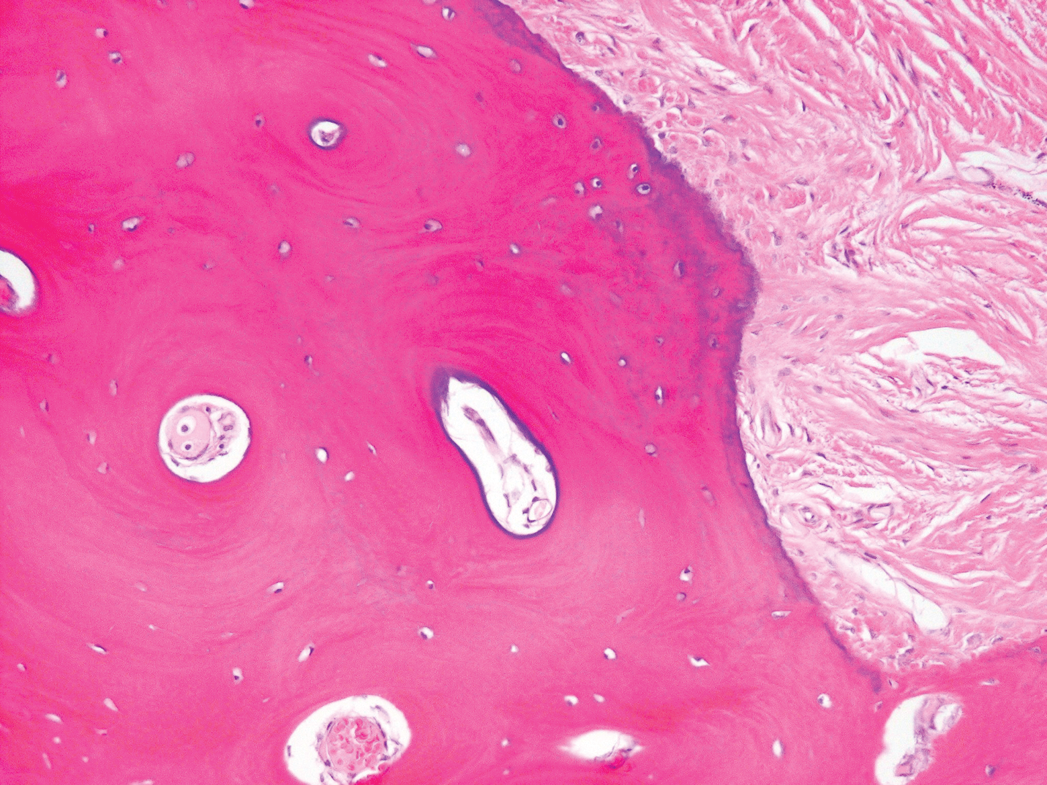
Figure 1: Bone Skull with Placenta
View Figure 1
Discussion
The management of severe preeclampsia at an extremely premature gestational age poses a dilemma to the obstetrician. One must always ask if the risks of continuing the pregnancy outweigh the risks of extreme prematurity to the fetus. We decided to admit the patient for magnesium sulfate prophylaxis with continuous monitoring in an attempt to prolong the pregnancy to betamethasone maturity. The risk of requiring an emergent cesarean delivery, however, was also discussed amongst the obstetric team and with the patient. Once decompensation of the fetal heart rate occurred, the decision to proceed with surgery was made.
Based on the review of patient�s verbal and documented medical and surgical history, intraoperative discovery of the skull remnant was an unexpected complication. When considering the location of her scars from the previous surgeries, it was thought that the area of the skull preservation was distant enough from the lower abdomen that adhesive disease would not interfere with a cesarean delivery. A second review of her operative notes post operatively confirmed removal of all skull fragments. Due to the possible fracture and migration of the skull remnant from its original location, it was most likely left behind by the neurosurgery team. By delaying delivery for fetal lung maturity and prolonging gestational age, the team ran the risk of necessitating an emergent surgery, which would not allow for time to deal with any complications encountered. Therefore, fetal lung maturity and perfect timing of surgery must be balanced with allowing for adequate time to deliver the fetus prior to fetal compromise.
References
-
Hatashita S, Koga N, Hosaka Y, Takagi S (1993) Acute subdural hematoma: severity of injury, surgical intervention, and mortality. Neurol Med Chir (Tokyo) 33: 13-18.
-
Wilberger JE Jr, Harris M, Diamond DL (1991) Acute subdural hematoma: morbidity, mortality, and operative timing. J Neurosurg 74: 212-218.
-
Cook RJ, Fearnside MR, McDougall P, McNeil RJ (1996) The Westmead head injury project: Outcome prediction in acute subdural haematoma. J Clin Neurosci 3: 143-148.
-
Bhaskar IP, Zaw NN, Zheng M, Lee GY (2011) Bone flap storage following craniectomy: a survey of practices in major Australian neurosurgical centres. ANZ J Surg 81: 137-141.
-
Baldo S, Tacconi L (2010) Effectiveness and safety of subcutaneous abdominal preservation of autologous bone flap after decompressive craniectomy: a prospective pilot study. World Neurosurg 73: 552-556.
-
Wilberger JE Jr, Harris M, Diamond DL (1991) Acute subdural hematoma: morbidity, mortality, and operative timing. J Neurosurg 74: 212-218.
