

Obstetrics and Gynaecology Cases - Reviews
Sclerosing Stromal Tumor of the Ovary in Postmenopausal Women: A Report of Two Cases
Chae Min Lee1, Soyi Lim1*, Hyun Yi Cho2, Seung Yeon Ha2 and Jin Woo Shin1
1Departments of Obstetrics & Gynecology, Gachon University Gil Hospital, Incheon, Korea
2Department of Pathology, Gachon University Gil Hospital, Incheon, Korea
*Corresponding author: Soyil Lim, M.D., Department of Obstetrics & Gynecology, Gachon University Gil Hospital, 1198 Guwol-dong, Namdong-gu, Incheon, Korea, Tel: 82-32-460-3254, Fax: 82-32-460-3290, E-mail: soyilim@gmail.com
Obstet Gynecol Cases Rev, OGCR-2-045, (Volume 2, Issue 3), Case Report; ISSN: 2377-9004
Received: April 29, 2015 | Accepted: June 16, 2015 | Published: June 19, 2015
Citation: Lee CM, Lim S, Cho HY, Ha SY, Shin JW (2015) Sclerosing Stromal Tumor of the Ovary in Postmenopausal Women: A Report of Two Cases. Obstet Gynecol Cases Rev 2:045. 10.23937/2377-9004/1410045
Copyright: © 2015 Lee CM, et al. This is an open-access article distributed under the terms of the Creative Commons Attribution License, which permits unrestricted use, distribution, and reproduction in any medium, provided the original author and source are credited.
Abstract
Sclerosing Stromal Tumor (SST) was first delineated as a distinct ovarian sex cord stromal tumor in 1973 by Chalvardjian and Scully. It is a benign neoplasm, distinguished from other ovarian stromal tumors by the production of collagen and a pseudolobular pattern, and it tends to occur in the second and third decades of life in diagnosed patients. We discovered two rare cases of SST in post-menopausal women which are the topic of this report. These case studies are accompanied by a brief review of the literature.
Keywords
Sex cord-stromal tumor, Ovarian neoplasm, Menopause
Introduction
Sclerosing stromal tumor (SST) is a rare benign ovarian neoplasm of the sex cord stromal category first delineated as a distinct ovarian sex cord stromal tumor entity in 1973 by Chalvardjian and Scully [1]. Most patients afflicted with SST present with nonspecific symptoms related to an adnexal mass. The tumor, with rare exceptions, is hormonally inactive. Diagnosis of SST can be confirmed by postoperative pathologic examination. It is important to perform differential diagnoses of SST relative to other sex cord stromal tumors including fibroma, thecoma and lipoid cell tumors [2]. It is distinguished from other ovarian stromal tumors by the production of collagen, a pseudolobular pattern, and it tends to occur in the second and third decades of patient life [3].
Herein, we present two cases of SST occurred in postmenopausal women with brief review of literature.
Case 1
A 63-year-old postmenopausal woman, gravida 5, para 2, experienced lower abdominal discomfort accompanied by a palpable abdominal mass for one month prior to her visit. Physical examination revealed a solid, non-tender tumor palpable up to the umbilicus level, and pelvic ultrasonography revealed a well-defined 14cm sized heterogeneous, predominantly cystic pelvic mass with solid portions. A computed tomography (CT) scan of the abdomen and pelvis revealed a 15cm sized tumor with an enhancing solid component, small amount of collected ascites and minimal peritoneal thickening. All clinical chemistry and tumor markers were below cut-off levels. Proceeding under the assumption that the finding was an ovarian torsion or cancer, the patient underwent a total abdominal hysterectomy including salpingo-oophorectomy, left external iliac lymph node sampling and washing cytology. We discovered an enlarged left ovary which had been torsioned clockwise twice and accompanied by necrotic changes. There was no evidence of metastases or peritoneal seeding.
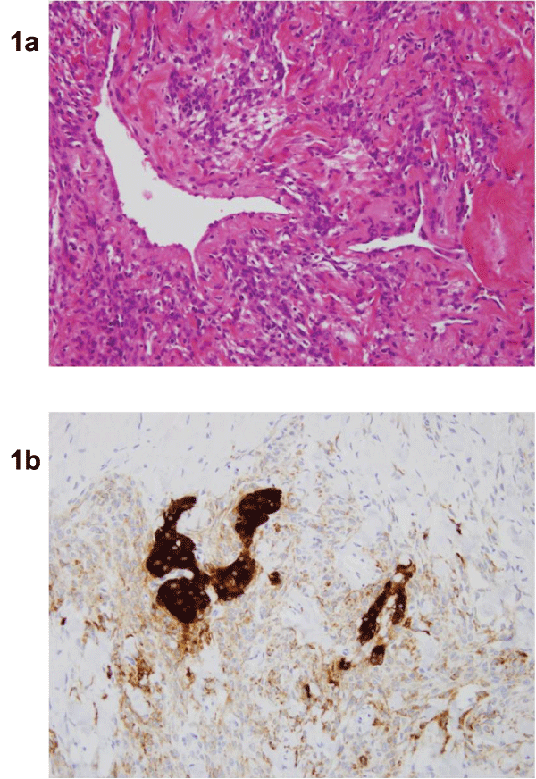
A gross examination revealed a 17.5 x 14.5 x 9.0cm mass weighing 1248g on the left ovary. Upon sectioning, the mass was found to be cystic with hemorrhagic fluid and a solid portion. Microscopically, the vessels contained a hemangiopericytoma-like staghorn pattern (Figure 1a). The luteinized cells were strongly immunoreactive for inhibin alpha (Figure 1b). The immunohistochemistry (IHC) results included Inhibin α: positive, CD99: positive, Desmin: positive, and Actin: positive. Based on these histomorphologic findings, the diagnosis was SST.
.
Figure 1: Microscopic features of the sclerosing stromal tumor from case 1
(a) The vessels have a hemangiopericytoma-like staghorn pattern (H&E x400).
(b) The luteinized cells are strongly immunoreactive for inhibin-alpha (x200).
View Figure 1
Case 2
A 59-year-old postmenopausal woman, gravida 3, para 3, presented with complaints of abdominal pain with dysuria for 3 days prior to her visit. Upon clinical examination, a tender tumor was palpable up to the umbilicus level and was accompanied by left costovertebral angle (CVA) tenderness.
Ultrasonography revealed a 11.8 x 11.9 x 9.4cm sized solid mass with a cystic component in the left adnexa. A CT scan of the abdomen and pelvis revealed a 14cm sized solid-cystic mass and bowel wall thickening. The patient's serum CA-125 level was 37.9U/mL (reference range <35U/mL). Proceeding with the assumption that this finding was a left adnexal mass, the patient underwent a total abdominal hysterectomy including both salpingo-oophorectomy and omental biopsy. A smooth, well-circumscribed, bosselated mass of approximately 15cm in diameter was enucleated. The uterus, right ovary and bilateral fallopian tubes were normal in appearance.
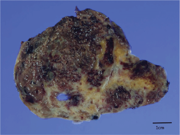
Gross examination revealed a 13.2 x 10.0 x 5.5cm mass weighing 470.1g on the left ovary. It was a cystic mass with a solid portion showing a diffuse hemorrhagic, variegated appearance. The cystic contents included dark brown serous fluid, blood clots and necrotic tissue. The solid components were white to yellow and fibrotic in appearance (Figure 2).
.
Figure 2: Gross finding from case 2
A cystic mass with a solid portion revealing a diffuse hemorrhagic, variegated appearance. The cystic contents include dark brown serous fluid, blood clots and necrotic tissue. The solid components were fibrotic and white to yellow in appearance.
View Figure 2
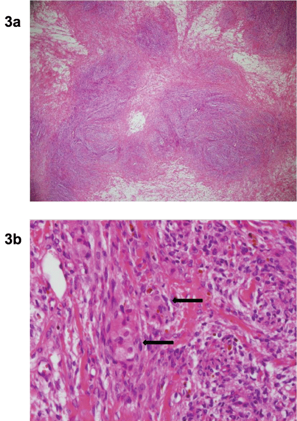
Microscopically evaluated, the solid area showed a pseudolobular pattern alternating between hypercellular and hypocellular areas (Figure 3a). The cellular areas were composed of dual cell populations: collagen-producing spindle cells and rounded weak lutein cells (Figure 3b). The results of the immunohistochemistry (IHC) assessment included Inhibin α: Focal positive, Calretinin: Positive, Smooth muscle actin: Positive, Desmin: Focal weak positive and CD34: Negative. The pathological diagnosis was SST of the left ovary.
.
Figure 3: Microscopic features of the sclerosing stromal tumor from case 2
(a) The solid area includes a pseudolobular pattern of alternating hypercellular and hypocellular areas (H&E x40).
(b) The cellular areas are composed of dual cell populations: collagen-producing spindle cells and rounded weak lutein cells (arrow) (H&E x400).
View Figure 3
Discussion
SST of the ovary is a distinct ovarian stromal tumor subtype [4]. Ovarian SST most commonly occurs during the second to third decades of life at an average age of diagnosis of 27.5 years. More than 80 % of SST cases occur in patients under 30 years of age [5]. It is rare that SST is diagnosed in postmenopausal women, however, recently it has been reported that a 80-year old woman had SST. I t is far more difficult to diagnose SST in elderly women for many reasons; the incidence is much lower in elderly women, symptoms related to menstruation cycle can be hidden due to patients' menopause, and various common conditions can cause non-specific abdominal pain in senior populations [6].
In children and adolescents, symptoms include premature menarche, menstrual irregularities, abdominal discomfort/pain, and rarely, ascites [6]. In post-menarcheal females, SST presents with menstrual irregularities and/or an abdominal mass. In our cases, the patients presented with abdominal pain and a palpable mass. SST sizes vary from 1.5 to 20cm in diameter [5].
In patients with SST, serum CA-125 levels have been found to be either elevated or within reference ranges [7]. The tumor in SST cases is usually hormonally inactive, although cases accompanied by irregular menses and genital bleeding have been reported. Peng et al. found 114 SST cases reported through 2003 [4]. In our two cases, we observed normal serum hormone levels with no clinical virilization. One patient's blood tumor marker was normal while the other was mildly elevated.
The sonographic findings associated with SST include a well-defined solid mass with hyperechoic honeycomb structures, which are also the characteristics of a mixed heterogenecity tumor without focal calcifications [8].
It is difficult to diagnose before surgery by imaging studies. It used be diagnosed by pathological examination during or after surgery. A preoperative diagnosis of SST is possible based on magnetic resonance imaging (MRI) findings that demonstrate pseudolobulation, which consist of low-intensity nodules set against high-intensity stroma on T2-weighted images [9]. The presence of tightly packed cellular areas associated with foci of sclerosis justifies the observed low density of these nodules on T2-weighted images. High-intensity areas found on the T2-weighted images correlated with poorly formed cellular tissue that was markedly edematous. However, the differentiation between SST and other stromal tumors and metastatic ovarian tumors based on MRI results needs further investigation. Upon the analysis of dynamic contrast enhanced images, the tumors revealed early peripheral enhancement with centripetal progression. Striking early enhancement reflects the cellular areas with their prominent vascular networks, and an area of prolonged enhancement in the inner portion of the mass represents the collagenous hypocellular area. These findings can be useful in differentiating SST from fibroma, as fibroma produces an absence of early enhancement and delayed accumulation of the contrast material [10,11].
Histologically, SST is characterized by cellular heterogeneity, prominent vasculature, and a pseudolobular structure composed of both cellular and hypocellular areas [12]. The name "sclerosing stromal tumor" was proposed because the cellular portions of the tumor tend to undergo collagenous sclerosis. SST has occasionally been confused with massive ovarian edema and Krukenberg's tumor [13]. The distinction between SST and Krukenberg's tumor can be made using immunohistochemistry staining [4]. SST is positive for desmin and smooth muscle action (SMA). Inhibin also has been shown to be positive and be a useful marker for ovarian sex cord stromal tumors [14,15]. In addition, both the cellular and edematous areas are positive for vascular endothelial growth factor. Other stromal tumors, i.e. thecoma and fibroma, tend to occur in the fifth and sixth decades of life of afflicted patients [13] whereas almost 80 % of SSTs occur in women under 30 years of age [14].
While most cases of SST have been reported to occur in the second and third decades of life, in this report, we present two cases of SST in postmenopausal women. We expect that this report will be helpful in the differential diagnosis of future SST cases that may occur in atypical patient populations [16,17].
References
-
Russell P, Farnsworth A (1997) Surgical Pathology of the Ovaries. (2nd edn). New York: Churchill Livingstone: 422.
-
Prat J (2004) Pathology of the Ovary. London: Saunders: 216-218.
-
Fox H, Wells M (2008) Haines and Taylor Obstetrical and Gynecological Pathology. (5th edn) New York: Cherchill Livingstone: 842-843.
-
Peng HH, Chang TC, Hsueh S (2003) Sclerosing stromal tumor of ovary. Chang Gung Med J 26: 444-448.
-
Saitch A, Tsutsumi Y, Osamura Y, Watanabe Y (1989) Sclerosing stromal tumor of the ovary. Immunohistochemical and electron-microscopic demonstration of smooth-muscle differentiation. Arch Pathol Lab Med 113: 372-376.
-
Kim TH, Lee HH, Hong JA, Park J, Jeon DS, et al. (2014) Sclerosing stromal tumor in an elderly postmenopausal woman. J Menopausal Med 20: 80-83.
-
Gwin K, Marino-Enriquez A, Martel M, Reyes-Mugica M (20009) Sclerosing stromal tumor: An important differential diagnosis of ovarian neoplasms in childhood and adolescence. Pediatr Dev Pathol 12: 366-370.
-
Marelli G, Carinelli S, Mariani A, Frigerio L, Ferrari A (1998) Sclerosing stromal tumor of the ovary. Report of eight cases and review of the literature. Eur J Obstet Gynecol Reprod Biol 76: 85-89.
-
Stylianidou A, Varras M, Akrivis C, Fylaktidou A, Stefanaki S, et al. (2001) Sclerosing stromal tumor of the ovary: a case report and review of the literature. Eur J Gynaecol Oncol 22: 300-304.
-
Ihara N, Togashi K, Todo G, Nakai A, Kojima N, et al. (1999) Sclerosing stromal tumor of the ovary: MRI. J Comput Assist Tomogr 23: 555-557.
-
Jung SE, Rha SE, Lee JM, Park SY, Oh SN, et al. (2005) CT and MRI findings of sex cord-stromal tumor of the ovary. AJR Am J Roentgenol 185: 207-215.
-
Mikami M, Tanaka K, Komiyama S (2003) Magnetic resonance imaging in sclerosing stromal tumor of the ovary. Int J Gynaecol Obstet 83: 319-321.
-
Kawauchi S, Tsuji T, Kaku T, Kamura T, Nakano H, et al. (1998) Sclerosing stromal tumor of the ovary: a clinicopathologic, immunohistochemical, ultrastructural, and cytogenetic analysis with special reference to its vasculature. Am J Surg Pathol 22: 83-92.
-
Ozdemir O, Sari ME, Sen E, Kurt A, Ileri AB, et al. (2014) Sclerosing stromal tumour of the ovary: A case report and the review of literature. Niger Med J 55: 432-437.
-
Zekioglu O, Ozdemir N, Terek C, Ozsaran A, Dikmen Y (2010) Clinicopathological and immunohistochemical analysis of sclerosing stromal tumours of the ovary. Arch Gynecol Obstet 282: 671-676.
-
Terauchi F, Onodera T, Nagashima T, Kobayashi Y, Moritake T, et al. (2005) Sclerosing stromal tumor of the ovary with elevated CA125. J Obstet Gynaecol Res 31: 432-435.
-
Young RH, Scully RE (2001) Differential diagnosis of ovarian tumors based primarily on their patterns and cell types. Semin Diagn Pathol 18: 161-235.
