

Obstetrics and Gynaecology Cases - Reviews
Uterine Tumour Resembling Ovarian Sex Cord Tumour (UTROSCT): Experience with a Rare Disease. Two Case Reports and Overview of the Literature
Brenda Hermsen1#*, Fabrizio Bogliatto2#, Maaike Bleeker3, Luciano Leidi2, Hans Trum4, Erika Comello5 and Christianne Lok4
1Department of Gynaecology and Obstetrics, Sint Lucas Andreas Hospital, The Netherlands
2Department of Gynaecology and Obstetrics, Chivasso Civic Hospital, Italy
3Department of Pathology, VU University Medical Centre, The Netherlands
4Department of Gynaecologic Oncology, Centre for Gynaecologic Oncology Amsterdam, The Netherlands
5Department of Pathology, Ivrea Hospital, Italy
#Contributed equal to this manuscript
*Corresponding author: Dr. B.B.J. Hermsen, Department of Gynaecology and Obstetrics, Sint Lucas Andreas Hospital, Jan Tooropstraat 161, 1064 AE, Amsterdam, The Netherlands, E-mail: b.hermsen@slaz.nl
Obstet Gynecol Cases Rev, OGCR-2-049, (Volume 2, Issue 4), Case Report; ISSN: 2377-9004
Received: May 31, 2015 | Accepted: July 06, 2015 | Published: July 09, 2015
Citation: Hermsen B, Bogliatto F, Bleeker M, Leidi L, Trum H, et al. (2015) Uterine Tumour Resembling Ovarian Sex Cord Tumour (UTROSCT): Experience with a Rare Disease. Two Case Reports and Overview of the Literature. Obstet Gynecol Cases Rev 2:049. 10.23937/2377-9004/1410049
Copyright: © 2015 Hermsen B, et al. This is an open-access article distributed under the terms of the Creative Commons Attribution License, which permits unrestricted use, distribution, and reproduction in any medium, provided the original author and source are credited.
Abstract
Background: Uterine tumour resembling an ovarian sex cord tumour (UTROSCT) is a rare neoplastic lesion. Patients are generally of reproductive age. Although conservative treatment to preserve fertility has been reported, literature on the optimal treatment on UTROSCT is scarce and only case reports or small case series are available.
Aim: To describe the difficulties and resemblances of two different cases of UTROSCT in a pre- and post-menopausal woman in order to classify this uncommon tumour.
Methods: Case reports and review of the literature.
Case 1: A 36-year-old woman presented with irregular blood loss. A transcervical resection of a fibroid was performed and histology showed an UTROSCT. At the time of the diagnosis she was 7 weeks pregnant. After conservative management, a caesarean section and postpartum hysterectomy were performed at 34 weeks and no residual disease was found. Follow up did not show any abnormalities.
Case 2: A 68 year old post-menopausal woman presented with abnormal uterine bleeding for which she underwent a curettage. Endometrial stromal sarcoma was suspected and an abdominal hysterectomy with bilateral salpingo-oophorectomy was performed. Revision of histology by expert pathologists showed UTROSCT. Follow up was uneventful.
Literature overview: Only case-reports and small case series are described in literature. Ages of the patients ranged from 24 years to 79 years old. In the majority of cases an abdominal hysterectomy and bilateral salpingo-oophorectomy was performed and only in a few cases conservative therapy was performed. Follow up is often unknown.
Conclusion: Histologic review by experts in multi-institutional panels is helpful in making this difficult diagnosis. In selected cases, conservative treatment of an UTROSCT can be considered. Follow up in fertility-sparing procedures is important. Although, recurrences after hysterectomy have not been described, it is still unknown whether follow up can be safely omitted.
Introduction
Uterine tumour resembling an ovarian sex cord tumour (UTROSCT) is a rare neoplastic lesion. It has been described for the first time in 1945 by Morehead [1]. This tumour often causes abnormal vaginal bleeding and uterine enlargement, suggesting leiomyomas or polyps [2].
Originally, tumours with sex cordlike elements were divided in two groups based on clinical and histopathological features [2]. Tumours showing a predominant endometrial stromal tumour like differentiation and focal ovarian sex cordlike areas (<50%) were called endometrial stromal tumours with sex cordlike elements (ESTSCLE). Tumours that predominantly composed of sex cordlike elements were called UTROSCT. The majority of this second type of tumours behave in a benign fashion [3] but loco regional spread or abdominal relapse have been reported in some patients. In contrast, ESTSCLE is more often associated with metastases and recurrences.
More recently, the WHO 2014 classifies UTROSCT in a miscellaneous group of tumours of the uterine corpus [3]. Cytogenetic investigations showed that in ESTSCLE fusion of two novel genes (JAZF1 and JJAZ1) occurs which is not observed in UTROSCT, emphasizing that UTROSCT is another entity than ESTSCLE.
UTROSCT should be differentiated from a.o. leiomyoma, low-grade endometroid carcinoma with sex-cordlike features, metastases of ovarian sex-cord stromal tumours and carcinosarcoma.
The aim of this paper is to describe two cases of UTROSCT and to give an overview of the literature with respect to the difficulties of diagnosing UTROSCT, the importance of expert pathology review and to its treatment and prognosis.
Case 1
A 36-year-old woman presented with complaints of irregular blood loss. Her medical history did not reveal any notable medical problems. Ultrasonography showed a submucosal fibroid. A transcervical resection (TCR) of the fibroid was performed. Because of persistent complaints a second TCR was performed, complicated by a perforation requiring laparoscopy. Histological examination of the tissue showed the possibility of an UTROSCT. Upon returning to the outpatient clinic to discuss the results of the histology, she was found to be 7 weeks pregnant. She was then referred to our tertiary clinic. A healthy woman without lymphadenopathy was seen. Ultrasound confirmed the pregnancy and gestational age. In the myometrium, a structure of three centimetres was found suggestive of recurrent tumour. Both ovaries appeared to be normal. Laboratory investigations showed an increased inhibine A (177ng/L) and inhibine B (36ng/L). MRI at 9 weeks showed a transmural hypodense abnormality of almost four centimetres, there were no signs of lymphadenopathy or metastases. Histopathology and immunohistochemistry suggested an UTROSCT (Figure 1). After regional and international pathological review an unusual UTROSCT with infiltrative pattern of the myometrial smooth muscle was diagnosed.
.
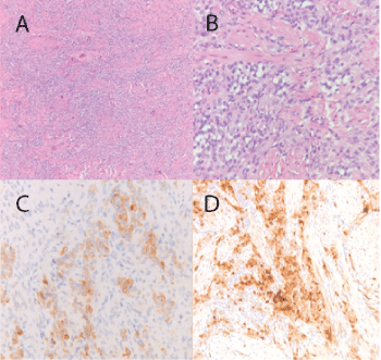
Figure 1: Microscopic images of case 1
Histopathology showed infiltrative pattern (A) and sex cord like elements (B). The tumour cells contained bland ovoid to spindle shaped nuclei and abundant cytoplasm, which ranged from eosinophilic to clear. Occasionally a more organoid growth pattern was present as well as focal pseudovascular or pseudoglandular spaces. The mitotic count was low. There was no network of small arteriole-like vascular channels. Immunohistochemical stains showed quite diffuse staining with AE1/3 and ER (not shown). There was focal staining with desmin and immunoreactivity with CD99, inhibin (C), calretinin (D) and CD10. H-caldesmon, HMB45 and CD56 were negative (not shown).
View Figure 1
While conservative treatment may be undertaken for UTROSCT if fertility preservation is a matter of debate, the presence of an infiltrative pattern in this case was disturbing. A multidisciplinary team discussed the appropriate treatment during this pregnancy. The patient and her partner expressed a strong wish to continue the pregnancy to a viable gestational age. She was counselled about the risks reported in the scarce information from the literature. It was decided to perform a MRI every 2 months for follow up of the lesion and for scientific interest (Figure 2). During the pregnancy, the abnormality seen on MRI did not increase in size. Ultrasound revealed no fetal abnormalities and normal fetal growth. A caesarean section followed by hysterectomy was performed at a gestational age of 34 weeks because it was felt that waiting 6 more weeks could be hazardous for the mother because of the infiltrative component of the tumour for which normally an immediate hysterectomy would have been performed. The procedure was uneventful. A healthy baby girl was born and was admitted to the neonatal care unit for prematurity. No abnormalities were found in the abdomen or retroperitoneum during surgery. The neonate did recover well from prematurity. Final histological examination showed no residual tumour. Adenomyosis was found in the myometrium and in the biopsies from the peritoneum. No rest of tumour was found. Follow up at 2 year did not show recurrent disease.
.
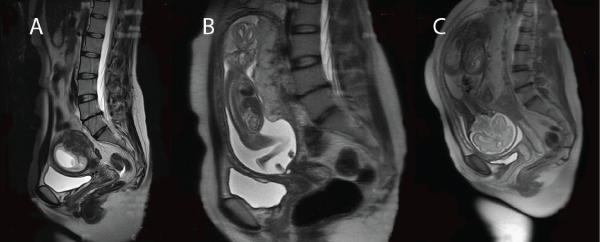
Figure 2: MRI scans during pregnancy week 8 and 15
A selection of MRI's from 8 weeks gestational age (A), 15 weeks (B) and 27 weeks (C) is shown.
View Figure 2
Case 2
A 68 year old postmenopausal woman presented with abnormal uterine bleeding. Her past medical history revealed no medical problems. Ultrasonography showed a 15mm irregular endometrial thickness. She underwent dilatation and curettage (D&C). Gross examination revealed multiples gelatinous grayish-brown endometrial fragments. The case was initially diagnosed as endometrial stromal sarcoma. No evidence of metastasis was found. CT-scan and MRI revealed a submucosal exophytic mass, but no lymph node enlargement. She underwent an uncomplicated abdominal hysterectomy and bilateral salpingo-oophorectomy.
On macroscopic examination, a polypoid mass was located at the fundus of uterus, protruding into the uterine cavity. Necrosis and hemorrhage were not found. The tumour border was demarcated. Myometrial infiltration was not grossly detected. Histopathology is shown in figure 3. Results of immunohistochemistry are described in the figure legend. Expert pathologists were consulted and only then UTROSCT was diagnosed.
.
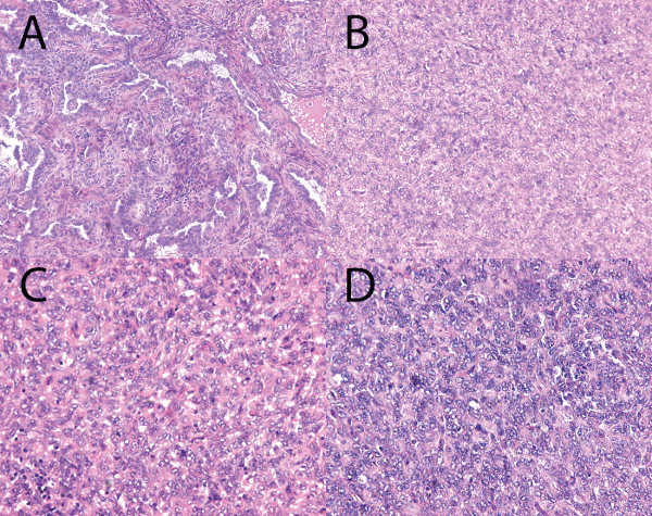
Figure 3: Microscopic images of case 2
Histopathology revealed monomorphic round to oval tumour cells, vesicular nuclei with inconspicuous moderate amount of pale cytoplasm. A variety of architectural patterns like anastomosing trabeculae (B, D), plexiform cords, and tubules were identified. Immunohistochemical evaluation revealed high diffuse positivity for vimentin, ER, PR in tumour cells. CD10, cytokeratin 8-18 and AE1-AE3 showed focal positivity (not shown). Ki67 showed a proliferation index of maximum 15%. Microscopic examination of the specimen was completed with testing CD99 and S100, with diffuse high positivity. Inhibin and CDX2 were found to be negative (not shown).
View Figure 3
Since effect of adjuvant chemotherapy in UTROSCT is unknown, it was decided to follow the patient with CT-scan and MRI every 6 months, without any adjuvant therapy. Follow up did not show any recurrences.
In table 1 the literature on UTROSCT is summarized. Ages of the patients ranged from 24 years to 79 years old. Patients often present with abnormal uterine blood loss. In the majority of cases an abdominal hysterectomy and bilateral salpingo-oophorectomy was performed and only in a few cases conservative therapy was performed. Follow up is often unknown or short, but no recurrence of disease has been reported.
Discussion
UTROSCT are rare tumours and only case-reports and small case series are described in literature (Table 1). However, because of the recent changed insights and the new WHO classification, it is difficult to compare or pool these different cases. The origin of UTROSCT remains uncertain. UTROSCTs do not have the JAZF1-SUZ12 fusion that characterized endometrial stromal tumours, suggesting that these tumours do not originate from the endometrial stroma [3]. UTROSCTs display different patterns and typically grow in sheets, cords, small nests, trabeculae and tubuli. The tumours show little atypia, mitotic figures are usually rare and cells are often small with round nuclei and indistinct eosinophilic cytoplasm [4]. The most reliable immunohistochemistry markers are calretinin, inhibin CD99 and melanin-A [5].
![]()
Table 1: Overview of the literature of uterine tumour resembling an ovarian stromal sex cord tumour
View Table 1
The diagnosis of an UTROSCT is often only made postoperatively when the uterus is examined, like in the second case. In the majority of cases described in literature a hysterectomy was performed. The largest series (N=14) of cases was published in 1976 [6,7] and all patients underwent a hysterectomy. UTROSCT is usually a well circumscribed lesion. However, occasional neoplasm occurs which exhibit both smooth muscle differentiation and features of UTROSCT and this might be possible in our first case. They could have originated from foci of adenomyosis, stromal myosis, endometriosis, or multipotential cells within the myometrium [7] in accordance to our case where adenomyosis was found in the uterus after hysterectomy.
The optimal treatment in the perimenopausal period is abdominal hysterectomy and bilateral salpingo-oophorectomy. However, when the patient is younger, preservation of fertility is an important consideration. A fertility conserving treatment has been reported in only three patients with UTROSCT [4-6]. Follow up of these cases until respectively 13 and 18 months was described with no evidence of disease, but in one case follow up was not stated.
When the tumour is well differentiated on microscopic examination, local excision with an adequate margin of adjacent uninvolved myometrium may be justified. Whatever treatment is chosen, all patients should be followed up at regular intervals. Although, fertility was not spared in our first case, treatment was postponed to allow the pregnancy to continue. In this case, a hysterectomy would have been performed if our patient would not have been pregnant because of the following reasons: 1) the tumour was regarded as an intrauterine recurrence because the first conservative treatment (TCR) failed 2) the infiltrative component of the tumour increased the risk of metastases and recurrences in the mother, 3) the second procedure (TCR) was complicated by perforation and incomplete resection, further increasing the risks for maternal health, 4) conservative treatment is described in only a few women presenting with the tumour for the first time and with uncomplicated surgical treatment. On the other hand, the parents had a very strong wish to continue the pregnancy despite the risks and cancer in pregnancy often can be treated conservatively in order to achieve a gestational age at which the unborn child has a change of survival.
It is estimated that 3000 to 5000 pregnant women are confronted with cancer across Europe each year [8]. Treatment is a challenge under such circumstances considering that doctors have to consider both the recovery of the mother as well as the wellbeing of the unborn child. In our case surgery was no option, the effect of chemotherapy in UTROSCT is unknown and radiotherapy of the uterus with a fetus in utero is impossible.
Van Calsteren et al reports that in 72% of pregnancies complicated by (suspicion of) cancer, delivery is induced and that elective caesarean section is performed in 35% of the women [9]. Thus, a major problem in the treatment of pregnant women with cancer is prematurity and if possible, an attempt should be made to postpone delivery. In the first case, after careful consideration with a multidisciplinary team it was decided to accept prematurity to decrease potential maternal risk due to the uncertainty on the presence of tumour cells in the uterine cavity and the fear for metastases and uterine rupture. In retrospect, the operation could have been performed at term.
In the second case, according to the literature regarding postmenopausal treatment, a total hysterectomy with bilateral salpingo-oophorectomy was performed.
Because of the rarity of this neoplasia, histological review by expert pathologists is essential to avoid misdiagnosis. One institution will never achieve high patients' numbers with this rare tumour. Therefore, a multi-institutional cooperation between gynaecologist and pathologist guarantees optimal diagnosis and treatment of patients affected by UTROSCT.
In conclusion, in selected cases, (temporarily) conservative treatment of an UTROSCT seems to be acceptable without compromising survival of the patient. Thus, in pregnancy postponement of definitive treatment can be considered to increase the chance of survival of the newborn. In peri- or postmenopause, a hysterectomy is advised. In both situations, follow up is advocated, since theoretically, UTROSCT may recur.
Acknowledgements
We thank doctor McCluggage, department of pathology, Belfast, for providing an expert opinion on the histology of the first case.
References
-
Morehead RP, Bowman MC (1945) Heterologous Mesodermal Tumors of the Uterus: Report of a Neoplasm Resembling a Granulosa Cell Tumor. Am J Pathol 21: 53-61.
-
Kurman RJ, Hedrick Ellenson L, Ronnett BM (2011). Blaustein's pathology of the female genital tract. (6th edn). Springer, USA (ISBN 978-1-4419-0488-1) 10: 455-496.
-
Pradhan D, Mohanty SK (2013) Uterine tumors resembling ovarian sex cord tumors. Arch Pathol Lab Med 137: 1832-1836.
-
Wang J, Blakey GL, Zhang L, Bane B, Torbenson M, et al. (2003) Uterine tumor resembling ovarian sex cord tumor: report of a case with t(X;6)(p22.3;q23.1) and t(4;18)(q21.1;q21.3). Diagn Mol Pathol 12: 174-180.
-
Irving JA, Carinelli S, Prat J (2006) Uterine tumors resembling ovarian sex cord tumors are polyphenotypic neoplasms with true sex cord differentiation. Mod Pathol 19: 17-24.
-
Nogales FF, Stolnicu S, Harilal KR, Mooney E, Garc�a-Galvis OF (2009) Retiform uterine tumours resembling ovarian sex cord tumours. A comparative immunohistochemical study with retiform structures of the female genital tract. Histopathology 54: 471-477.
-
Clement PB, Scully RE (1976) Uterine tumors resembling ovarian sex-cord tumors. A clinicopathologic analysis of fourteen cases. Am J Clin Pathol 66: 512-525.
-
Amant F, Van Calsteren K, Halaska MJ, Gziri MM, Hui W, et al. (2012) Long-term cognitive and cardiac outcomes after prenatal exposure to chemotherapy in children aged 18 months or older: an observational study. Lancet Oncol 13: 256-264.
-
Van Calsteren K, Heyns L, De Smet F, Van Eycken L, Gziri MM, et al. (2010) Cancer during pregnancy: an analysis of 215 patients emphasizing the obstetrical and the neonatal outcomes. J Clin Oncol 28: 683-689.
-
Tang CK, Toker C, Ances IG (1979) Stromomyoma of the uterus. Cancer 43: 308-316.
-
Fekete PS, Vellios F, Patterson BD (1985) Uterine tumor resembling an ovarian sex-cord tumor: report of a case of an endometrial stromal tumor with foam cells and ultrastructural evidence of epithelial differentiation. Int J Gynecol Pathol 4: 378-387.
-
Fukunaga M, Miyazawa Y, Ushigome S (1997) Endometrial low-grade stromal sarcoma with ovarian sex cord-like differentiation: report of two cases with an immunohistochemical and flow cytometric study. Pathol Int 47: 412-415.
-
Franco A, Aquino NM, Malik SL, Navarro C (1999) Sonographic presentation of uterine sex cord-stromal tumor. J Clin Ultrasound 27: 199-201.
-
Hauptmann S, Nadjari B, Kraus J, Turnwald W, Dietel M (2001) Uterine tumor resembling ovarian sex-cord tumor--a case report and review of the literature. Virchows Arch 439: 97-101.
-
Okada S, Uchiyama F, Ohaki Y, Kamoi S, Kawamura T, et al. (2001) MRI findings of a case of uterine tumor resembling ovarian sex-cord tumors coexisting with endometrial adenoacanthoma. Radiat Med 19: 151-153.
-
Suzuki C, Matsumoto T, Fukunaga M, Itoga T, Furugen Y, et al. (2002) Uterine tumors resembling ovarian sex-cord tumors producing parathyroid hormone-related protein of the uterine cervix. Pathol Int 52: 164-168.
-
Franco S, Andrade MJ, Silva TS, Guerra C, De Oliveira C (2003) Uterine tumor resembling ovarian sex-cord tumor. Acta Med Port 16: 365-367.
-
Kuruvila S, Samarasinghe D, Thomas S (2003) Uterine neoplasm resembling an ovarian sex cord tumor. Saudi Med J 24: 221-223.
-
Kabbani W, Deavers MT, Malpica A, Burke TW, Liu J, et al. (2003) Uterine tumor resembling ovarian sex-cord tumor: report of a case mimicking cervical adenocarcinoma. Int J Gynecol Pathol 22: 297-302.
-
Wang J, Blakey GL, Zhang L, Bane B, Torbenson M, et al. (2003) Uterine tumor resembling ovarian sex cord tumor: report of a case with t(X;6)(p22.3;q23.1) and t(4;18)(q21.1;q21.3). Diagn Mol Pathol 12: 174-180.
-
Hillard JB, Malpica A, Ramirez PT (2004) Conservative management of a uterine tumor resembling an ovarian sex cord-stromal tumor. Gynecol Oncol 92: 347-352.
-
Czernobilsky B (2008) Uterine tumors resembling ovarian sex cord tumors: an update. Int J Gynecol Pathol 27: 229-235.
-
Garuti G, Gonfiantini C, Mirra M, Galli C, Luerti M (2009) Uterine tumor resembling ovarian sex cord tumors treated by resectoscopic surgery. J Minim Invasive Gynecol 16: 236-240.
-
de Leval L, Lim GS, Waltregny D, Oliva E (2010) Diverse phenotypic profile of uterine tumors resembling ovarian sex cord tumors: an immunohistochemical study of 12 cases. Am J Surg Pathol 34: 1749-1761.
-
Carta G, Crisman G, Margiotta G, Mastrocola N, Di Fonso A, et al. (2010) Uterine tumors resembling ovarian sex cord tumors. A case report. Eur J Gynaecol Oncol 31: 456-458.
-
Abdullazade S, Kosemehmetoglu K, Adanir I, Kutluay L, Usubutun A (2010) Uterine tumors resembling ovarian sex cord-stromal tumors: synchronous uterine tumors resembling ovarian sex cord-stromal tumors and ovarian sex cord tumor. Ann Diagn Pathol 14: 432-437.
-
Stefanovic A, Jeremic K, Kadija S, Mitrovic M, Filimonovic D, et al. (2013) Uterine tumor resembling ovarian sex cord tumor. Case report and review of literature. Eur J Gynaecol Oncol 34: 275-277.
-
Umeda S, Tateno M, Miyagi E, Sakurai K, Tanaka R, et al. (2014) Uterine tumors resembling ovarian sex cord tumors (UTROSCT) with metastasis: clinicopathological study of two cases. Int J Clin Exp Pathol 7: 1051-1059.
-
Bakula-Zalewska E, Danska-Bidzinska A, Kowalewska M, Piascik A, Nasierowska-Guttmejer A, et al. (2014) Uterine tumors resembling ovarian sex cord tumors, a clinicopathologic study of six cases. Ann Diagn Pathol 18: 329-332.
-
Abid N, Mnif H, Mellouli M, Charfi S, Khabir A, et al. (2014) Uterine tumour resembling ovarian sex cord tumours presenting as multiple endometrial and cervical uterine polyps: a case report. Pathologica 106: 73-76.
-
Hashmi AA, Faridi N, Edhi MM, Khan M (2014) Uterine tumor resembling ovarian sex cord tumor (UTROSCT), case report with literature review. Int Arch Med 7: 47.
