

Obstetrics and Gynaecology Cases - Reviews
Cervical Varices Presenting as Vaginal Bleeding: A Description of Two Cases and A Management Plan
Kathy Chyjek1*, Catherine Hutz2, Charles Macri3, Jeffrey Berger4, Anthony Venbrux5, Nadia Khati6, Dorothy Bulas7 and John Larsen3
1Department of Obstetrics, Gynecology and Reproductive Science, Icahn School of Medicine at Mount Sinai, USA
2Department of Obstetrics and Gynecology, Mayo Clinic, USA
3Department of Obstetrics and Gynecology, The George Washington University Hospital, USA
4Department of Anesthesiology and Critical Care Medicine, The George Washington University Hospital, USA
5Department of Radiology, Division of Interventional Radiology, The George Washington University Hospital, USA
6Department of Radiology, The George Washington University Hospital, USA
7Department of Diagnostic Imaging and Radiology, Children's National Medical Center, USA
*Corresponding author: Kathy Chyjek, M.D, Department of Obstetrics, Gynecology and Reproductive Science at Icahn School of Medicine at Mount Sinai New York, NY 10029, USA, E-mail: kathy.chyjek@mountsinai.org
Obstet Gynecol Cases Rev, OGCR-2-053, (Volume 2, Issue 5), Case Report; ISSN: 2377-9004
Received: April 20, 2015 | Accepted: July 28, 2015 | Published: August 01, 2015
Citation: Chyjek K, Hutz C, Macri C, Berger J, Venbrux A, et al. (2015) Cervical Varices Presenting as Vaginal Bleeding: A Description of Two Cases and A Management Plan. Obstet Gynecol Cases Rev 2:053. 10.23937/2377-9004/1410053
Copyright: © 2015 Chyjek K, et al. This is an open-access article distributed under the terms of the Creative Commons Attribution License, which permits unrestricted use, distribution, and reproduction in any medium, provided the original author and source are credited.
Abstract
Background: Varicose veins are common in pregnancy. However, potentially life-threatening uterine cervical varices have rarely been reported and there are currently no guidelines for their management.
Cases: We present two cases of cervical varices found in the setting of second and third trimester bleeding, report on the clinical outcomes and propose a management plan. Both of our patients were delivered between 38-39 weeks gestation and required interdisciplinary meetings between Obstetrics and Gynecology, Anesthesiology, Neonatology and Interventional Radiology to prepare for delivery.
Conclusion: For patients in which varices are found inside the cervix, we recommend consideration of second trimester cerclage followed by serial ultrasound and scheduled cesarean delivery. Vascular embolization should also be considered.
Introduction
Varicose veins are a common finding in up to 15% of pregnancies [1]. However, potentially life-threatening uterine cervical varices have only rarely been reported [2-11]. In a retrospective review, O'Brien et al. suggest that the incidence of cervical varices may be significantly higher than previously thought, particularly in association with placenta previa [11]. Given that they can present with second or third trimester hemorrhage, uterine cervical varices should be considered as part of the differential diagnosis when evaluating vaginal bleeding in pregnancy and should be documented at the time of routine ultrasound, if present [8]. There are currently no specific guidelines for managing pelvic varicose veins. We present two cases of uterine cervical varices found in the setting of second and third trimester bleeding, report on the clinical outcomes and propose a management plan.
Case 1
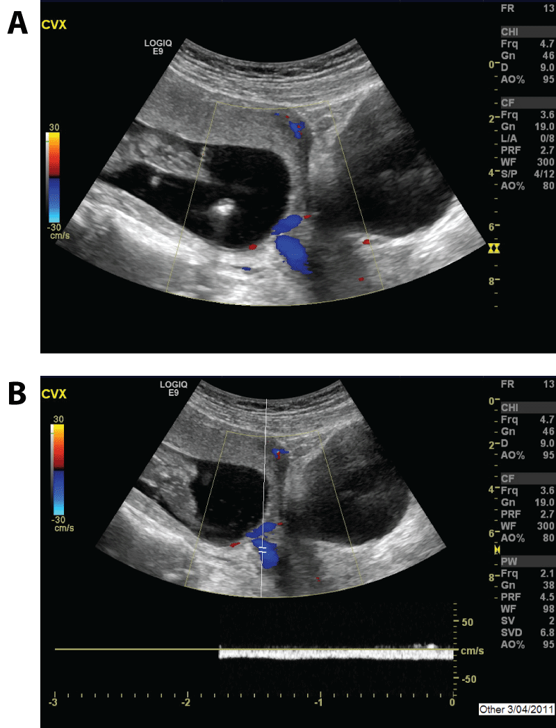
A 31-year-old, gravida 1 para 0 abortus 0, presented to her obstetrician at 15 weeks gestation timed by intrauterine insemination, complaining of severe abdominal cramping. Transabdominal ultrasound showed a single live intrauterine pregnancy and a low lying placenta. A complex echogenic area within the endocervical canal suggestive of a mucus plug was noted. Two days later, the patient experienced severe vaginal bleeding with passage of numerous blood clots with an estimated blood loss of 200 milliliters. Repeat imaging at that time revealed a hypoechoic area distending the endocervix to 1.5cm. Color and pulsed Doppler showed markedly increased vascularity indicative of cervical varices versus a venous angioma or other arteriovenous malformation (AVM). Active flow extended into the anterior body of the cervix, near the internal os and given the low velocity venous pressure, cervical varices were confirmed (Figure 1). There was no apparent placenta previa at this time. Although this acute bleeding episode resolved, the patient presented the following day to the emergency department with profuse, painless, bright red vaginal bleeding of approximately 600 milliliters. She remained hemodynamically stable with minimal change in hemoglobin. Conservative management with vaginal packing and observation was implemented. Sterile speculum exam revealed a 1-2cm dilated cervix with decreased bleeding and tortuous appearing vessels at the level of the internal os. No contractions were observed on tocometry. An emergent McDonald cerclage was placed and modified bed rest and pelvic rest was enforced. Follow-up ultrasound examinations continuously demonstrated varices at the level of the internal os. An MRI at 26 weeks gestation was performed and demonstrated intermediate signal material in the internal os with a normal appearing anterior placenta. The prenatal course was otherwise uneventful.
.
Figure 1: Intracervical varices in a 31-year-old gravid woman at 15 weeks gestation. A, Color Doppler transabdominal ultrasound of the cervix and lower uterine segment showing an intrauterine pregnancy, intracervical varices, and bladder in the sagittal plane. B, Color and pulsed Doppler transabdominal ultrasound at the same level showing a venous waveform consistent with intracervical varices.
View Figure 1
In order to avoid the possibility of uncontrollable bleeding with vaginal delivery, a primary cesarean delivery was scheduled for 39 weeks gestation. Multi-disciplinary meetings between obstetrician, anesthesiologist, interventional radiologist, neonatologist, and nursing staff allowed for proper planning and communication of concerns. Following epidural placement in the Interventional Suite, bilateral uterine artery stent and balloon placement was performed by the interventional radiologist to help control any intraoperative or postpartum hemorrhaging. The cesarean delivery was performed in the Main Operating Room with fluoroscopy readily available. Intraoperatively, multiple varices, including a midline 2cm wide anomalous vein running vertically from the cervix to the fundus on the anterior surface of the uterus, were found. Uterine artery balloons were inflated during delivery to allow for better hemostasis.
A viable male infant was delivered in cephalic presentation, weighing 7 pounds 13 ounces with Apgar scores of 8 and 9 at 1 and 5 minutes, respectively. The placenta was extracted manually and the uterus was cleared of any retained products. Uterine artery balloons were deflated and hemostasis confirmed prior to closure. After inspecting for bleeding, the uterus was closed and the cerclage was removed without incident. Estimated total blood loss was 1000 milliliters. At the conclusion of the cesarean delivery, the patient was taken back to the Interventional Suite where, under epidural anesthesia, uterine artery embolization with Gelfoam was performed.
Case 2
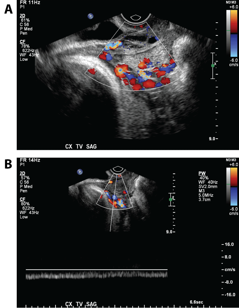
A 36-year-old, gravida 1 para 0 abortus 0, who was followed by an obstetrician after in vitro fertilization, presented to triage at 30 weeks and 5 days gestation with vaginal spotting. Transabdominal ultrasound showed a single live intrauterine pregnancy and a low lying placenta 1.7cm from the cervix. Five days later, the patient experienced two episodes of heavy vaginal bleeding, of approximately 100cc, which again brought her into triage. Evaluation of the vaginal bleeding using transabdominal and transvaginal color and pulsed Doppler ultrasound revealed prominent varices in the region of the lower uterine segment and internal os (Figure 2). The patient was advised to meet with an interventional radiologist, who discussed prophylactic placement of balloon occlusion catheters in the bilateral internal iliac arteries in case hemorrhage ensued during cesarean delivery. Using a multi-disciplinary approach, a unified plan was conveyed to the patient. The prenatal course was otherwise unremarkable with a primary cesarean delivery scheduled for 38-39 weeks gestation. Prior to delivery, the patient had epidural anesthesia placed in the Interventional Suite. Bilateral internal iliac artery stent and balloon placement was performed by the interventional radiologist. The cesarean delivery was performed in the Main Operating Room with fluoroscopy readily available. Intraoperatively, multiple dilated veins were noted in the lower uterine segment extending into the cervix.
.
Figure 2: Intracervical varices in a 36-year-old gravid woman at 30 weeks and 5 days gestation. A, Color Doppler transvaginal ultrasound of the cervix showing multiple intracervical dilated vascular structures in the sagittal plane. B, Color and pulsed Doppler transvaginal ultrasound at the same level showing a venous waveform consistent with intracervical varices.
View Figure 2
A viable male infant was delivered in cephalic presentation, weighing 8 pounds 6 ounces with Apgar scores of 9 and 9 at 1 and 5 minutes, respectively. The placenta was extracted manually, though with notable adherence in the lower uterine segment. Several bleeding veins were encountered and sutures were placed in the lower uterine segment to obtain hemostasis. There was no need for balloon inflation. After inspecting for retained products and any residual bleeding, the uterus was closed. Estimated total blood loss was 1000 milliliters. After the cesarean delivery, the interventional radiologist performed bilateral internal iliac arteriograms in the Interventional Suite with delayed imaging. Both anterior division arterial inflow branches were embolized using Gelfoam.
Discussion
Varices are a relatively common finding in pregnancy [1]. Increased blood volume, compression of the inferior vena cava by an enlarging uterus, and hormonal influences all contribute to this finding [2]. The legs, vulva and hemorrhoidal plexus are the most common locations of varices [1].
Although perhaps not as rare as previously thought, there are only 20 reported cases of cervical varices in the literature [3-6,8-11]. Most present with profuse vaginal bleeding in the second trimester, though one diagnosis occurred at 13 weeks without bleeding [3]. The diagnosis of one of our patients at 15 weeks gestation is the earliest documented case presenting with bleeding. The other presented with third trimester bleeding, similar to those currently documented in the literature. The precise etiology of cervical varices is unknown. In previous cases, 14 were associated with placenta previa, four with DES exposure, one with multi-fetal pregnancy, and one with polyhydramnios. Both of our patients had a low lying placenta identified by early ultrasound. Neither of our patients had any thrombi complicating their pregnancy, as was documented in a case presented by Sammour et al. [9].
When evaluating a gravid patient, it is important to map out and document whether varices are located within the cervical canal or outside the cervix. Color and pulsed Doppler ultrasound examination of the cervix plays a key role in establishing the diagnosis of uterine cervical varices. Transabdominal ultrasound may be used to visualize cervical varices; however, it may be necessary to use a transvaginal approach to fully evaluate vaginal bleeding during pregnancy [8], as was used in one of our cases. It is now an accepted practice to screen for abnormal vasculature when measuring cervical length and placental location using a transvaginal ultrasound.
There is currently no clear management protocol for cervical varices. Acute bleeding episodes have been immediately addressed with vaginal packing, transfusion, and emergent cerclage. Bed rest and pelvic rest are routinely implemented [3-6,8-10]. Prophylactic tocolytics were used in two instances when associated with placenta previa [5,6]. We emphasize the importance of ultrasound in mapping out the location of varices, particularly using color and pulsed Doppler; carefully identifying placental location; and finding any vascular connections to varices. Kusanovic et al. [8] similarly underscore the value of ultrasound imaging using Doppler in diagnosing cervical varices. Although considered in the case reported by Kazuaki et al. [5], a cerclage was ultimately not placed. We, however, utilized a McDonald cerclage in one of our patients with good effect, with the idea of carefully preventing the tortuous cervical varices from collapsing downward and bursting. In contrast to the approach used by Kumazawa et al. [6], we purposely avoided repetitive speculum exams in both of our patients in an attempt to decrease the risk of bleeding. We prepared our patients with pre-admission counseling about intravascular catheters, stent and balloon placements as well as arterial embolization, including its implications in future pregnancies. We informed our patients that arterial embolization may be utilized prophylactically even in the setting of normal blood loss. In both of the cases we present, bleeding was controlled and hysterectomy was avoided. Additionally, there was no need for blood transfusions.
This underscores the importance of being ready for any intraoperative bleeding using a multi-disciplinary approach between Obstetrics and Gynecology, Anesthesiology, Neonatology and Interventional Radiology.
To prevent severe intrapartum hemorrhage, scheduled cesarean delivery is indicated in patients with documented uterine cervical varices. Only one reported case was delivered vaginally without complications [3]. One patient in the literature required hysterectomy secondary to uncontrollable cervical bleeding [4]. Therefore, radiological intervention, specifically uterine artery or internal iliac artery embolization should be considered, as was in our patients. Research suggests that uterine artery embolization is a safe, minimally invasive procedure with few reported complications, reduction in blood loss and minimal effects on future fertility [12]. Although it was used prophylactically in both of our patients, it may have even more value in patients who are actively bleeding and should be strongly considered in those situations.
Another important consideration is how to address future pregnancies. We suggest individualizing management of future pregnancies based on both clinical and ultrasound findings. If large varices are visualized and documented within the cervix, we would strongly recommend a repeat cesarean delivery.
In conclusion, for similar cases in which varices are found inside the cervix, we recommend consideration of second trimester cerclage followed by serial ultrasound and scheduled cesarean delivery. Vascular embolization should also be considered.
References
-
Evans CJ, Allan PL, Lee AJ, Bradbury AW, Ruckley CV, et al. (1998) Prevalence of venous reflux in the general population on duplex scanning: the Edinburgh vein study. J Vasc Surg 28: 767-776.
-
Cunninghan FG, MacDonald PC, Gant NF (1989) Williams Obstetrics. (18th edn) East Norwalk, CT, Appleton & Lange 133: 147.
-
Follen MM, Fox HE, Levine RU (1985) Cervical vascular malformation as a cause of antepartum and intrapartum bleeding in three diethylstilbestrol-exposed progeny. Am J Obstet Gynecol 153: 890-891.
-
Hurton T, Morrill H, Mascola M, York C, Bromley B (1998) Cervical varices: an unusual etiology for third-trimester bleeding. J Clin Ultrasound 26: 317-319.
-
Yoshimura K, Hirsch E, Kitano R, Kashimura M (2004) Cervical varix accompanied by placenta previa in twin pregnancy. J Obstet Gynaecol Res 30: 323-325.
-
Kumazawa Y, Shimizu D, Hosoya N, Hirano H, Ishiyama K, et al. (2007) Cervical varix with placenta previa totalis. J Obstet Gynaecol Res 33: 536-538.
-
Scheer B, Perel A, Pfeiffer UJ (2002) Clinical review: complications and risk factors of peripheral arterial catheters used for haemodynamic monitoring in anaesthesia and intensive care medicine. Crit Care 6: 199-204.
-
Kusanovic JP, Soto E, Espinoza J, Stites S, Gonçalves LF, et al. (2006) Cervical varix as a cause of vaginal bleeding during pregnancy: prenatal diagnosis by color Doppler ultrasonography. J Ultrasound Med 25: 545-549.
-
Sammour RN, Gonen R, Ohel G, Leibovitz Z (2011) Cervical varices complicated by thrombosis in pregnancy. Ultrasound Obstet Gynecol 37: 614-616.
-
Dilbaz S, Atasay B, Bilgic S, Caliskan E, Oral S, et al. (2001) A case of conservative management of cervical pregnancy using selective angiographic embolization. Acta Obstet Gynecol Scand 80: 87-89.
-
O'Brien B, Smoleneic J (2013) Cervical varicosities and placenta praevia. Aust N Z J Obstet Gynaecol 53: 451-454.
-
Hunter LA (2010) Exploring the role of uterine artery embolization in the management of postpartum hemorrhage. J Perinat Neonatal Nurs 24: 207-214.
