

Trauma Cases and Reviews
Traumatic Bilateral Renal Artery Thrombosis: A Case of Kidney Salvage with Local Lytic Therapy and Systemic Anticoagulation in the Setting of Poly-trauma
Allan Capote1, Martin Goldman2, Kevin Kemp1 and Ruby Skinner1*
1Division of Trauma and Acute Care Surgery, Kern Medical Center, USA
2Department of Radiology, Kern Medical Center, USA
*Corresponding author: Ruby Skinner, Kern Medical Center, Division of Trauma and Acute Care Surgery, Bakersfield, California, USA, E-mail: ruby.skinner@aol.com
Trauma Cases Rev, TCR-1-006, (Volume 1, Issue 1), Case Report; ISSN: 2469-5777
Received: July 24, 2015 | Accepted: August 26, 2015 | Published: August 28, 2015
Citation: Capote A, Goldman M, Kemp K, Skinner R (2015) Traumatic Bilateral Renal Artery Thrombosis: A Case of Kidney Salvage with Local Lytic Therapy and Systemic Anticoagulation in the Setting of Poly-trauma. Trauma Cases Rev 1:006. 10.23937/2469-5777/1510006
Copyright: © 2015 Capote A, et al. This is an open-access article distributed under the terms of the Creative Commons Attribution License, which permits unrestricted use, distribution, and reproduction in any medium, provided the original author and source are credited.
Abstract
We present a case of acute blunt traumatic bilateral renal artery thrombosis in the setting of multisystem trauma. Catheter-directed thrombolysis with tissue plasminogen activator afforded kidney salvage with return of glomerular filtration rate to normal values at the time of patient discharge.
Introduction
With the advent of advances in endovascular techniques and interventional radiology, the treating physician has added very useful and relatively less invasive methods of managing otherwise complex traumatic injuries compared to open surgical techniques. One such application is in the treatment of acute traumatic solid organ injuries, and some of the earliest applications related to the management of blunt spleen and liver injuries [1,2]. Acute traumatic renal artery occlusion is a rare pattern of kidney injury. Methods of treatment for kidney salvage described in the past included observation, open surgical revascularization, and endovascular techniques of revascularization. Catheter-directed thrombolysis for acute renal artery thrombosis has also been described including endovascular renal artery stent placement combined with catheter-directed thrombolysis [3,4]. Success rates and kidney salvage rates vary considerably and may be related to the sparse data available with such a rare injury pattern.
This case illustrates the usefulness of interventional radiology with endovascular methods in the treatment of acute traumatic renal artery thrombosis due to blunt trauma.
Case Presentation
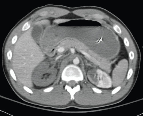
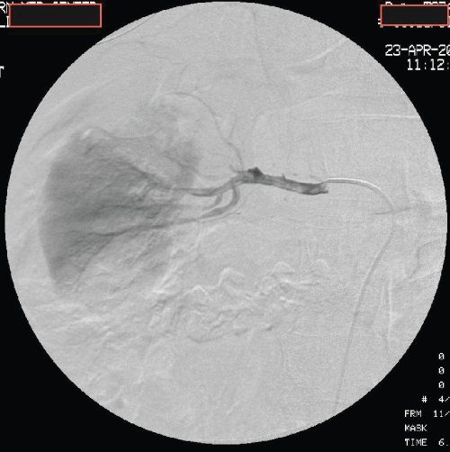
A 20-year-old male arrived as a level 1 trauma code after being struck by a pick-up truck traveling about 50 mph. Intubation in the emergency department occurred for airway protection and hemodynamic instability. A left chest tube yielded moderate blood egress and bilateral lower extremity deformities with open fractures were present. Findings on computed tomography (CT) imaging included a small left subdural hematoma, a left hemothorax and lung contusion. Bilateral renal infarctions with traumatic renal vascular thrombosis, and a right renal pelvis tear with minimal contrast extravasation was demonstrated. The remaining abdominal viscera were without acute injury. The right renal infarct was extensive compared to the left and involved the majority of the right kidney (Figure 1). A visceral angiogram confirmed bilateral renal infarctions, greater in the right kidney. The thrombus on the right renal artery was large and extending to several interlobar arteries (Figure 2). A catheter was placed in the right main renal artery for infusion of tissue plasminogen activator (TPA) at 2 mg/hr for four hours after an initial bolus of 8 mg for a total dose of 16 mg. The time from injury to the treatment with TPA was approximately 5 hours. The patient was also placed on systemic heparin drip due to the bilateral nature of the renal artery thrombosis. This decision was made following consultation with a neurosurgeon as the patient also had an acute subdural bleed. A collaborative decision was reached to proceed with the systemic heparin as the risk of bilateral renal ischemia without intervention seemed to outweigh the benefits of withholding the heparin given the non-operative pattern of the subdural bleed. The systemic heparin was also applied at approximately five hours post-injury. During the time of heparin infusion, the patient remained on mechanical ventilation and was monitored closely with one hour neurologic exams for signs of deterioration. A second head CT scan was performed 12 hours post-injury which showed a stable subdural bleed with no progression.
.
Figure 1: CAT scan image demonstrating extensive right kidney ischemia from acute renal artery thrombosis and patchy ischemia of the left kidney.
View Figure 1
.
Figure 2: Angiography of right kidney showing large thrombus (black solid arrow) in the right renal artery extending into several interlobar arteries.
View Figure 2
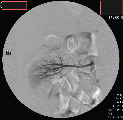
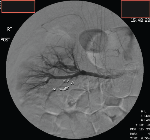
The TPA was stopped at 9 hours post-injury for a total dose of 16 mg and the heparin was to continue with the plan for 24 hours of treatment at which time a follow-up angiogram was to be performed to evaluate renal perfusion. The heparin was stopped prematurely at 18 hours because the patient developed marked hematuria. The follow-up angiogram at that time showed resolution of the clot in the right main renal artery but there was an area of contrast extravasation in the inferior pole of the right kidney (Figure 3). This area of active hemorrhage was then successfully controlled by using microcoil embolization into two segmental arteries. Additionally, the kidney appeared to have restored perfusion (Figure 4). The patient was then managed with prophylactic heparin.
.
Figure 3: Repeat angiography after TPA infusion showing resolution of thrombus but with evidence of contrast extravasation (solid arrow) in the lower pole of the right kidney.
View Figure 3
.
Figure 4: Angiography after successful embolization with microcoils of two inferior segmental branches of the right renal artery. Perfusion of the right kidney is demonstrated with patency of the main renal artery.
View Figure 4
The patient's renal function gradually improved, as there was an initial profound metabolic acidosis and hyperkalemia, which was managed medically. Renal replacement therapy was not required and his glomerular filtration rate (GFR) improved from 30 ml/min on admission to normal at the time of discharge. His lower extremity fractures were repaired in a staged fashion after the resolution of metabolic derangements related to acute kidney injury. A decompressive laparotomy was also required at 72 hours due to a secondary abdominal compartment syndrome related to massive resuscitation requirements. The traumatic subdural bleed did not progress and no surgical intervention was required. The patient was discharged to a rehabilitation facility at three weeks with normal renal function. The open abdomen was managed with skin closure, and his neurologic injury showed near complete resolution. At a three month clinic follow up the patient was neurologically normal, his renal indices and GFR were within normal limits and he was tolerating outpatient physical therapy.
In the setting of polytrauma aggressive local and systemic anticoagulation was successfully and safely applied for renal salvage due to bilateral traumatic renal artery thrombosis.
Discussion
Traumatic kidney injuries account for about 1% to 3% of trauma admissions with up to 85% due to blunt mechanisms [5]. Other associated injuries may be present in 60-80% of cases and mortality ranges between 20-40% [6]. The severity of the injury is graded using the American Association for the Surgery of Trauma (AAST) Organ Severity Score varies from grades 1-5 [7]. Renal vascular injuries are classified as either grade 4 or grade 5. Grade 4 renal vascular injuries refer to injuries involving the segmental veins or segmental arteries while Grade 5 renal vascular injuries involve injury to the main renal vein or artery. These vascular injuries may be transections or occlusions.
Spirnak and colleagues list two possible explanations for the occurrence of blunt traumatic renal artery occlusion(s) [8,9]. The first mechanism is thru abnormal stretching of the renal artery during a severe blunt trauma involving deceleration, leading to a tear in the inelastic intima with subsequent hemorrhage into the vessel wall and thrombosis. The second mechanism is thru compression of the renal artery between the body wall and the vertebral bodies in severe blunt trauma resulting in vessel wall contusion and thrombosis.
The diagnosis of traumatic vascular renal injuries is usually initially made by computed tomography scanning, as in our case the CT scan accurately revealed the bilateral thrombosis. Angiography still remains the gold standard for confirmation and provides an opportunity for interventions, such as embolization, stenting, or catheter-directed thrombolysis.
The decision to treat the patient using open surgical techniques when there is isolated renal vascular thrombosis in the absence of other reasons for surgical exploration is controversial versus other less invasive methods (e.g., endovascular or interventional techniques). Large modern databases have confirmed that complex renal vascular injuries are best managed with observation or in selected cases minimally invasive techniques, as the risk of surgical complications outweigh the benefits of open surgery [10]. Endovascular intervention has the advantage of fewer serious complications that are primarily due to local injury to the renal vessels. The incidence of such complications is low and most are of low clinical significance [11].
In our case, the patient had bilateral renal artery involvement. There was a left accessory renal artery visualized on angiography and there was no accessory renal artery on the right. The right infarct was noted to be more extensive compared to the left side. There were also no extra-renal injuries demonstrated on CAT scan that required surgical exploration. Therefore the decision was made to proceed to rapid endovascular methods of attempted revascularization of the right kidney to preserve renal function and avoid the need for long-term renal replacement therapy.
Thrombolytic agents commonly used for acute renal artery occlusions described in past literatures are tissue plasminogen activator, recombinant tissue-type plasminogen activator (rt-PA), streptokinase, urokinase, and reteplase with varying success and kidney salvage rates [12-17]. Common to all of these agents is the risk of hemorrhage. In our case, after the successful catheter-directed lysis using TPA, a source of hemorrhage was seen in one of the inferior segmental branches to the right kidney as evidenced by contrast extravasation. We hypothesize that the clot that was blocking the laceration to this segmental artery was dissolved during the infusion of the TPA and, hence, became the source of bleeding in this patient. This source of bleeding was successfully treated by embolization.
Factors associated with poor outcome commonly cited in the past are delays in diagnosis, delay in treatment, presence of other associated injuries, and hemodynamic instability. Likewise, other factors such as blunt mechanisms, high-grade injuries (i.e. grade 5), and an attempted arterial repair are also associated with a poor prognosis [3]. In a retrospective review of renal injuries admitted over a 12-year period to a Level 1 trauma center, high injury severity score, high renal injury grade, hemodynamic instability, and transfusion requirements were predictive of the need for nephrectomy [17]. These variables were all present in our case. Furthermore, the surgical and endovascular expertise available is thought to impact outcomes as well [18].
Our case illustrates the usefulness of interventional methods of treating blunt injuries to the renal arteries. This method is becoming an increasingly important armamentarium for the treating physician when faced with a complex patient with hemodynamic instability and polytrauma that would not otherwise tolerate the additional stresses of general anesthesia. The use of systemic and local anticoagulation was well tolerated despite the presence of multiple injuries, and ultimately the patient had return of normal renal function and did not require any short or long term renal replacement therapy.
References
-
Rosati C, Ata A, Siskin GP, Megna D, Bonville DJ, et al. (2015) Management of splenic trauma: a single institution's 8-year experience. Am J Surg 209: 308-314.
-
Sivrikoz E, Teixeira PG, Resnick S, Inaba K, Talving P, et al. (2015) Angiointervention: an independent predictor of survival in high-grade blunt liver injuries. Am J Surg 209: 742-746.
-
Haas CA, Spirnak JP (1998) Traumatic renal artery occlusion: a review of the literature. Tech Urol 4: 1-11.
-
Arabi M, Vellody R, Cho K (2011) Acute Renal Artery Occlusion with Prolonged Renal Ischemia: A Case of Successful Treatment with Stent Placement and Catheter-directed Thrombolysis. J Clin Imaging Sci 1: 11.
-
Malaeb B, Figler B, Wessells H, Voelzke BB (2014) Should blunt segmental vascular renal injuries be considered an American Association for the Surgery of Trauma Grade 4 renal injury? J Trauma Acute Care Surg 76: 484-487.
-
Lopera JE, Suri R, Kroma G, Gadani S, Dolmatch B (2011) Traumatic occlusion and dissection of the main renal artery: endovascular treatment. J Vasc Interv Radiol 22: 1570-1574.
-
Buckley JC, McAninch JW (2011) Revision of current American Association for the Surgery of Trauma Renal Injury grading system. J Trauma 70: 35-37.
-
Spirnak JP, Resnick MI (1987) Revascularization of traumatic thrombosis of the renal artery. Surg Gynecol Obstet 164: 22-26.
-
Dinchman KH, Spirnak JP (1995) Traumatic renal artery thrombosis: evaluation and treatment. Semin Urol 13: 90-93.
-
Sangthong B, Demetriades D, Martin M, Salim A, Brown C, et al. (2006) Management and hospital outcomes of blunt renal artery injuries: analysis of 517 patients from the National Trauma Data Bank. J Am Coll Surg 203: 612-617.
-
Liu FY, Wang MQ, Fan QS, Wang ZJ, Duan F, et al. (2009) Emergency intervention therapy for renal vascular injury. Chin J Traumatol 12: 81-86.
-
Mügge A, Gulba DC, Frei U, Wagenbreth I, Grote R, et al. (1990) Renal artery embolism: thrombolysis with recombinant tissue-type plasminogen activator. J Intern Med 228: 279-286.
-
Rangel-Abundis A, Olvera R, Cordero J, Cordero E, Ramos MA (1991) [Postangioplasty renal artery thrombosis treated intraluminally with thrombolysis. A case report]. Gac Med Mex 127: 253-256.
-
Piffaretti G, Riva F, Tozzi M, Lomazzi C, Rivolta N, et al. (2008) Catheter-directed thrombolysis for acute renal artery thrombosis: report of 4 cases. Vasc Endovascular Surg 42: 375-379.
-
Kushimoto S, Shiraishi S, Miyauchi M, Tanabe S, Fukuda R, et al. (2011) Traumatic renal artery occlusion treated with an endovascular stent--the limitations of surgical revascularization: report of a case. Surg Today 41: 1020-1023.
-
Knudson MM, Harrison PB, Hoyt DB, Shatz DV, Zietlow SP, et al. (2000) Outcome after major renovascular injuries: a Western trauma association multicenter report. J Trauma 49: 1116-1122.
-
Davis KA, Reed RL 2nd, Santaniello J, Abodeely A, Esposito TJ, et al. (2006) Predictors of the need for nephrectomy after renal trauma. J Trauma 60: 164-169.
-
Buckley JC, McAninch JW (2006) Selective management of isolated and nonisolated grade IV renal injuries. J Urol 176: 2498-2502.
