
Trauma Cases and Reviews
A Rare Cause of Lower Airway Obstruction Due to Inhalation of Illicit Drug Packet
P Ruggiano, MC Ferraro, A Franci, N Stanflin, M Bonizzoli, A Peris and Filippo Socci*
Emergency and Trauma Intensive Care, Careggi Teaching Hospital, Florence, Italy
*Corresponding author:
Filippo Socci, Emergency and Trauma Intensive Care, Careggi Teaching Hospital, Florence, Italy, E-mail: f.socci@sast.it
Trauma Cases Rev, TCR-1-017, (Volume 1, Issue 3), Case Report; ISSN: 2469-5777
Received: October 17, 2015 | Accepted: November 20, 2015 | Published: November 23, 2015
Citation: Ruggiano P, Ferraro MC, Franci A, Stanflin N, Bonizzoli M, et al. (2015) A Rare Cause of Lower Airway Obstruction Due to Inhalation of Illicit Drug Packet. Trauma Cases Rev 1:017. 10.23937/2469-5777/1510017
Copyright: © 2015 Michele M, et al. This is an open-access article distributed under the terms of the Creative Commons Attribution License, which permits unrestricted use, distribution, and reproduction in any medium, provided the original author and source are credited.
Case Report
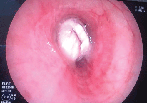
We report a case of a previously healthy 29-year-old male who experienced a severe traumatic brain injury with right temporal extracerebral intracranic hematoma and splenic injury after massive trauma sustained after a car accident. The patient was on his bike, he missed a red traffic light, and a car invested him and threw him 4 meters away. In the prehospital setting, the patient was found with GCS 3, with pupils equals, round and reactive to light with good hemodynamics. The medical emergency team who operated on the scene found difficulty in providing advanced airway management. The patient arrived in the emergency department with GCS 7 (E1, M5, V1,) without alteration of the pupil size, ventilated with bag-mask, hemodynamically stable but with metabolic acidosis (BE - 4, 4; lat 0.6) and poor respiratory exchanges (Pa02/Fi02: 135); he was promptly intubated without any difficulty and ventilated mechanically. The cerebral CT scan found a right fronto-temporal sphenoid fracture, a right temporal extracerebral intracranic hemorrhage of 16 mm that compressed the lateral ventricle; the abdominal CT scan revealed an intraparenchymal lesion of the medial part of the spleen. The thoracic CT scan revealed in the main left bronchus a round object of an unknown nature that occupied the entire origin of the left superior pulmonary lobe. The injury severity score was calculated to be 36 and the patient was brought to the operating room for a splenectomy and for a temporal craniotomy with drainage of the extracerebral intracranic hemmorrhage and the positioning of an intraparenchimal probe for intracranial pressure monitoring. In consideration of presence of an unknown object in the lower airway, documented by CT scan, a flexible fiberoptic bronchoscopy (FOB) was performed in presence of hemodynamic, respiratory and cerebral perfusion stable conditions. A white round object was visualized within the main left bronchus completely occupying the origin of the superior left bronchus (Figure 1). The removal of the foreign object was attempted with a Dormia basket® which was advanced beyond the unknown object, opened and closed around the object. The basket with the object and the endotracheal tube were removed together and rapid sequence re intubation was performed immediately without complications. After recruitment maneuvers and adequate ventilation, respiratory exchange progressively improved. The white round object was delivered to the police who analyzed it and identified it as a packet of cocaine. The clinical and laboratory data documented absence of cocaine intake. He was extubated two days after surgery and was subsequently discharged from ICU without complications after three days.
Discussion
Proper airway management represents a critical element in the treatment of trauma. In a traumatized patient, many factors may contribute to the impairment of gas exchange and, in particular hypoxia. Pulmonary contusion, pneumothorax, hemothorax and rib fractures are the most common causes of respiratory failure in a patient with chest trauma. In a trauma center is routinely deal with this type of injury by putting in place all the necessary maneuvers to improve the oxygenation of the patient. After the correct positioning of an endotracheal tube and the resolution of all the possible removable causes of hypoxemia caused by direct trauma, respiratory gas exchange generally improves but in rare cases, despite the efforts we do not get an improvement of hypoxia and is vitally importance considers other causes of respiratory failure, although uncommon. Additional failure of gas exchange resulting from lower airway obstruction compromises tissue oxygenation and causes systemic acidosis exposing to the risk of multiple organ failure. Lower airway obstruction due to foreign bodies can be a rare cause of respiratory insufficiency in trauma. Lung computed tomography (CT) scan is of considerable support in morphological and extent evaluation in course of trauma and also for the documentation of inhaled foreign bodies of significant volume that is a rare event as a major cause of post-traumatic respiratory failure; the event is even more rare and less likely if the foreign body that causes obstruction is represented by a container full of illegal narcotic substance kept in the mouth at the time of a car accident. The simultaneous presence of post-traumatic lung lesions and one or more obstructions to the first branch of the bronchial airways contribute to make very difficult the clinical interpretation and the cause of hypoxia refractory to conventional treatment. The description of this particular case of airway obstruction has the purose not only to document a rare mechanism of airway obstruction in the course of chest trauma, but also to emphasize the importance of considering other cause of hypoxia, when the clinical picture does not fit the data, and also to emphasize the importance of evaluation of subglotic airway patency to find a cause to a post-traumatic hypoxia; hence the need for the availability of advanced skills in bronchoscopy in a trauma center.
Lower airway obstruction as a consequence of trauma is a rare cause of respiratory failure in trauma. A foreign body aspirated into the respiratory tract constitutes a serious condition for respiratory function, characterized by a considerable variability of effects and prognosis. In the majority of cases, the problem of the aspiration of a foreign body into the respiratory system affects children of 1-3 years old and it is less common in adults were the peak incidence of foreign body aspiration occurred during the sixth decade of life [1,2] normally in general population inhaled solid materials derived from vomit or consist of teeth, dentures or fragments of prostheses. This case describes a rare situation in which the patient was a young man and the foreign body was a drug packet stuck in a main bronchus. The act to maintain inside the mouth packets containing illegal drugs for the purpose of smuggling has been termed "bodypacking" [3]. A variant of this practice is seen in drug dealers and users who hastily ingest packets of drugs to hide the evidence when confronted by police. This syndrome has been termed "bodystuffing" [4]. The quantity of drug contained in bodystuffing packets (grams) is far less than for bodypackers (kilograms). Many bodystuffers are asymptomatic or moderately symptomatic at presentation. It is also important to consider the type of packing material the bodystuffer uses. There is a difference in the liberation of cocaine from the various containers used to wrap it [5]. These containers can include plastic wrap, plastic bags, cellophane, paper, aluminum foil, glassine crack vials and condoms [6,7]. There is evidence that there may be delayed liberation of the drug depending on the type of wrapping used. In our case, it is likely that the man aspirated the drug packet that was in his mouth while he was ventilated with the bag mask. Since the drug packet was well crafted, it is probably the cause of an absence of symptoms associated with cocaine abuse. The drug was densely packed into a latex sheath, such as a condom or balloon. This layer was tied at the open end, covered with several other layers of latex, and sealed with a hard wax coating; a plastic food wrap was incorporated. Usually drug packets are swallowed and can be localized in the gastro-intestinal tract and can have toxic effects or can determine bowel obstruction, bowel perforation, esophageal obstruction, and esophageal perforation.
To the best of our knowledge, this is the first case of inhalation of a cocaine packet and subsequent respiratory failure due to obstruction of the left main bronchus. The presence of unresolved hypoxia despite the implementation of maneuvers designed to improve it must make us consider the presence of other causes, which can be highlighted with diagnostic tests targeted and resolved with appropriate equipment and skills.
The diagnostics and treatment of a patient with a foreign body in the respiratory tract is a complex process, which requires close cooperation between intensivist and specialists. In this case we confirm early the presence of a foreign body in the airway through the CT scan and we proceeded to his removal through appropriate bronchoscopic techniques.
The fundamental role in the algorithm of the therapeutic procedure is played by the CT scan and by bronchoscopy. This case above all highlights that in trauma the use of computed tomography can be extremely useful in the evaluation of subglottic airway patency and to individuate rare causes of lower airway obstruction. Moreover the removal of foreign bodies from the airway is essential in order to avoid complications such as atelectasis, pneumonia, bronchiectasis and foreign body migration. Furthermore this case demonstrates the need in a trauma center of a multidisciplinary team who has advanced skills in operative bronchoscopy; the outcome associated with these procedures improves when the operator is experienced, the removal of tracheobronchial foreign objects should be performed in medical centers that are capable of acquiring and maintaining the necessary expertise.
References
-
Zerella JT, Dimler M, McGill LC, Pippus KJ (1998) Foreign body aspiration in children: value of radiography and complications of bronchoscopy. J Pediatr Surg 33: 1651-1654.
-
Baharloo F, Veyckemans F, Francis C, Biettlot MP, Rodenstein DO (1999) Tracheobronchial foreign bodies: presentation and management in children and adults. Chest 115: 1357-1362.
-
June R, Aks SE, Keys N, Wahl M (2000) Medical outcome of cocaine bodystuffers. J Emerg Med 18: 221-224.
-
Roberts JR, Price D, Goldfrank L, Hartnett L (1986) The bodystuffer syndrome: a clandestine form of drug overdose. Am J Emerg Med 4: 24-27.
-
Pidoto RR, Agliata AM, Bertolini R, Mainini A, Rossi G, et al. (2002) A new method of packaging cocaine for international traffic and implications for the management of cocaine body packers. J Emerg Med 23: 149-153.
-
Traub SJ, Hoffman RS, Nelson LS (2003) Body packing--the internal concealment of illicit drugs. N Engl J Med 349: 2519-2526.
-
Aks SE, Vander Hoek TL, Hryhorczuk DO, Negrusz A, Tebbett I (1992) Cocaine liberation from body packets in an in vitro model. Ann Emerg Med 21: 1321-1325.
