

Trauma Cases and Reviews
Symptomatic Yuxta-articular Facet Cyst: A Case Report
J Miguel Rodríguez Solera*, Ana M Cerván de la Haba, Miguel S Hirschfeld León and Enrique Guerado Parra
Department of Orthopaedic Surgery, Traumatology and Rehabilitation, Hospital Universitario Costa del Sol, University of Malaga, Spain
*Corresponding author: José Miguel Rodríguez Solera, Hospital Universitario Costa del Sol, Marbella, España, Autovía A7 km 187 C.P. 29603, Spain, Tel: 951976224, E-mail: jmyl09@icloud.com
Trauma Cases Rev, TCR-1-023, (Volume 1, Issue 4), Case Report; ISSN: 2469-5777
Received: October 31, 2015 | Accepted: December 28, 2015 | Published: December 30, 2015
Citation: Solera JMR, de la Haba AMC, León MSH, Parra EG (2015) Symptomatic Yuxtaarticular Facet Cyst: A Case Report. Trauma Cases Rev 1:023. 10.23937/2469-5777/1510023
Copyright: © 2015 Solera JMR, et al. This is an open-access article distributed under the terms of the Creative Commons Attribution License, which permits unrestricted use, distribution, and reproduction in any medium, provided the original author and source are credited.
Abstract
We report a case of 66 years-old male with left leg pain that began six months earlier. The pain was aggravated by sitting or climbing stairs but was relieved in the standing position. The physical examination was consistent with a left radiculopathy without sensory or motor deficit. His initial MRI lumbar spine demonstrated a cystic mass with T1 and T2 hyperintensity on the L3-L4 level with considerable thecal sac and nerve root compression. The lesion originated next to the left L3-4 facet joint and extended centrally under the lamina of L3 and L4. A surgical treatment was offered to the patient and subsequently underwent a left L3 hemilaminectomy, medial facetectomy, left ganglion excision and L3, L4 fusion, with a very satisfactory outcome.
Spine ganglions are grouped within yuxta-articular facet cysts, along with synovial cysts. Etiology is unknown, but it is related with repeated trauma or microtrauma as a part of degenerative spinal disease. It is an uncommon pathology and 90% are located in the lumbar region. The most common symptom is pain and the radiculopathy, which is unusual; it will be due to compression of adjacent structures. Initial treatment is conservative. Surgery is indicated in cases where a neurological deficit exists or where conservative treatment fails in pain relief. Generally good results are described. In our case the patient had a very satisfactory outcome.
Keywords
Ganglion, Synovial Cyst, Lumbar Ganglion, Yuxta-articular facet cyst
Case Report
A 66 year-old male with no past medical history presented with low back pain and left leg pain and numbness that began six months earlier, with no recent history of injury. After three months of progressive worsening of his pain and conservative management recommended by his community doctor, the patient came at our clinic, describing low back pain and left leg and buttock pain.
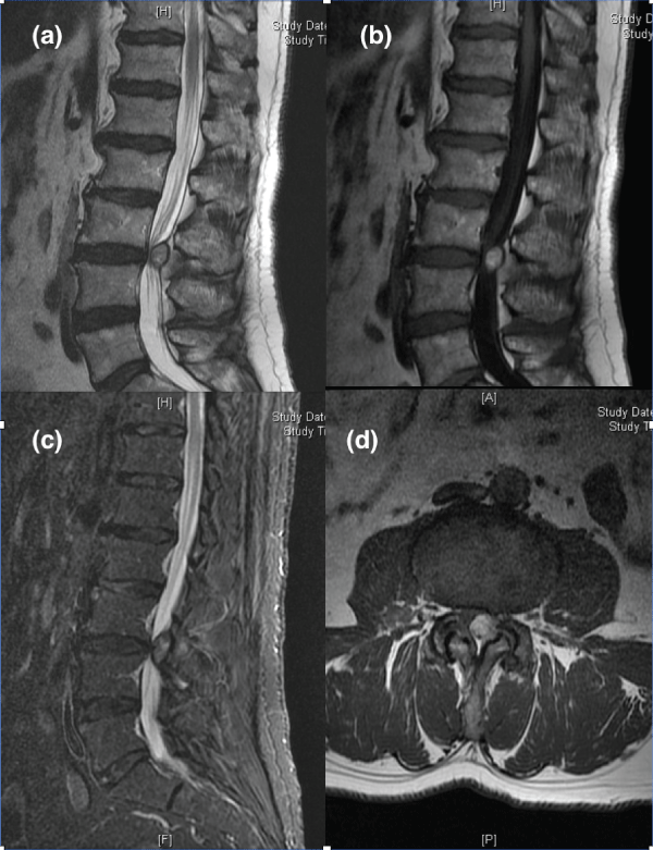
Patient only complained of low back pain. Lumbar physical examination only showed a Lasegue and Bragard tests positives, the left patellar reflex was absent and both strength and sensitivity were preserved. On radiographs standing degenerative signs are observed, on dynamic radiographs instability is not appreciable. Lumbar spine MRI was performed reporting degenerative signs with endplate osteophytes, signs of widespread dehydration of the discs without evidence of herniated discs, important hypertrophic degenerative changes at posterior joints, identifying at the level of subsequent L3-L4 left interapofisaria joint, an image of intraspinal ganglion of 11 mm, which compresses and displaces the nerve roots and influences canal stenosis at that level (Figure 1).
.
Figure 1: (a) Sagittal T2- weighted; (b) sagittal T1-weighted; (c) Sagittal STIR-weighted; (d) axial T2-weighted. MR images demonstrate a 11-mm maximum diameter, ovoid cystic lesion, posterolateral to the thecal sac, on the left L3-L4 facet joint. The cyst projects off the more cephalab aspect of the involved facet joint.
View Figure 1
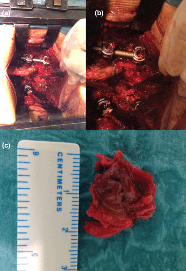
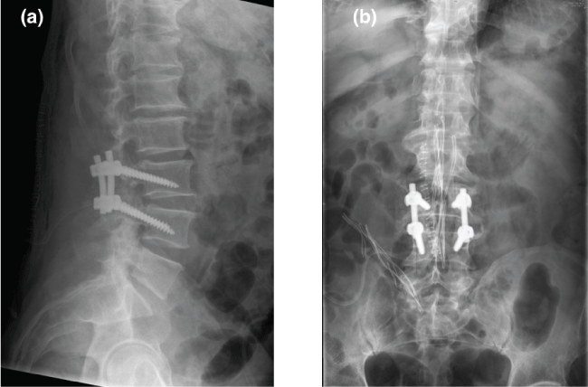
As conservative treatment does not work surgical we decided to do surgical treatment, performing hemilaminectomy L3, left yuxtafacetaria excision of tumor (Figure 2), foraminotomy, release the left L4 root and fusion of L3-L4 (Figure 3).
.
Figure 2: (a,b) Intraoperative photographs demonstrating the cyst in situ; (c) Cyst.
View Figure 2
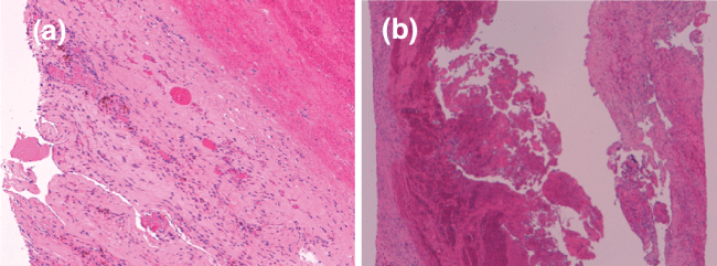
Histopathological study was carried out finding components in connection with hemorrhagic cyst (Figure 4). The patient evolved satisfactorily, being asymptomatic nowadays.
.
Figure 4: (a,b) On histologic examination was observed soft tissue fragments measuring between 8-23 mm, they were fragments of fibrous tissue and fibrocartilage with recent bleeding, abundant haemosiderinic macrophages, hematoidin and granulation tissue with little presence of mixed inflammatory component observed in connection with hemorrhagic cyst.
View Figure 4
Discussion
Yuxta-articular facet cyst was first described by Kao et al. [1]. Its pathogenesis is nowadays well known, relating to repeated traumas or microtraumas, rheumatoid arthritis, spinal degeneration and segmental instability [2,3]. Previous traumatisms are recognized as the "primum movens" in their pathogenesis. This theory initially suggested by clinical data has now found experimental evidence, being confirmed also by immunohistochemical analysis on the pathological specimens [4]. In a literature review conducted in 2008 they were related to spondylolisthesis (43%), disc degeneration (13%), scoliosis (1%) and stenosis (1%) [2]. It is more common in women and usually appears after the fourth decade [5,6]. It has a prevalence of 2.3% in symptomatic patients who will perform a MRI [7]. Its most common site is the lumbar spine and the most common levels are L4 L5, L3 L4 and L5S1 in that order [2,8]. Usually is unilateral, although there are bilateral cases described [9]. Patients generally have a history of low back pain with radiculopathy or back claudication that increases with standing or walking [10]. The study of Martha et al. reports successful cyst ruptures in 81% of cases. However, 54% of patients required subsequent surgery because of inadequate symptom relief [11].
There are some articles that relate to 52% of the dural sac adhesions [12]. They have been described as bleeding complications in the epidural space [2,8-13] or cauda equina syndrome [4].
The differential diagnosis includes all lesions that can potentially produce radicular pain. These injuries include: extruded disc fragments, primary or metastatic bone lesions, meningiomas, arachnoid cysts or Schwannomas or dermoid cysts [14]. It is also necessary to make a differential diagnosis with many other benign cystic lesions that can be found intradurally in the lumbar spine, as cystic dilatation of the ventriculus terminalis, ependymal cyst or arachnoidal cysts [15], and extradurally including synovial cysts, ligamentous cyst and ganglion cysts [16]. Ganglion versus synovial cyst should be differentiated: gel vs. synovial fluid, connective tissue vs. synovial line cells, and ligament/tendon connection vs. articular/tenosynovial connection. For it will require an anatomopathological study.
The diagnosis begins with clinical suspicion, scanning and imaging tests. In plain radiography is appreciable the presence of spondylolisthesis in 70% of patients. The gold standard is MRI, where we can see a usually unilateral soft tissue injury adjacent to the joint, well-circumscribed, bulging the flavum ligament into the posterolateral epidural space. Communication with the facet joint is rarely visible and signal characteristics are to be those of a cyst with a hypointense on T1-weighted and hyperintensity on T2-weighted , although sometimes you can see a hyperintense signal on T1-weighted due to increased protein contained therein [8].
Initial treatment of spinal stenosis caused by synovial cyst is the same as any low back pain. Usually it is recommended exercises, weight reduction and smoking cessation. Medical regimens for pain management include combinations of non-steroidal anti-inflammatory agents, narcotics, and muscle relaxants antidepressants. Injection therapies have been described to include epidural steroid injections, facet blocks, and direct aspirations of the cysts [17]. Mixed results have been reported, but are all very reasonable first-line therapies to obtain symptomatic relief before surgery. Most series report that nonsurgical treatment methods are not as effective as decompressive surgery [18].
When non-operative treatments fail, surgical decompression should be considered. The clinical indications for surgery include myelopathy, motor weakness, painful radiculopathy, or back pain not responsive to non-operative management. The procedure currently recognized is total excision of the tumor with laminotomy, laminectomy or hemilaminectomy. Sometimes, depending on the size of the cyst, you must perform only a facectomy. In planning the surgery, for the tactics of the surgery, Ross R et al. [18] recommended the three following questions: 1. what is required to decompress the effected neurological structure; 2. is spinal instability present preoperatively; and 3. will the decompression lead to instability? If the answers to either question 2 or 3 are yes, then primary fusion at the time of the decompression should be strongly considered.
If there is documented instability or deformity before the decompression, then a primary fusion is performed. If the decompression leads to significant facet disruption, then primary fusion must be performed [19]. There are several publications advocating fusion, although spinal fusion also increased the perioperative morbidity compared with decompression only, including incidental durotomies, wound infection, and deep venous thrombosis, decreasing cyst recurrence and unfavorable clinical outcome in the fusion group [19-21]. We always prefer to perform a vertebrae fusion in order to treat the segment instability and spinal degeneration which increase the chance of developing a yuxta-articular facet cyst.
Conflicts of Interest
All authors have no financial support or relationships that may pose a conflict of interest to disclose for this work.
References
-
Kao CC, Uihlein A, Bickel WH, Soule EH (1968) Lumbar intraspinal extradural ganglion cyst. J Neurosurg 29: 168-172.
-
Boviatsis EJ, Staurinou LC, Kouyialis AT, Gavra MM, Stavrinou PC, et al. (2008) Spinal synovial cysts: pathogenesis, diagnosis and surgical treatment in a series of seven cases and literature review. Eur Spine J 17: 831-837.
-
Hostalot C, Mozas M, Bilbao G, Pomposo I, Aurrekoetxea J, et al. (2001) Lumbar root compression secondary to juxtafacet cysts: review of 10 cases. Neurocirugia (Astur) 12: 308-315.
-
Ganau M, Ennas F, Bellisano G, Ganau L, Ambu R, et al. (2013) Synovial cysts of the lumbar spine--pathological considerations and surgical strategy. Neurol Med Chir (Tokyo) 53: 95-102.
-
Banning CS, Thorell WE, Leibrock LG (2001) Patient outcome after resection of lumbar juxtafacet cysts. Spine (Phila Pa 1976) 26: 969-972.
-
Epstein NE (2004) Lumbar synovial cysts: a review of diagnosis, surgical management, and outcome assessment. J Spinal Disord Tech 17: 321-325.
-
Doyle AJ, Merrilees M (2004) Synovial cysts of the lumbar facet joints in a symptomatic population: prevalence on magnetic resonance imaging. Spine (Phila Pa 1976) 29: 874-878.
-
Trummer M, Flaschka G, Tillich M, Homann CN, Unger F, et al. (2001) Diagnosis and surgical management of intraspinal synovial cysts: report of 19 cases. J Neurol Neurosurg Psychiatry 70: 74-77.
-
Bozzao A, Floris R, Fraioli C, Ticca L, Simonetti G (2001) "Relapsing-remitting" bilateral synovial cysts of the lumbar spine. A case report. Neuroradiology 43: 1076-1078.
-
Brown C, Stambough JL (2005) Epidural hematoma secondary to a rupture of a synovial cyst. Spine J 5: 446-450.
-
Martha JF, Swaim B, Wang DA, Kim DH, Hill J, et al. (2009) Outcome of percutaneous rupture of lumbar synovial cysts: a case series of 101 patients. Spine J 9: 899-904.
-
Scholz C, Hubbe U, Kogias E, Klingler JH (2015) Incomplete resection of lumbar synovial cysts - Evaluating the risk of recurrence. Clin Neurol Neurosurg 136: 29-32.
-
Cannarsa G, Clark SW, Chalouhi N, Zanaty M, Heller J (2015) Hemorrhagic lumbar synovial cyst: case report and literature review. Nagoya J Med Sci 77: 481-492.
-
Greenberg M (2006) Handbook of Neurosurgery. (6a Edition) Thieme, New York.
-
Ganau M, Talacchi A, Cecchi PC, Ghimenton C, Gerosa M, et al. (2012) Cystic dilation of the ventriculus terminalis. J Neurosurg Spine 17: 86-92.
-
Ganau M, Ennas F, Ambu R, Faa G, Maleci A (2013) Excision of synovial cysts: pathology matters. J Neurosurg Spine 19: 266-267.
-
Cambron SC, McIntyre JJ, Guerin SJ, Li Z, Pastel DA (2013) Lumbar facet joint synovial cysts: does T2 signal intensity predict outcomes after percutaneous rupture? AJNR Am J Neuroradiol 34: 1661-1664.
-
Ross R Moquin (2006) Synovial Cysts of the Spine: Clinical Presentation and Treatment. Semin Spine Surg 18: 175-179.
-
Xu R, McGirt MJ, Parker SL, Bydon M, Olivi A, et al. (2010) Factors associated with recurrent back pain and cyst recurrence after surgical resection of one hundred ninety-five spinal synovial cysts: analysis of one hundred sixty-seven consecutive cases. Spine (Phila Pa 1976) 35: 1044-1053.
-
Knafo S, Page P, Pallud J, Roux FX, Abi-Lahoud G (2015) Surgical Management of Spinal Synovial Cysts: A Series of 23 Patients and Systematic Analysis of the Literature. J Spinal Disord Tech 28: 211-217.
-
Mattei TA, Rodriguez AH (2013) True synovial cysts of the lumbar spine: an epiphenomenon of instability of the functional spine unit? Neurosurg Rev 36: 495-500.
