Intra-articular foreign bodies especially in the knee joint are well reported. Knee joint is commonly involved due to the large joint area and also due to it being a superficial joint. Foreign bodies enter the joint either by direct inoculation or migration from nearby tissues. Potential complications of intra articular foreign bodies include infection, damage to articular surface and locking of joint. This is a report on a penetrating injury to the knee resulting in an intra capsular but extra synovial foreign body. A 10-year-old boy fell on to railing and sustained injury to the right knee. The railing broke and part of it was protruding out of the knee. Radiographs showed that the piece of railing had lodged into the lateral compartment of the knee. At surgery the part of railing was found to between the capsule and synovium of the knee joint. This was successfully removed. Synovial lining was found to be intact with intraoperative saline test of the joint that showed no leakage of fluid in to the wound. Capsule was closed and rest of the left open. Patient underwent a further debridement and delayed closure at 48 hours. Patient made an uneventful recovery. This report is presented to show that all intra articular foreign bodies are not intrasynovial.
Foreign body, Joint, Knee, Open wound, Penetrating wound, Extra synovial
Limb injuries from penetrating foreign bodies are common. Intra-articular foreign bodies often enter the joint due to trauma, either as a direct injury or by migration from either nearby soft tissues [1]. Intra-articular foreign bodies are a variety ranging from plastic bottle cap [2], varying types of projectiles [3-5], broken instruments [6], broken implants [7], broken guide wire [8], glass piece [9,10], lead pencil [11], gear shift [12] broken needle [13], thorn [14-16] and mercury [17]. These can be either present acutely or after some time has passed [18].
Intra-articular foreign bodies require removal to reduce risk of infection and avoid secondary damage to articular cartilage. Occasionally, smaller foreign bodies could become loose bodies causing mechanical symptoms.
A 10-year-old boy while playing on a wall fell on the railing and sustained an injury to his right knee. He presented to casualty with a piece of metal protruding out of his flexed right knee. Primary and secondary survey did not reveal any other injuries. On examining the right knee there was a transverse laceration of about 4 cm at the level of lateral joint line with a rusted metal rod protruding out of the laceration (Figure 1a and Figure 1b). The knee was flexed at about 140° and no movement could be achieved due to pain. There was no distal neurovascular deficit. Rest of the right lower limb was normal. Radiographs showed a bent metal rod that appeared to be in the lateral compartment of the right knee (Figure 2). Patient underwent exploration of the wound and removal of the foreign body under general anaesthesia within 4 hours of the incident. Per-operative findings were show a dirty wound which was extended longitudinally, and the metal rod followed deep into the wound and removed. The metal rod had gone through the potential space between capsule and synovial lining of the knee joint. Exploration of this space did not show any breach of synovium (Figure 3). Thirty mL of Normal saline was infiltrated into the right knee from the superomedial aspect and there was no leakage of fluid into the space where the foreign body was found. The wound was thoroughly washed out and found to be clean. Intra-operative fluoroscopy showed no residual foreign body. The wound extension was closed. Patient was on intravenous broad-spectrum antibiotics for 48 hours and taken to theatre at 48 hours for a second debridement when it was found to be clean and a delayed wound closure was performed. Patient was allowed full weight bearing and range of movement exercises as tolerated. At 6 months follow up there was no evidence of infection in the wound. Patient had full range of movements in his right knee and no instability or features of meniscal pathology. He was back to playing football with no difficulties and hence, discharged from clinical care.
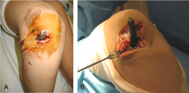 Figure 1: a) Part of rusted railing protruding out of the right knee; b) Per-operative photograph showing the rusted railing. View Figure 1
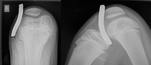 Figure 2: Radiographs of right knee. View Figure 2
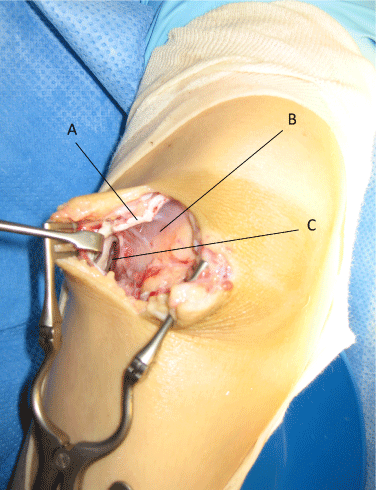 Figure 3: Per-operative photograph showing the potential space between capsule and synovium of the right knee. A - capsule; B - Synovium; C - Space between capsule and synovium. View Figure 3
Intra-articular foreign bodies can occur either due to direct or indirect injury and can be delayed presentation due to migration from local soft tissues. Most of these are caused by trauma and occasionally by self-harm.
Acute joint penetrating wounds require urgent debridement. If there is a foreign body that has entered the joint, then removal of foreign body reduces the risk of infection and prevents potential damage to the articular cartilage [19,20]. Radiographic examination show radio-opaque foreign bodies but not non-opaque substances like plastic. Computerised Tomography (C.T.) in radio-opaque foreign bodies is generally not recommended due to artefacts. Magnetic Resonance Imaging (M.R.I.) is contra-indicated due to potential for foreign body moving and causing more damage. C.T. and/or M.R.I. could assist in locating, assessing size and numbers of foreign bodies. Often, these penetrating injuries require wound extensions to assess and debride the wound. The wound extensions should be such that it would allow extensile exposures if required. The path of the foreign body should be followed carefully to avoid any iatrogenic injuries and removed completely, intact or otherwise. Often when the foreign body is inert and not involving articular cartilage, it does not cause any major problems. If the foreign body behaves as a third body, irritating the articular cartilage, it could lead to significant articular damage. However, in case of intra articular lead bullet, there is a possibility of lead toxicity occurring soon after the bullet injury or several years after the injury when the bullet is expected to be walled off by fibrous tissue [21,22].
Further investigations like Computerised Tomography could provide detailed imaging regarding site of the foreign body. Though Magnetic Resonance Imaging (M.R.I.) could provide more information regarding soft tissues and joint involvement, ferrous nature of most metallic objects precludes this investigation and in an acute situation, it is often difficult to assess the nature of the foreign body safely to consider M.R.I. In the case presented, possible mechanism of the foreign body tracking through the potential space between capsule and synovium include blunt foreign body (acting like an obturator) and the pliability of the thick synovium of the child’s knee joint. This case is presented as a reminder that all foreign bodies that appear intra articular are not always intra articular and careful intra-operative assessment of the wound following the track of the foreign body is essential to prevent iatrogenic damage and prevent opening tissue planes unnecessarily.
The author confirms that he does not have any competing interests in preparation of this article.