The existence of a vertebral body deformity is considered a relevant finding in order to start a secondary therapeutic prevention of osteoporosis in a population at risk. However, not all vertebral body deformities are properly identified and registered. In our experience, those, less striking, can be unnoticed. One of the most common radiographic studies of the axial skeleton is the lumbar and dorsal acute pain. In this group of patients, clinicians are particularly willing to order axial x-ray studies. Due to that, they are particularly suitable for establishing proportions of any grade of vertebral body deformities according to the Genant's scale. The aim of this study is to determine the prevalence of any vertebral body deformities in postmenopausal patients radiologically assessed due to lumbar or dorsal non-traumatic related pain. To reach our goal we performed a randomization of the registries of female patients with more than 65-years-old who asked for medical assessment due to dorsal or lumbar non-traumatic acute pain, between January 2014 and December 2015. All included registries should have had a radiological evaluation. Vertebral body measures were performed according to Genant's scale recommendations from the seventh thoracic to the fifth lumbar vertebral body. The demographic profile of patients and the information of medical radiologic interpretation were also collected. After the randomization and excluding incomplete registries, 275 randomised registries of dorsal and lumbar pain were included (total = 550). Among patients with dorsal pain, we identified 62 (22.5%), 30 (10.9%) and 18 (6.5%) vertebral deformities grade I, II and III respectively. Among patients with lumbar pain, we identified 31 (11.2%), 49 (17.8%) and 33 (12%) vertebral deformities grade I, II and III respectively. Prevalence of any grade of dorsal vertebral deformity was 40.00% (CI 34.39 - 45.89%), and lumbar was 41.09% (CI 95% 35.44 - 46.99). Lumbar vertebral body deformities grade II and III summed 17.4% while dorsal grade II and III summed 29.8%. From the 93 vertebral body deformities grade I, identified towards this study, 6.4% had been recognised and described in their corresponding clinical history during his assessment. Although our population sample is circumscribed to symptomatic patients, our results contribute knowing the prevalence of vertebral body deformities in postmenopausal symptomatic patients grade I and II and who were mostly unnoticed. Some studies have questioned the real value of Genant's grade I deformities considering them real vertebral body collapses or the effect of axial osteoarthritis. Inspite of Kellgren and Lawrence grading of osteoarthritis was homogeneous in our sample, we can not exclude this factor as a confounding one when the clinical assessment was performed during the usual clinical practice. Nevertheless, proper identification of vertebral body deformities in patients with osteoporosis is crucial to decide treatment strategies in patients with known osteoporosis.
Vertebral osteoporotic fracture, Lumbar pain, Dorsal pain
The clinical assessment of patients with suspect of postmenopausal osteoporosis includes a radiographic evaluation of the dorsal and lumbar skeleton in order to establish the presence or absence of vertebral body collapses [1,2]. In patients in risk of osteoporosis fracture, this study is particularly relevant to distinguishing primary from secondary prevention behaviours [3,4]. Despite the existence of many methods of quantification, the traditional semiquantitative classification method developed by Genant, et al. is still one of the most popular and accepted for establishing the presence of vertebral body collapse [5,6]. Genant's scale of vertebral body deformities allows a reproducible and easy way to classify vertebral collapses. However, mild grade of deformity is still considered a controversial finding when assessing a patient at risk of osteoporosis fracture due to its relationship with degenerative processes such as osteoarthritis in both genders [7,8]. Due to that, decisions about therapeutic interventions on such patients are made only with grade II or III spinal body wedges.
In Spain, two important barriers are in front of our capability to predict a number of people in need of an osteoporotic secondary prevention. First, in our national setting, there is a lack of information about the prevalence of spinal body wedges of any grade because the natural access to radiologic studies is reduced to an episode of lumbar or dorsal pain as long as the patient is female and over 65-years-old. Second, the knowledge of the relevance of clinical spinal body wedges among clinicians at emergency units is widely variable. The existence of a vertebral body deformity is considered a relevant finding in order to start a secondary prevention therapy of osteoporosis in the population at risk. However, not all vertebral body wedges are correctly noticed and registered. In our experience, those, less stunning, can be certainly unnoticed. In general, when a female patient older than 65-years-old complaints by lumbar or dorsal pain, even without a traumatic process related, the diagnostic behavior in our Emergency Departments (ED) is to order a radiological study of the spine.
The purpose of the present study is to focus on this last group of patients who almost always underwent a radiological examination of the spine in order to know the prevalence of different kinds of vertebral body wedges according to the Genant's classification.
This study is a sub-analysis performed from another study conducted to determine the impact of rheumatic diseases in an emergency department, which has not been publishing yet. We performed a randomization of registries of patients who asked for medical assessment in our ED due to lumbar of dorsal non-traumatic pain from January 2014 to December 2015. According to previous studies performed in our institution, 13.7% of all our patient's primary complaints are related to the musculoskeletal system and from them, about 25% due to lumbar or dorsal axial pain [9]. The randomization was performed systematically dividing the year by seasons and collecting the same amount of patients from each one. The prevalence of vertebral fractures in small cohorts studies conducted in our country is variable, from 4.3% to 17% including women older than 50-years-old [10-13]. We performed the randomization using or electronic records of clinical registries from the ED until we reach a 120% of the estimated sample size for a non-finite theoretical population, a precision of 3% and a hypothetical estimated pessimists prevalence of 7% based on those local studies. We performed one randomization for lumbar and another for dorsal pain with the same sample size. Only registries with complete information (including topographic characteristics of pain and radiological studies) were included.
After randomization, we collected epidemiological and demographic data from clinical charts. The clinical description of the radiologic study was also included translating it to categorical values according to the presence or absence of vertebral body fractures or deformities. Finally, a single expert (CG) reviewed all radiologic studies and performed all measurements to establish Genant's classification of all vertebral bodies. Radiologic data used to establish the Genant's grade of deformity was the measures of the vertebral height of the anterior and posterior wall, and the height of the middle part of the vertebral body view from its lateral aspect. The rules followed to perform the classification were those exposed by Genant, et al. [5]. The expert was blinded to other clinical information and radiological reports. Inter-observer average kappa index of 95% - to classify in the same Genant category twelve different patients at two separate times - was reached in three different attempts in a previous pilot study (data not showed). All data was included into a single database to process and analysis.
Epidemiological and demographic data were analysed using central and dispersion-tendency statistics: average and Standard Deviation (SD). Prevalence of Genant's vertebral body fractures was expressed in percentages terms and comparing the numeric value of the fractures clinically recognised by the clinicians and those radiologically demonstrated by the expert's review.
This study is a sub-analysis of the data of another study which was approved by the Ethical Scientific Committee of our institution. Authors declare not have any conflicts of interest regarding these studies. Also, we have not had any financial support from any source to perform this sub-analysis or the main study. Finally, all the data showed in this study have not been published before.
A total of 550 registries of dorsal and lumbar pain were randomised (275 records for each kind of complaint). Patients who consulted due to dorsal non-traumatic pain had a mean age of 75.3 SD 4.1-years-old. Patients who consulted due to non-traumatic lumbar pain had a mean age of 78.1 SD 5.2-years-old (P < 0.001). Time since onset of symptoms was 5.2 SD 2.0 and 6.3 SD 1.9 days, respectively (P < 0.001). History of pharmacological osteoporosis treatment was present in 3.2% and 4.0% of patients, respectively (P = 0.64). History of previous and diagnosed vertebral body collapse was present in 4.3% and 7.6% (P = 0.8). Distribution of chronic diseases among both groups did not show differences statistically significant (Table 1).
Table 1: Epidemiological and demographic features of patients which registries were included. View Table 1
Among patients with dorsal pain, we identified 62 (22.5%), 30 (10.9%) and 18 (6.5%) vertebral deformities grade I, II and III, respectively (Table 2). Among patients with lumbar pain, we identified 31 (11.2%), 49 (17.8%) and 33 (12%) vertebral deformities grade I, II and III respectively. In both cases, the difference between the proportions of Class I wedges and the others were statistically significant (P < 0.001). Prevalence of any grade of dorsal vertebral deformity was 40.00% (CI 34.39 - 45.89), and lumbar was 41.09% (CI 95% 35.44 - 46.99). The concordance between the detection of vertebral deformities by our study and those registered in the clinical histories of patients included in this study was 6.4%, 20.2% and 92.1% for grades I, II and III, respectively. All these accuracy proportions showed differences statistically significant (P < 0.001 in all combinations). Lumbar vertebral body deformities grade II and III summed 17.4% while dorsal grade II and III summed 29.8% (P < 0.0006).
Table 2: Radiologic findings after expert assessment and comparison with clinician interpretation during formal evaluation. View Table 2
As shown in Figure 1, the distribution of mild Genant's deformities was slightly more prevalent in the second lumbar vertebral body and the eleventh dorsal vertebral body while Genant's deformity grade II was more common in the first lumbar vertebra (6.55%) and second lumbar vertebra (5.82%). Genant's deformity grade III was not detectable in the fourth and fifth lumbar vertebrae.
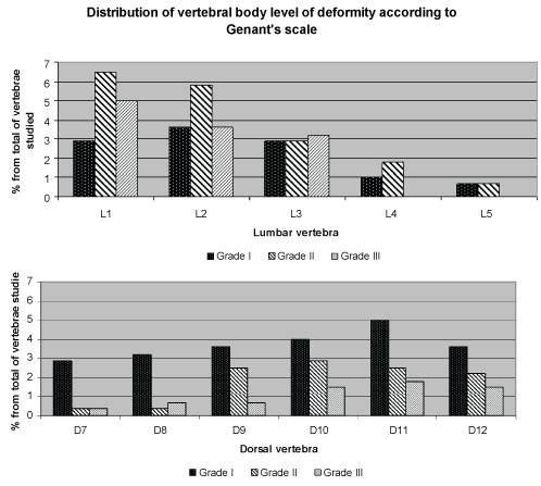 Figure 1: Distribution of vertebral body level of deformity according to Genant's scale. View Figure 1
Although our population sample is circumscribed to symptomatic patients, our main results contribute to knowing the prevalence of vertebral body deformities in postmenopausal patients which were mostly unnoticed.
The lack of accuracy showed by the low ratio of concordance between the expert opinion and the radiological interpretation of the clinicians who assessed patients also highlight the need for educative campaigns among emergency unit physicians in order to improve their capability to detect even the mildest vertebral body wedges for purposes of proper registration.
Nowadays the relevance of moderate or severe vertebral wedges is clear; however, there is some scepticism about the aetiology of the mildest level of a vertebral wedge, and for now, we do not consider it as a factor to start a secondary osteoporosis prevention therapy [14]. Prospective studies of follow up of patients with mild vertebral wedges to detect how many of them progress to significant vertebral deformities could be suitable to assess the real value of these Genant's grade I wedges.