Arthropathy induced by monosodium salt of uric acid [C5H4N4O3] (MSU) (gout), by calcium pyrophosphate dihydrate [Ca2P2O7.2H2O] (CPPD) crystals (chondrocalcinosis, pseudogout, pyrophosphate arthropathy) and arthropathy induced by hydroxyapatite [Ca5(PO4)3(OH)] (HA) crystals (apatite rheumatism, hydroxyapatite arthritis, calcifying tenosynovitis, Milwaukee syndrome, calcific tendinitis, calcific periarthritis) are regarded as distinct clinical entities. The solubility of MSU, CPPD and HA crystals in conventional fixatives (aqueous formaldehyde solution), in alcohol, acetone, and xylene or in solutions of dyes vary. The crystals in tissues may dissolve during fixation in aqueous formaldehyde solution, embedding in paraffin or during staining. Only those minerals or crystals can be detected microscopically with stains or histochemical reactions which remain in tissue sections after fixation, paraffin embedding or staining. The probability of identification of crystals is much higher in unstained sections viewed under polarized light than in traditionally stained ones. The aim of this study was to compare the "non-staining" technique according to Bély and Apáthy (2013) with worldwide accepted stains and histochemical methods: hematoxylin-eosin (H-E) staining, Gömöri's methenamine silver method, Schultz staining, Alizarin red S staining, and von Kossa's reaction in tissue samples of patients with clinically diagnosed gout, chondrocalcinosis and apatite rheumatism in order to demonstrate the effectivity of crystal detections by these standard methods in comparison with the non-staining technique.
One hundred and five (105) tissue samples of 47 patients with clinically diagnosed gout, 25 tissue samples of 16 patients with clinically diagnosed chondrocalcinosis, and 19 tissue samples of 4 patients with clinically diagnosed apatite rheumatism were studied. The tissue blocks were fixed in an 8% aqueous solution of formaldehyde [CH2(OH)2] at pH 7.6 for > 24 hours at room temperature (20 ℃) and embedded in paraffin. Serial tissue sections (5 microns thin) were cut. Using Bély and Apáthy's "non-staining" technique, the fixation of tissue blocks, and embedding were the same as with the standard stainings and reactions. Unstained tissue sections were deparaffinized, mounted and cover slipped with Canada balsam. The standard and unstained sections were examined with a professional polarizing light microscope (Olympus BX51).
Bély and Apáthy's non-staining technic was more effective: MSU was demonstrated in 83 (79.05% of 105) tissue samples of 37 (78.72% of 47) patients; CPPD in 15 (60.00% of 25) tissue samples of 10 (62.50% of 16) patients. HA crystals were detected exclusively with this method: in all tissue samples (in 19 of 19; 100.0%) of all patients (in 4 of 4; 100.0%), none with the traditional stains and reactions. The non-staining technique is a simple and very effective method to demonstrate crystal deposits in tissue samples. Handbooks of histologic methods and histochemistry do not mention this simple technique. In case of clinically or histologically suspected metabolic or crystal-induced diseases it is advisable to analyze unstained tissue sections as well, supplemented with the traditional hematoxylin-eosin stained ones. This approach may also be useful in other crystal deposition induced diseases or identification of foreign bodies and refractile artefacts.
HE: Hematoxylin Eosin; MSU: Monosodium Urate- [C5H4N4O3]; CPPD: Calcium Pyrophosphate Dehydrate- [Ca2P2O7.2H2O]; HA: Calcium Hydroxyapatite- [Ca5(PO4)3(OH)]
Non-staining technique, Tradicional stainings and histochemical methods, Gout, Chondrocalcinosis, Apatite rheumatism
Metabolic diseases and crystal induced arthropathies are characterized by deposits of crystal and/or non-crystalline (amorphous) calcium phosphates in synovial membranes (synovium) and periarticular soft tissues. Bone and cartilage may also be involved, and crystals may be present in the synovial fluid as well. Foreign bodies and refractile artefacts may vary the histopathological findings. Different crystals (with or without foreign bodies and/or refractile artefacts) may exist simultaneously in synovial fluid, synovium or adjacent bone and cartilage. Most common crystals and foreign bodies are summarized in Table 1 [1-3].
Table 1: Characteristics of most common crystals and foreign bodies in synovial fluid or synovium*. View Table 1
Arthropathy induced by monosodium salt of uric acid [C5H4N4O3] (MSU) (gout), by calcium pyrophosphate dihydrate [Ca2P2O7.2H2O] (CPPD) crystals (chondrocalcinosis, pseudogout, pyrophosphate arthropathy) and arthropathy induced by hydroxyapatite [Ca5(PO4)3(OH)] (HA) crystals (apatite rheumatism, hydroxyapatite arthritis, calcifying tenosynovitis, Milwaukee syndrome, calcific tendinitis, calcific periarthritis) are regarded as distinct clinical entities [4-14].
The solubility of MSU, CPPD and HA crystals in conventional fixatives (aqueous formaldehyde solution), in alcohol, acetone, and xylene or in solutions of dyes vary. The crystals in tissues may dissolve during fixation in aqueous formaldehyde solution, embedding in paraffin or during staining [15,16]. Only those minerals or crystals can be detected microscopically by staining or histochemical reactions which remain in tissue sections after fixation, paraffin embedding and staining [17-21]. The probability of identification of crystals is much higher in unstained sections viewed under polarized light than in traditionally stained ones [22-25,26].
The aim of this study was to compare the "non-staining" technique according to Bély and Apáthy (2013) [22] with worldwide accepted stains and histochemical methods such as hematoxylin-eosin (H-E) staining, Gömöri's methenamine silver method, Schultz staining, Alizarin red S staining, and von Kossa's reaction [17-21] in tissue samples of patients with clinically diagnosed gout, chondrocalcinosis and apatite rheumatism in order to demonstrate the effectivity of crystal detection with these standard methods in comparison with non-staining technique [22].
One hundred and five (105) tissue samples of 47 patients with clinically diagnosed gout, 25 tissue samples of 16 patients with clinically diagnosed chondrocalcinosis, and 19 tissue samples of 4 patients with clinically diagnosed apatite rheumatism were studied. Demographics are summarized in Table 2.
Table 2: Sex, average age (range) of patients with gout, chondrocalcinosis and apatite rheumatism. View Table 2
The biopsy material (tissue samples, surgical specimens) was varied: articular and periarticular soft tissues of different joints (elbow, knee, carpal synovium, subcutaneous nodules etc.); analysis of clinical relationships, localization, symptoms, radiological signs etc., were not considered. The tissue blocks were fixed in an 8% aqueous solution of formaldehyde [CH2(OH)2] at pH 7.6 for > 24 hours at room temperature (20 ℃) and embedded in paraffin. Tissue samples of gout fixed in alcohol (in clinically recognized cases) were excluded from this study, in order to compare the results under the same circumstances.
Using Bély and Apáthy's "non-staining" technique, the fixation of tissue blocks in 8% aqueous solution of formaldehyde at pH 7.6 for > 24 hours at room temperature (20 ℃) and embedding in paraffin were the same as with the standard stainings and reactions. Unstained tissue sections were deparaffinized, mounted and cover slipped with Canada balsam.
Serial sections (5 micron thin) were stained: in case of gout with H-E [20], according to Gömöri [18,20], and Schultz [17], and in case of chondrocalcinosis, and apatite rheumatism with H-E [20], Alizarin red S [19,21], and von Kossa's reaction [17,21]. The results with these standard stainings and reactions were compared in serial sections with the unstained ones [22,25].
The standard and unstained sections were examined with a professional polarizing light microscope (Olympus BX51). In selected cases the nature of crystals was confirmed by a JEM 100CX electron microscope and electron diffraction as well (Figure 1a, Figure 1b, Figure 1c and Figure 1d).
 Figure 1: a) Urate crystals, surface electron micrograph, x1600; b) Urate crystals, transmission electron micrograph, x6000; c) CPPD crystals, surface electron micrograph, x10000; d) Hydroxy apatite crystals, surface electron micrograph, x50000; e) Cholesterol crystals, surface electron micrograph, x1300, Original magnifications correspond to the 600 × 900 mm negative. The printed size may be different; therefore, it is necessary to indicate the original magnifications corresponding to a fixed size (in case of electron micrographs this is the 6 × 9 cm analogue negative).
View Figure 1
Figure 1: a) Urate crystals, surface electron micrograph, x1600; b) Urate crystals, transmission electron micrograph, x6000; c) CPPD crystals, surface electron micrograph, x10000; d) Hydroxy apatite crystals, surface electron micrograph, x50000; e) Cholesterol crystals, surface electron micrograph, x1300, Original magnifications correspond to the 600 × 900 mm negative. The printed size may be different; therefore, it is necessary to indicate the original magnifications corresponding to a fixed size (in case of electron micrographs this is the 6 × 9 cm analogue negative).
View Figure 1
The average age of patients with gout was significantly lower than the average age of patients with chondrocalcinosis (p < 0.050). There was no significant difference between the average age of patients with chondrocalcinosis and with apatite rheumatism (p < 0.129).
In sections stained with H-E: MSU crystals were present in 24 (22.86% of 105) tissue samples of 16 (34.04% of 47) patients; CPPD in 11 (44.00% of 25) tissue samples of 8 (50.00% of 16) patients; HA crystals were not detected. In sections with Gömöri's methenamine silver method: MSU was present in 59 (56.19% of 105) tissue samples of 25 (34.19% of 47) patients; CPPD or HA crystals were not detected. In sections stained according to Schultz: MSU was present in 66 (62.86% of 105) tissue samples of 27 (57.45% of 47) patients; CPPD or HA crystals were not detected. CPPD crystals were detected in sections stained with Alizarin Red S in 7 (29.17% of 24) tissue samples of 4 (25.00% of 16) patients; with von Kossa reaction in 2 (8.33% of 24) tissue samples of 2 (12.50% of 16) patients; with these methods HA crystals were not detected.
In contrast with these classic stains and reactions Bély and Apáthy's non-staining technic was more effective: MSU was demonstrated in 83 (79.05% of 105) tissue samples of 37 (78.72% of 47) patients; CPPD in 15 (60.00% of 25) tissue samples of 10 (62.50% of 16) patients. HA crystals were detected exclusively with this method: in all tissue samples (in 19 of 19; 100.0%) of all patients (in 4 of 4; 100.0%).
The prevalence of MSU, CPPD and HA crystals in tissue samples (Ts) of patients (Pts) with gout, chondrocalcinosis and hydroxyapatite arthritis (in case of standard stains and reaction in comparison with Bély and Apáthy's "non-staining" technique) is summarized in Table 3.
Table 3: The prevalence of MSU, CPPD, HA crystals or crystal aggregates in tissue samples of patients with gout, chondrocalcinosis or with apatite rheumatism. View Table 3
Comparing the classical staining methods and histochemical reactions, there was a significant difference between their effectivity (sensitivity). The Gömöri or Schultz stains were more effective in detection of MSU than with H-E, and the non-staining technique of Bély and Apáthy's was much more sensitive than all of these (Table 4). The differences between these methods, regarding the effectivity and sensitivity, were significant, except for the number of patients. Though more patients were positive for urate crystals with the none staining technique than with Schultz's stain, but this difference was statistically not significant. The statistical correlations ("p" values of significance) are summarized in Table 4, comparing tissue samples and involved patients with gout with different stains and techniques.
Table 4: The statistical correlations ("p" values of significance) are summarized. View Table 4
CPPD crystals were not detected in combination with MSU crystals in our patient cohort's. The HE staining was more effective in detection of CPPD crystals than Alizarin red S staining or von Kossa reaction, and Bély and Apáthy's non-staining technique detected many more CPPD crystals than HE staining (Table 5). Using these methods there was a difference in the number of detected crystals, but regarding the effectivity (sensitivity) of these methods these differences were not significant statistically in most cases (Table 5). The statistical correlations ("p" values of significance) are summarized in Table 5, comparing tissue samples and involved patients with chondrocalcinosis with different stains and techniques.
Table 5: The statistical correlations ("p" values of significance) are summarized. View Table 5
Clusters of HA crystals and aggregates were detected only in unstained sections according to Bély and Apáthy in combination with more or less CPPD crystals together (significance was not calculated; there were no comparable values). Clusters of HA crystals and aggregates of clusters were not detected in combination with MSU crystals.
Cholesterol [C27H46O] crystals were present only in tissue sections stained according to Schultz, and in unstained sections according to Bély and Apáthy; cholesterol crystals were not detected with H-E, Gömöri, and Alizarin Red S staining or with von Kossa's reaction. In tissue sections stained according to Schultz, cholesterol was detected in case of gout in 10 (21.27%) of 47 patients (with or without with MSU) and in case of chondrocalcinosis or apatite rheumatism, in 14 (77.77%) of 18 patients without CPPD or HA crystals (CPPD or HA crystals do not stain according to Schultz). In unstained tissue section according to Bély and Apáthy, cholesterol was present with variable prevalence in the entire patient group, but exact analysis was not possible in patients with the clinical diagnosis of gout or chondrocalcinosis, because of large amounts of MSU or CPPD crystals. Figures 2, Figure 3 and Figure 4 demonstrate the presence of MSU, CPPD and HA crystals with traditional staining and reaction in comparison with the non-staining technique. The cholesterol crystals are demonstrated with Schultz staining (Figure 5) in comparison with non-staining technique (Figure 5). (The original magnification corresponds to the 24 × 36 mm transparency slide; the correct height: width ratio is 2:3). Cholesterol [C27H46O] and lipid crystals stained according to Schultz and viewed under polarized light. The cholesterol [C27H46O] and lipid crystals were associated in this patient with HA and CPPD crystals. The HA and CPPD crystals are not visible in tissue sections stained according to Schultz.
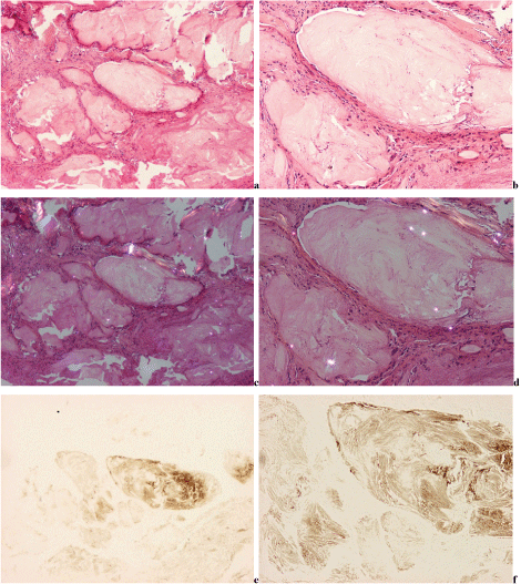 Figure 2A-F: Gouty arthritis (tophaceous gout), monosodium salt of uric acid [C5H4N4O3] (MSU) crystal deposits, viewed with the light microscope and under polarized light, respectively.
(a) H-E, viewed with the light microscope, x40; (b) same as (a) x100; c) H-E, viewed under polarized light, same as (a) x40; (d) same as (c) x100 MSU crystals dissolved in 8% formaldehyde fixed specimens, and hematoxylin-eosin stained sections MSU crystals are not present (birefringent fragments are artefacts of remained paraffin debris); e) Unstained section, with the light microscope, same field as (a) x40; f) same as (e) x100 MSU crystals of swart natural colour are retained in unstained sections, and are arranged in characteristic bundles;
View Figure 2A-F
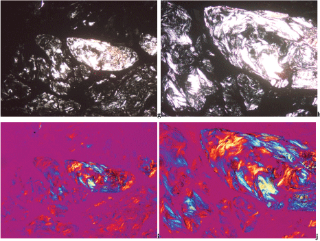 Figure 2G-J: g) Unstained section, viewed under polarized light, same field as (a) x40; (h) same as (g) x100 MSU crystals are demonstrable in 8% formaldehyde water solution fixed specimens in unstained sections. MSU crystals with intensive birefringence are arranged in characteristic bundles; i) Unstained section, Red I compensator, viewed under polarized light, same field as (a) x40; j) same as (i) x100 MSU crystals show a strong negative birefringence with Rot I. compensator.
View Figure 2G-J
Figure 2G-J: g) Unstained section, viewed under polarized light, same field as (a) x40; (h) same as (g) x100 MSU crystals are demonstrable in 8% formaldehyde water solution fixed specimens in unstained sections. MSU crystals with intensive birefringence are arranged in characteristic bundles; i) Unstained section, Red I compensator, viewed under polarized light, same field as (a) x40; j) same as (i) x100 MSU crystals show a strong negative birefringence with Rot I. compensator.
View Figure 2G-J
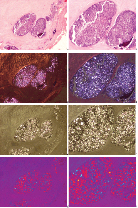 Figure 3A-H: Chondrocalcinosis (pseudogout, pyrophosphate arthropathy, calcium pyrophosphate dihydrate [Ca2P2O7.2H2O] (CPPD) crystal induced arthropathy), viewed with the light microscope and under polarized light, respectively.
(a) H-E viewed with the light microscope x100; (b) same as (a) x200 CPPD crystals and crystal fragments are accompanied by amorphous calcium phosphate, or calcium carbonate deposits of blue-violet colour; (c) H-E, viewed under polarized light, same as (a) x100; (d) same as (c) x200; (e) Unstained section, viewed under polarized light, same field as (a) x100; (f) same as (e) x200; (g) Unstained section, Red I compensator, viewed under polarized light, same field as (a) x100; (h) same as (g) x200 Under polarized light CPPD crystals show positive birefringence (parallel to the long axis of the crystals analogous to the birefringence of collagen fibers, see: Figure 3c and Figure 3g).
View Figure 3A-H
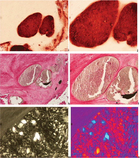 Figure 3I-N: (i) Alizarin red S, viewed with the light microscope, same as (a) x100; (j) same as (c) x200
Non-crystalline calcium containing mineral deposits are staining with calcium specific Alizarin red S. Alizarin red S does not stain the CPPD crystals, and the masses of amorphous calcium phosphate and carbonate may mask the crystals, with no detectable birefringence; (k) von Kossa's reaction, viewed with the light microscope, same as (a) x100; (l) same as (c) x200 Non-crystalline phosphate or carbonate containing mineral deposits show a positive reaction according to von Kossa. The CPPD crystals are negative with von Kossa's reaction, and the masses of amorphous calcium phosphate and carbonate may mask the crystals, with no detectable birefringence.
(m) Intact CPPD crystals and fragments, unstained section, viewed under polarized light, same field as (e-f) x600
The intact CPPD crystals have a rhomboid shape, they range in size is from 5 to 40μm and show a strong birefringence; (n) Unstained section, Red I compensator, viewed under polarized light, same field as (g-h and m) x600 Axis parallel direction of birefringence of CPPD crystals is positive.
View Figure 3I-N
Figure 3I-N: (i) Alizarin red S, viewed with the light microscope, same as (a) x100; (j) same as (c) x200
Non-crystalline calcium containing mineral deposits are staining with calcium specific Alizarin red S. Alizarin red S does not stain the CPPD crystals, and the masses of amorphous calcium phosphate and carbonate may mask the crystals, with no detectable birefringence; (k) von Kossa's reaction, viewed with the light microscope, same as (a) x100; (l) same as (c) x200 Non-crystalline phosphate or carbonate containing mineral deposits show a positive reaction according to von Kossa. The CPPD crystals are negative with von Kossa's reaction, and the masses of amorphous calcium phosphate and carbonate may mask the crystals, with no detectable birefringence.
(m) Intact CPPD crystals and fragments, unstained section, viewed under polarized light, same field as (e-f) x600
The intact CPPD crystals have a rhomboid shape, they range in size is from 5 to 40μm and show a strong birefringence; (n) Unstained section, Red I compensator, viewed under polarized light, same field as (g-h and m) x600 Axis parallel direction of birefringence of CPPD crystals is positive.
View Figure 3I-N
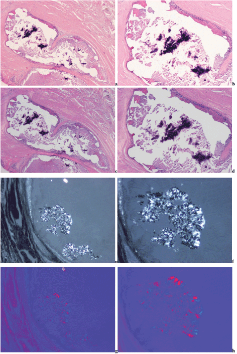 Figure 4A-H: Hydroxyapatite arthropathy (Milwaukee syndrome, apatite rheumatism) induced by hydroxyapatite [Ca5(PO4)3(OH)] (HA) crystals (crystal clusters and aggregates of clusters) in association with a few CPPD crystals, viewed with the light microscope and under polarized light, respectively.
(a) H-E viewed with the light microscope x20; (b) same as (a) x40 The HA (and CPPD) crystals are accompanied by amorphous calcium phosphate, or calcium carbonate deposits of blue-violet colour. The absence of an inflammatory reaction is characteristic [27]. (c) H-E viewed under polarized light, same as (a) x20; (d) same as (c) x40 In traditionally fixed tissue specimens the HA crystals (crystal clusters and aggregates of clusters) dissolved and are not demonstrable (the sporadic CPPD crystals or fragments are also not visible). (e) Unstained section viewed under polarized light, same field as (a) x100; (f) same as (e) x200 The individual HA crystals are small, 50-500 nm, rod-shaped and are arranged typically in 1-5 μm spheroid microaggregates, which are not in visible (detectable) range with polarizing microscopy [2]. The Figures m-p demonstrate crystal clusters and aggregates of clusters of 6.5 and 20 mm size, which may appear under plain light microscopy, but according to Forster, et al. [28] without birefringence. Using a professional polarizing microscope with high brightness, the clusters show of a week birefringence. Under polarized light the direction of birefringence is positive according to the long axis of HA crystals, like that of collagen fibers. The HA crystal clusters (microaggregates) and aggregates of clusters are associated sporadically with much larger and partially fragmented CPPD crystals. The CPPD crystals are less soluble in comparison with HA crystals or crystal aggregates. The birefringence of CPPD crystals is stronger than that of HA crystals or crystal aggregates. The masses of amorphous calcium phosphate and carbonate may mask the crystals, with no detectable birefringence, even in in synovial fluid "identifying individual calcium hydroxyapatite crystals can be elusive" [28]. (g) Unstained section, Rot I compensator, viewed under polarized light, same field as (a and e) x100, (h) same as (g) x200. Under polarized light HA and CPPD crystals show positive birefringence parallel to the long axis of the crystals, but the intensity of birefringence of HA is much weaker than that of CPPD crystals.
View Figure 4A-H
 Figure 4I-P: (i) Alizarin red S, viewed with the light microscope, same as (a) x20, (j) same as (i) x40 Massive non-crystalline (amorphous), calcium containing mineral deposits are staining with calcium specific Alizarin red S [19-21]. Alizarin Red S stain is considered by several authors as a non-specific but effective and sensitive screen method for HA crystals or crystal aggregates [3,28-31]. Alizarin red S does not stain the HA or CPPD crystals [11-21], but the masses of amorphous calcium phosphate and carbonate may mask the crystals, with no detectable birefringence [3,28-31]. (k) von Kossa's reaction, viewed with the light microscope, same as (a) x20, (l) same as (c) x40), Non-crystalline (amorphous) phosphates or carbonate containing mineral deposits show positive reaction according to von Kossa [17-21]. The HA or CPPD crystals are negative with von Kossa's reaction, but the masses of amorphous calcium phosphate and carbonate may mask the crystals, with no detectable birefringence. (m) Unstained section viewed under polarized light, same field as (e) x200, (n) same as (m) x600 The 50-500 nm small, rod-shaped individual HA crystals are arranged typically in 1-5 μm spheroid clusters (microaggregates) and larger aggregates of clusters [28], sporadically associated with fragmented CPPD crystals. The CPPD crystals are larger, have a rhomboid shape, and show a strong birefringence in comparison with HA. (o) Unstained section, Red I compensator, viewed under polarized light, same field as (m) x200, (p) same as (o) x600 Axis parallel direction of birefringence of HA (and CPPD) crystals are positive.
View Figure 4I-P
Figure 4I-P: (i) Alizarin red S, viewed with the light microscope, same as (a) x20, (j) same as (i) x40 Massive non-crystalline (amorphous), calcium containing mineral deposits are staining with calcium specific Alizarin red S [19-21]. Alizarin Red S stain is considered by several authors as a non-specific but effective and sensitive screen method for HA crystals or crystal aggregates [3,28-31]. Alizarin red S does not stain the HA or CPPD crystals [11-21], but the masses of amorphous calcium phosphate and carbonate may mask the crystals, with no detectable birefringence [3,28-31]. (k) von Kossa's reaction, viewed with the light microscope, same as (a) x20, (l) same as (c) x40), Non-crystalline (amorphous) phosphates or carbonate containing mineral deposits show positive reaction according to von Kossa [17-21]. The HA or CPPD crystals are negative with von Kossa's reaction, but the masses of amorphous calcium phosphate and carbonate may mask the crystals, with no detectable birefringence. (m) Unstained section viewed under polarized light, same field as (e) x200, (n) same as (m) x600 The 50-500 nm small, rod-shaped individual HA crystals are arranged typically in 1-5 μm spheroid clusters (microaggregates) and larger aggregates of clusters [28], sporadically associated with fragmented CPPD crystals. The CPPD crystals are larger, have a rhomboid shape, and show a strong birefringence in comparison with HA. (o) Unstained section, Red I compensator, viewed under polarized light, same field as (m) x200, (p) same as (o) x600 Axis parallel direction of birefringence of HA (and CPPD) crystals are positive.
View Figure 4I-P
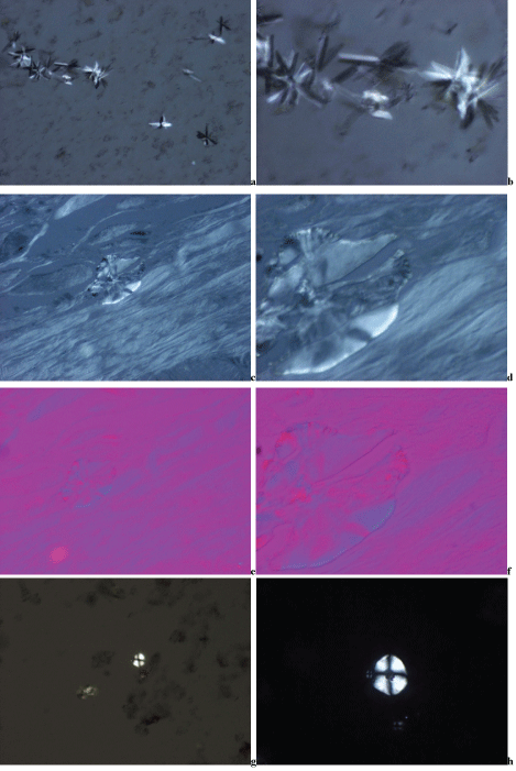 Figure 5A-H: Same tissue samples of a patient with clinical diagnosis of apatite rheumatism demonstrated on Figure 4a-n, stained according to Schultz or Bély and Apáthy's non-staining technique.
Cholesterol [C27H46O] and lipid crystals stained according to Schultz and viewed under polarized light. The cholesterol [C27H46O] and lipid crystals were associated in this patient with HA and CPPD crystals. The HA and CPPD crystals are not visible in tissue sections stained according to Schultz. The size of cholesterol crystals is 5-40 μm [3], rhomboidal, notched, needle-shaped cloven and are present as separate sheets or typically arranged in clusters. A "semi-liquid" appearance is also characteristic. The birefringence of cholesterol crystals is positive or negative; the needle-shaped or cloven crystal fragments rotating around the axis may show in the same position (direction) positive or negative birefringence. The lipid crystals are small 0.5-30 μm spherules, with positive Maltese cross birefringence.
(a) Needle-shaped (0.5-30 μm) cholesterol crystals arranged in typical clusters. The birefringence in the same direction is negative (black) and positive (white), Schultz staining, viewed under polarized light x200; (b) same as (a) x600; (c) Cholesterol crystals with "semi-liquid" appearance, Bély and Apáthy's non-staining technique, viewed under polarized light x200; (d) same as (c) x600; (e) Cholesterol crystals with "semi-liquid" appearance. The birefringence is positive; analog like by agreement positive birefringence of collage fibers. Bély and Apáthy's non-staining technique, Red I compensator, viewed under polarized light, same field as (c) x200; (f) same as (d and e) x600; (g) Lipid crystal spherules with intensive Maltese cross birefringence, Schultz staining, viewed under polarized light x200; (h) same as (g) x600;
View Figure 5A-H
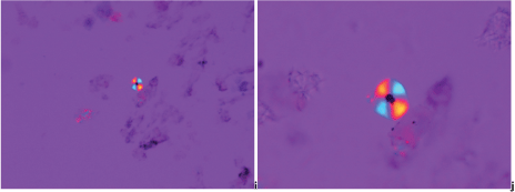 Figure 5I-J: (i) Lipid crystal spherule with positive Maltese cross birefringence (in contrast to talc or starch, which have negative Maltese cross birefringence), Schultz staining, Red I compensator, viewed under polarized light, same field as (g) x200; (j) same as (h and i) x600.
View Figure 5I-J
Figure 5I-J: (i) Lipid crystal spherule with positive Maltese cross birefringence (in contrast to talc or starch, which have negative Maltese cross birefringence), Schultz staining, Red I compensator, viewed under polarized light, same field as (g) x200; (j) same as (h and i) x600.
View Figure 5I-J
The histological diagnosis of metabolic disorders is based on the presence crystals in tissue sections with or without non-crystalline (amorphous), calcium phosphate and/or carbonate containing mineral deposits. There is a difference in the shape, size, intensity of birefringence, and optical breaking direction of MSU, CPPD, HA and cholesterol crystals. The solubility of these crystals in conventional fixatives (aqueous formaldehyde solution), in alcohol, acetone, and xylene or in solutions of dyes is also different. The crystals in tissues may dissolve during fixation in aqueous formaldehyde solution (formalin), embedding in paraffin or during staining.
In clinically known or suspected cases of gout the surgical tissue specimens should be fixed in absolute ethyl alcohol, because urate crystals are soluble in 8% formaldehyde solution [17,18,20,21]. To quote McManus and Mowry "since urates are slightly soluble in water, alcohol fixation is preferable" [21], but we found that in gout most urate crystals dissolve during the hematoxylin-eosin staining procedure [15,16,32-34]. In present study the alcohol fixed cases of gout were excluded, in order to compare the traditional staining and reaction with Bély and Apáthy's non-staining technique under the same circumstances.
CCPD crystals are less soluble than urate and are likely to be detected in traditionally processed tissue sections. The small and soluble HA or the highly soluble cholesterol crystals are not detected in traditionally fixed, embedded and stained tissue samples. In case of cholesterol deposition, the absence of crystals and characteristic empty spaces accompanied by a typical inflammatory reaction of macrophages and multinucleated giant cells are a reminder of the dissolved crystal deposits.
Our results indicate that the very simple "not-staining" technique is a most effective method to demonstrate crystal deposits in tissue samples [15,16,22,25]. Theoretically the largest amounts of crystals may be best preserved in unstained frozen sections. Indeed, large amounts of cholesterol or fatty acid crystals may be visualized in frozen sections under polarized light [35]. The frozen sections are not suggested for diagnosis of metabolic disorders in everyday practice, because large amounts of cholesterol crystals may conceal other crystals.
A disadvantage of unstained sections is that parallel (serial) tissue sections have to be stained traditionally, since detailed histology cannot be studied adequately in unstained sections with the light microscope or under polarized light. Another disadvantage (or advantage) is that in unstained sections other crystals can be found which differ in shape, size, arrangement or quality of birefringence from the well-known crystals, and their identification would require further specific (electron microscopic, electron diffraction, etc.) studies.
Major textbooks of histochemistry discuss many techniques and staining methods to demonstrate preserved crystals and mineral deposits in tissue, but none mention the simplest method, namely viewing of unstained tissue sections with polarized light [17-21].
In his book Mohr demonstrated crystals in unstained tissue sections (independent of us) but does not mention the advantage of this method in comparison with traditional stainings [9]. According to our best knowledge a detailed analysis or comparative study of our non-staining technique and its comparison with traditional stainings and reactions has not been available in the literature.
In case of suspected metabolic or crystal induced disorders, we suggest analyzing the tissue samples with unstained tissue sections as well, supplemented with traditional stainings and reactions. Crystals remain detectable in unstained sections viewed under polarized light in the great majority of cases which appear negative with H-E staining [22-24,32-34].
Bély and Apáthy's non-staining technique is a simple and sensitive method and may help in the microscopic demonstration and analysis of crystalline deposits.
The probability of identifying crystals is much higher in unstained sections viewed under polarized light than in haematoxylin-eosin stained ones. Textbooks of histologic methods and histochemistry do not mention this simple technique.
In case of clinically or histologically suspected metabolic or crystal induced diseases the analysis of tissue samples is suggested with unstained tissue sections as well, supplemented with the traditional hematoxylin-eosin staining. This approach may also be useful in other crystal deposition induced diseases or identification of foreign bodies and refractile artefacts.
There is no conflict of interest.
This work did not receive financial support from any source.
Authors contribution is equal.
The manuscript has been changed according to the required order of guidelines.