

Clinical Medical Reviews and Case Reports
Consultant, Respiratory and General Medicine, UK
*Corresponding author:
Dr. Avinash Aujayeb, Consultant, Respiratory and General Medicine, Elysium HealthCare Ltd, Cyber City, Mauritius, UK, Tel: 00230-58592078, E-mail: aujayeb@doctors.org.uk
Clin Med Rev Case Rep, CMRCR-4-155, (Volume 4, Issue 2), Case Report; ISSN: 2378-3656
Received: December 19, 2016 | Accepted: February 10, 2017 | Published: February 14, 2017
Citation: Aujayeb A (2017) A Missed Diagnosis. Clin Med Rev Case Rep 4:155. 10.23937/2378-3656/1410155
Copyright: © 2017 Aujayeb A. This is an open-access article distributed under the terms of the Creative Commons Attribution License, which permits unrestricted use, distribution, and reproduction in any medium, provided the original author and source are credited.
Case Report
A 64-year-old female patient presented with vomiting, abdominal pain and constipation.
Her past medical history included squamous cell carcinoma of lung origin T2BN0M0 treated in 2012 with a right lower lobectomy via video assisted thoracoscopy with a wound infection preventing adjuvant chemotherapy. She then developed local chest wall recurrence in 2014 which was resected. In June 2016, there was further recurrence on the chest wall, which was confirmed on PET scan. She had received one cycle of chemotherapy and presented as above.
She was febrile with a temperature of 38.7 degrees Celsius, tachycardic at a rate of 112 beats per minute but normotensive. Her oxygen saturations were 95% on air. She had reduced air entry at the right base of her chest, and her abdomen was generally tender with quiet bowel sounds. A rectal examination showed hard stool in the rectum.
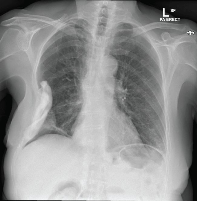
Her white cell count was 6.7 × 10⁹/L (normal range 4-11), C reactive protein raised at 257 mg/L (normal < 5) and her kidney function as well as bone panel was normal. Her chest x-ray was interpreted as no obvious consolidation on a background of post -surgical change (Figure 1). Her urine dip was negative.
.
Figure 1: X-ray at initial presentation which was interpreted as no obvious consolidation on a background of post surgical change.
View Figure 1
A presumptive diagnosis of undifferentiated sepsis in an immunosuppressed patient was made, along with vomiting and constipation.
Case Discussion
She was treated with broad spectrum antibiotics, fluids, analgesia, anti-emetics and laxatives. Blood cultures at 24 hours grew a gram-negative bacterium called Eubacterium limosum which is a gastrointestinal commensal [1]. This prompted a CT scan of her abdomen which showed that there was heavy faecal loading of the colon, which made assessment difficult but that there was a perforated colonic diverticulum at the hepatic flexure where there is a prominent paracolic gas locule and several adjacent free air locules. She was reviewed by surgery and managed conservatively with fluids, further antibiotics and analgesia. She recovered after a prolonged period of hospitalisation.
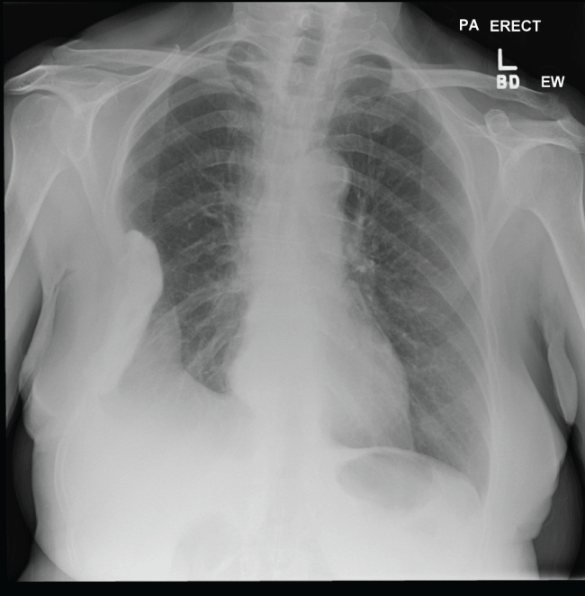
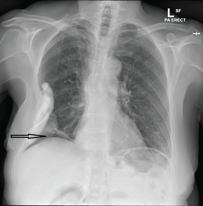
Her case was reviewed and air under the diaphragm was missed on her first chest X-ray. It was felt it was quite a subtle sign and there was a lot of post-surgical change as well but reinforces the teaching that X-ray interpretation is a vital tool and everyone should be systematic about it. Figure 2 shows a previous X-ray where it can appreciated that there is no air seen. Figure 3 is figure 1 again with an arrow depicting the air. It could be argued that no harm was done, and the consultant involved (myself) explained all the above to the patient who gracefully accepted apologies. An earlier CT scan would have secured the diagnosis earlier, but the treatment would have been the same.
A further discussion point is when to perform a CT scan in the emergency department (ED). There are clear guidelines relating to CT scanning in trauma cases and clear evidence that CT has a high sensitivity and specificity for a wide variety of injuries [1]. However, should there be criteria for scanning such patients in the ED itself? Li, et al. [2] found that CT enabled to triage patients adequately if they were finally hospitalised, but mildly prolonged ED length of stay in cases of patients discharged from the ED. Zwank, et al. [3] also found that only 25% patients were aware that radiation from CT can increase overall lifetime risk of cancer compared with previous surveys but it is widely known that CT usage is increasing [4]. I personally firmly believe that she was treated and investigated appropriately. If the air under the diaphragm had been recognised, she would have had an earlier CT, in all likelihood in the ED itself.
References
-
https://www.nice.org.uk/guidance/ng39.
-
Li C, Syue Y, Lin Y, Cheng H, Cheng FJ, et al. (2016) Influence of CT utilisation on patient flow in the emergency department: a retrospective 1-year cohort study. BMJ Open 6: e010815.
-
Zwank MD, Leow M, Anderson CP (2014) Emergency department patient knowledge and physician communication regarding CT scans. Emerg Med J 31: 824-826.
-
Kocher KE, Meurer WJ, Fazel R, Scott PA, Krumholz HM, et al. (2011) National Trends in Use of Computed Tomography in the Emergency Department. Ann Emerg Med 58: 452-462.
