Primary tumors of trachea are usually malignant (90%). Hamartomas are the most frequently seen benign lung tumors. Hamartomas are usually located peripherally but they can be present as endobronchial localization in 8-10% of cases. Squamous-cell papilloma, fibroma and haemangioma are the commonest benign tracheal tumors but hamartomas are rare.
Transbronchial endoscopic surgery is usually performed for patients with a small endobronchial hamartoma. Here we are presenting a case of tracheal hamartoma which diagnosed during the lung cancer investigating. The lesion completely removed with rigid bronchoscopy.
Bronchoscopy , Hamartoma, Tracheal lesion
Primary tumors of trachea are usually malignant (90%) [1]. Hamartomas are the most frequently seen benign lung tumors. 77% of all benign tumors of the lung are hamartomas [2,3]. Hamartomas are usually solitary, although sometimes they're multiple. Hamartomas are usually located peripherally but they can be present as endobronchial localization in 8-10% of cases [3]. Histologically, they are composed of cartilage, fat, bone and connective tissue, and smooth muscle cells. Most patients have asymptomatic parenchymal hamartomas, which defined incidently by radiology. Endobronchial hamartomas usually becomes symptomatic when they cause obstruction. We now describe a case of hamartoma located in trachea which diagnosed incidentally, while patient was been investigated for neuromotor diseases.
A 61-year-old male patient was admitted to our hospital with complaint of weakness in his hands for one month. The patient didn't have any pulmonary symptoms. Physical examination revealed blood pressure: 125-75 mmHg, pulse: 75 per minute, finger tip saturation: 98% and temperature was 36.5 degrees. Other systems were normal. He had a 40 pack-years of smoking history. His family history was unremarkable. While neuromotor diseases were investigating, Computed Tomography (CT) of thorax was performed to exclude pulmonary malignancy. CT showed a low density lesion in the trachea which extending from the posterior to 3 cm proximal of the carina (Figure 1). Fiber optic bronchoscopy revealed a polypoid and vesicular lesion which obstructed 10% of the tracheal lumen attached to the posterior tracheal wall with a broad base (Figure 2). Sampling with by fiber optic bronchoscopy avoided because of bleeding propensity. Patient referred to thoracic surgery department to perform rigid bronchoscopy under general anesthesia. The lesion completely removed with rigid bronchoscopy (Figure 3). Histopathological examination of lesion revealed hamartoma.
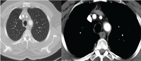 Figure 1: Image from CT scan. View Figure 1
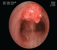 Figure 2: Bronchoscopic view of lesion. View Figure 2
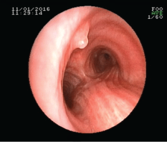 Figure 3: Post-treatment view of lesion. View Figure 3
Primary tracheal tumors were reportedly seen in only 0.1% of cancer patients [4]. Squamous-cell papilloma, fibroma and haemangioma are the commonest benign tracheal tumors. However endotracheal hamartomas are rare and 1.4-10% of hamartomas are located endobronchially as in the our case [1,3]. According to the autopsy results from the Mayo Clinic, pulmonary hamartomas were found in 2 of 7,972 cases (0.025%) [5]. Hamartomas are expressed as a combination of sequencing abnormal mature mesenchymal cells of the lung. It consists of a mixture of undifferentiated mesenchymal cells, cartilage and fat by simple cuboidal orciliated epithelium [5]. It is 2-4 times more common in men and is most commonly seen in 6th decade [2,6]. Our case was a man and he was 61-years-old. Hamartomas mostly appears as solitary lesions. Multiple lesions are rarely observed. The respiratory infection or pulmonary atelectasis due to mechanical obstruction of the bronchus could be encountered in part of patients with pulmoary hamartomas. Coughing, shortness of breath, sputum and fever are the most common symptoms for endobronchial hamartomas [4,7]. Peripheral pulmonary hamartomas are generally asymptomatic. However, based on its localization, they can even cause death through airway obstruction. For this reason, early diagnosis is crucial. Our case was asymptomatic even though its central localization.
Pulmonary hamartomas are smooth, lobular, well-defined peripheral lesions and incidentally diagnosed as abnormal chest radiograph [6,8,9]. Diameteris generally less than 2.5 cm [5,10]. Lobulation of the edge of nodules indicates that the tumor grows in to enviroment. This finding is also reflected in primary and metastatic cancer [5]. In 15% of cases, calcification occurs in the form of popcorn and these findings are pathognomonic [6]. Punctate calcifications can be observed in hamartomas as can be seen in many benign and malignant lesions.
Usually, the pulmonary hamartomas has non-specific clinical manifestations and was detected by chest radiographic examination occasionally. Radiographically, the tumor is usually 1 to 2 cm in size and is located mostly in the periphery of the lung [9]. CT and MRI are useful for demonstrating the outline, location and extent of the tumor and may also indicate spread to adjacent organs and localized lymph node enlargment. The internal structure of the lesion is beter evaluated by CT. Main CT finding of the endobronchial hamartoma is endobronchial mass with or without obstructive pneumonia or atelectasis [3]. CT can be useful when the lesion contains an abundance of fat. Intranodular fat is reported as a diagnostic CT finding, with a frequency of around 50% [11]. Although "popcorn" calcification is a well-known finding of pulmonary hamartoma, typical popcorn calcification is reported at a low frequency of only 5% to 15% [5,6,11].
Hamartomas shows low growth [5,10]. Nodular lesion growth for more than two years shows that it's good-natured. Other benign and malignant lung tumors should be considered in the differential diagnosis of hamartomas [5,6]. Cancer was in the differential diagnosis in our patients.
The treatment method of benign primary tracheal tumors varies, depending on the size and localization of the lesion. In benign tracheal lesions, more conservative treatments such as laser or endoscopic resection, are recommended. However, in the case where a mass invading the tracheal wall is present, surgical resection should be the initial choice. Bronchoscopy may directly reveal the tumor and permit biopsy, which are important for surgical planning and preoperative preparation. Biopsy was not performed in our patient, because of the perceived risk of bleeding. Most clinical studies about PH focused on the difference to malignant lesions and the diagnosis measures with effectiveness and specificity. Today, even with the advancement in medical therapy, pulmonary resection remains the most important measure of treatment of patient with hamartoma of lung. Rare cases of hamartomas can show malignant transformation, therefore surgical resection is recommended [3-6,12]. Resection is the definitive treatment and recurrence is extremely rare [6]. Bronchoscopic excision of the lesion can be made in appropriate cases. In our case, the lesion completely removed with rigid bronchoscopy.
Only 15% of endobronchial hamartomas are diagnosed preoperatively. As a result, lobectomy or pneumonectomy is performed in 47% of endobronchial hamartomas, even though the tumor is benign [9,13]. Cosio and colleagues [14] performed complete resection of the masses by transbronchial endoscopic surgery using Neodymium: Yttrium Aluminium Garnet (Nd:YAG) laser in 17 of 36 patients diagnosed with endobronchial hamartoma. Endoscopic surgery including Nd:YAG laser and electrosurgical snare are good therapeutic choices for a small resectable endobronchial hamartoma.
When literature is examined, it will be seen that the patients are generally male between 40-60 years old. The demographic characteristics of our case are consistent with the literature. The fact that our case is asymptomatic distinguishes it from other cases in the literature. Our patient did not have any complaints for 6 months follow up.
In conclusion, endotracheal hamartomas are benign tumors and quite rare. Hamartomas can show malignant transformation. Also, malignancy is the most important disease in the differential diagnosis, therefore surgical resection is recommended .