T-Cell Large Granular Lymphocyte Leukemia (T-LGL Leukemia) is a lymphoproliferative disease that usually presents with an indolent behavior, even though in some cases it is complicated by cytopenias and recurrent infections. Felty Syndrome (FS) is a subtype of Rheumatoid Arthritis (RA) characterized by the association of RA, neutropenia and splenomegaly. SF and T-LGL Leukemia have a few common aspects, especially immunogenetic and clinical ones. 30 to 40% of the patients with SF have T-LGL cell expansion in peripheral blood. These similarities, amongst others make some authors believe that these two pathologies may be two presentations of the same disease process. Because of that, the T-LGL Leukemia associated with RA is also called Pseudo-Felty. In this paper we report the case of an 82-year-old woman with severe RA diagnosed with SF. Due to the persistent neutropenia and the absence of response in several treatments, including two anti-TNFs, she had a bone marrow biopsy with immunophenotyping which was compatible with T-LGL Leukemia.
T-Cell large granular lymphocite leukemia, Felty syndrome, Rheumatoid arthritis, Methotrexate, Neutropenia
T-Cell Large Granular Lymphocyte Leukemia (T-LGL Leukemia) is a rare lymphoproliferative disease in which a T Cytotoxic Lymphocyte CD3+ expansion occurs [1]. This disease is more prevalent among adults, mainly between 55 and 60-years-old, and affects equally both genders. The risk of development is higher in populations with autoimmune diseases [2,3]. The diagnosis is made by the presence of T-LGL lymphocytes for more than 6 months, with infiltration of the bone marrow and the reticuloendothelial system with no other identifiable cause [1,2].
More than 30% of the patients are asymptomatic at diagnosis, and they are often diagnosed during routine exams [4]. Even though the disease is commonly indolent, 60%-70% of the patients are symptomatic and develop neutropenia and recurrent infections [4]. Symptoms of autoimmune diseases, such as fatigue, strength loss, low fever and night sweats predate the leukemia manifestations [5]. C
Felty's Syndrome (FS) is a Rheumatoid Arthritis (RA) subcategory that associates AR, neutropenia and splenomegaly [5]. This syndrome is found in less than 1% of RA cases, more frequently in patients with long-term disease. It is also related with more severe articular manifestations and higher titres of Rheumatoid Factor's (RF) [6].
T-LGL Leukemia and FS share clinical and immunogenetic features [5], such as T-LGL cell expansion in peripheral blood, the Haplotype (HLA) DR4 and mutations such as in JAK2/STAT3 and in the Fas/Fas-L system [4,7]. As a result, some authors believe these are part of the same disease process. When T-LGL leukemia is associated with RA without FS, it is called Pseudo-Felty [6].
D.M.C, 82-years-old female wheedle, from Curitiba, Brazil. Presents herself to the first medical appointment in 2009 with 77 years of age and a RA diagnosis made 15 years before. Her disease was rated class I/II according to the American College of Rheumatology (ACR) criteria. At time, she complained of daily joint pain with morning stiffness of few minutes. During physical exam, she presented swallowing in her fingers - as well as deformities - toes, wrists and elbows in which 1 rheumatoid nodule in each side, with 0.5 × 0.5 cm were verified. At the time, she began therapy with Methotrexate (MTX) 10 mg/week.
A month later, she presented new rheumatoid nodules, 1 in each side of her elbows and an increase in size of the older ones to 0.7 × 0.7 cm and 0.9 × 0.9 cm. Due to that, MTX therapy was interrupted and a treatment with Leflunomide 10 mg/day and Prednisone 5 mg/day was introduced.
During the first semester of 2010, she presented leucopenia (2260 cells/µL) with eosinophilia of 18%, and normal neutrophil, and lymphocyte percentages. She also had a chronic infection in her toe and a weight loss of 6 kg without loss of appetite. She had also superior airway infections without fever or persistent cough, but no splenomegaly. The DAS28 was 4.55. Serology for HBV, HCV, HIV, and Parvovirus B19 Were negative. Serum dosage of Vitamin B12 was normal. Due to these findings, we made the hypothesis of RA with FS, prescribed a therapy with Etanercept (ETC) 50 mg/week, and raised the Leflunomide dose to 20 mg/day.
One year after the ETC therapy began, no satisfactory response was observed. In august of 2011 the patient had DAS28 of 4.78, Erythrocyte Sedimentation Rate (ESR) of 44 mm (anterior of 23 mm) and a C Reactive Protein (CRP) of 8.61 mg/L (anterior of 8.09 mg/L), RF of 106 UI/mL, normocytic normochromic anemia, persistency of the leucopenia (with 2650 leukocytes/cells/µL) and low platelets (122,000 cells/µL). Thanks to these findings, therapy with Rituximab 1 g was introduced at week 0 and 2, and repeated after 6 months.
As example of the ETC therapy, the patient had no response with the Rituximab treatment. A small reduction of the ESR and RF was noted, but the CRP rose to 22.24 mg/L. She had no major changes in white cells count: leukocyte count 3,020/µL, with neutropenia (278 cells/µL), eosinophilia (8%) and lymphocytosis (2084 cells/µL). At the time, the patient referred lower limb edema and oral candidiasis. The therapy was maintained, but as the patient had no changes in the CRP rates in the following weeks, a bone marrow aspirate was made to investigate the leucopenia. Due to the persistent low white cell count, an investigation for myelodysplastic syndrome was also performed. In the trephine biopsy, reticulin Stain and the pearl's Stain were negative. No myelodysplastic evidence was detected in morphological Analysis.
The immunophenotyping of peripheral blood showed a population of lymphocytes composed by 55% of T cells with CD2 and CD3 expression, as well as CD5 and CD7 and negative for CD25. Another 45% were T cells with CD2 and CD3 expression but negative for CD5 and with a diminished expression of CD7, negative for CD25 and CD38. There was also an aberrant expression of the NK antigen associated with CD57 in 32% of the T cell sample. According to the technical report, this finding matched T-LGL leukemia. The bone marrow aspirate performed in the same period demonstrated that the bone marrow had a moderate hypocellularity with a dilution component. It was composed by 15% of erythroblasts with preserved maturation, 18% of granulocytes with a retard in maturation, 52% of mature lymphocytes, 2% of plasma cells, 8% of monocytes, frequent macrophages and a megakaryocyte series severely hypo cellular. The bone marrow biopsy demonstrated only hypocellularity with no evidence, thus to confirm LGL-lymphocyte infiltration. Bone marrow karyotype did not provide suitable material for interpretation.
After these findings, we implemented treatment with MTX 10 mg/week with folic acid that resulted in important improvement of the articular symptoms and remission of the rheumatic disease. Later laboratory exams demonstrated an increase in leukocytes to 3770 cells/µL with a lymphocyte count of 1018 cells/µL. No adverse events no MTX were neither observed nor new rheumatoid nodules, nor an increase in size of the older ones (Figure 1).
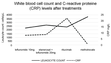 Figure 1: White blood cell count and C-Reactive Protein (CRP) levels after treatments. View Figure 1
D.M.C, 82-years-old female wheedle, from Curitiba, Brazil. Presents herself to the first medical appointment in 2009 with 77 years of age and a RA diagnosis made 15 years before. Her disease was rated class I/II according to the American College of Rheumatology (ACR) criteria. At time she complained of daily joint pain with morning stiffness of few minutes. During physical exam, she had impairment of finger and toes, wrists and elbow with several nodules. At the time, she began therapy with Methotrexate (MTX) 10 mg/week.
A month later, she presented new rheumatoid nodules and an increase in size of the older ones. Due to that, MTX therapy was interrupted and a treatment with Leflunomide 10 mg/day and Prednisone 5 mg/day was introduced. During the first semester of 2010, she presented leucopenia (2260 cells/µL) with eosinophilia of 18%, a chronic infection in her toe and weight loss of 6 kg without loss of appetite. She had also superior airway infections without fever or persistent cough. Due to these findings, we made the hypothesis of RA with FS, prescribed a therapy with Etanercept (ETC) 50 mg/week, and raised the Leflunomide dose to 20 mg/day.
One year after the ETC therapy began, no satisfactory response was observed. In august of 2011 the patient had DAS28 of 4.78, Erythrocyte Sedimentation Rate (ESR) of 44 mm (anterior of 23 mm) and a C Reactive Protein (CRP) of 8.61 mg/L (anterior of 8.09 mg/L), RF of 106 UI/mL, normocytic normochromic anemia, persistency of the leucopenia (with 2650 leukocytes/cells/µL) and low platelets (122,000 cells/µL). Thanks to these findings, therapy with Rituximab 1 g was introduced at week 0 and 2, and repeated after 6 months.
As example of the ETC therapy, the patient had no response with the Rituximab treatment. A small reduction of the ESR and RF was noted, but the CRP rose to 22.24 mg/L. She had no major changes in white cells count, and the leucopenia persisted. At the time, the patient referred lower limb edema. The therapy was maintained, but as the patient had no changes in the CRP rates in the following weeks, a bone marrow aspirate was made to investigate the leucopenia. Due to the persistent low white cell count, an investigation for myelodysplastic syndrome was also performed.
The immunophenotyping of peripheral blood showed a population of lymphocytes composed by 55% of T cells with CD2 and CD3 expression, as well as CD5 and CD7 and negative for CD25. Another 45% were T cells with CD2 and CD3 expression but negative for CD5 and with a diminished expression of CD7, negative for CD25 and CD38. There was also an aberrant expression of the NK antigen associated with CD57 in 32% of the T cell sample. According to the technical report, this finding matched T-LGL leukemia. The bone marrow aspirate performed in the same period demonstrated that the bone marrow had a moderate hypocellularity with a dilutional component. It was composed by 15% of erythroblasts with preserved maturation, 18% of granulocytes with a retard in maturation, 52% of mature lymphocytes, 2% of plasma cells, 8% of monocytes, frequent macrophages and a megakaryocyte series severely hypo cellular. The bone marrow biopsy demonstrated only hypocellularity with no evidence, thus to confirm LGL-lymphocyte infiltration.
We than implemented treatment with MTX 10 mg/week with folic acid with important improvement of the articular symptoms and remission of the rheumatic disease. Later laboratory exams demonstrated an increase in leukocytes to 3770 cells/µL with a lymphocyte count of 1018 cells/µL. No adverse events no MTX were neither observed nor new rheumatoid nodules, nor an increase in size of the older ones.
All the authors declare to have no conflicts of interest in the present paper.