Aberrant right subclavian artery (ARSA) or arteria lusoria is one of the most common congenital aortic arch anomaly with prevalence ranging from 1-2% [1-3]. However, it is mostly asymptomatic and consequently often found sporadically during various diagnostic procedures with prevalence of 0.45% during coronary angiography [4]. We report a case of a 67-year-old male with bifurcation stenosis of proximal left anterior descending artery and first diagonal branch (Medina 1,0,1). We performed an elective percutaneous coronary intervention (PCI) using specific culotte stenting technique using right transradial access through ARSA. To the authors best knowledge, this is the first described case of PCI of the bifurcation stenosis via the ARSA. Subsequent 64-slice computed tomography angiography confirmed the angiographic findings of ARSA. Although percutaneous coronary intervention, especially of the bifurcation stenosis, is difficult to perform using transradial approach through ARSA, the procedure could be done successfully in high-volume PCI centres dedicated to transradial access.
Aberrant right subclavian artery, Bifurcation, Culotte, Stenting, Transradial, Access
Aberrant right subclavian artery (ARSA) or arteria lusoria is one of the most common congenital aortic arch anomaly with prevalence ranging 1-2% [1,2]. The first case of ARSA was described in 1735 [3]. ARSA originates from aortic arch as most distal branch and has its own aberrant pathway in mediastinum, most commonly retroesophageal [2]. Patients are mostly asymptomatic (> 90%) and therefore ARSA is often found sporadically during various diagnostic procedures [2,3]. It is also a rare finding during percutaneous coronary angiography with prevalence of 0.45% [4]. According to international guidelines, methods of choice in diagnosing ARSA are computed tomography (CT) or magnetic resonance (MR) angiography [5,6]. There is no need for its active treatment except in cases of Kommerell's diverticulum and aneurysmal dilatation of ARSA due to risk of aortic dissection [3]. We report a percutaneous coronary intervention (PCI) using culotte stenting technique through ARSA in a 67-year-old man with a left anterior descending artery (LAD) bifurcation lesion. Success of this procedure was optimal resulting with normal TIMI flow grade and no complications [7,8].
67-year-old man with history of arterial hypertension and type 2 diabetes mellitus was admitted to hospital due to elective coronary angiography. Six weeks before he was admitted due to acute ST elevation myocardial infarction (STEMI) of inferior wall. Coronary angiography, using left transradial access, revealed occlusion of proximal right coronary artery (RCA) and primary PCI with implantation of two drug-eluting stents (DES) was done. Coronary angiography also revealed significant bifurcation stenosis in proximal LAD and ostial first diagonal branch (D1) (Medina 1,0,1) [9] whereas left circumflex artery (LCx) had no stenosis (Figure 1a and Figure 1b). During procedure right transradial access was used due to pathological Allen's test on the left arm as well as weaker pulses bilaterally in the groins.
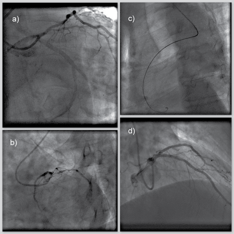 Figure 1a: Coronary angiography - initial recording (left anterior oblique 15° + caudal 40° projection) Bifurcation stenosis of left anterior descending artery (LAD) and first diagonal branch (D1) - classification Medina 1,0,1.
Figure 1b: Coronary angiography - initial recording (Left anterior oblique 40° + caudal 20° projection)
Bifurcation stenosis of left anterior descending artery (LAD) and first diagonal branch (D1) - classification Medina 1,0,1.
Figure 1c: Coronary angiography - initial recording (left anterior oblique 30°).
Positioning of guiding cathether revealed aberrant right subclavian artery (ARSA)
Figure 1d: Coronary angiography - final result after percutaneous coronary intervention (Right anterior oblique 40° + caudal 30° projection).
Final result after percutaneous coronary intervention with placement of one stent in left anterior descending artery (LAD) and two stents in first diagonal branch (D1) achieving TIMI III flow grade and myocardial blush grade 3 both in main and side branch. View Figure 1
Vascular access was obtained with regular radial 6Fr sheath (Radiofocus introducer II transradial Terumo, Tokyo, Japan) and then we advanced into the descending aorta with standard J-type guidewire 0.035 (BBraun). The advancement of the guiding catheter into the ascending aorta was difficult, so we performed aortography which revealed ARSA (Figure 1c). Due to peripheral vascular status, the procedure was continued without changing arterial access. Reaching ascending aorta was attempted with several cathethers, but we succeeded with Internal Mammary cathether 5Fr (Optitorque, Terumo Tokyo, Japan) and hydrophilic 0.035 wire (Radiofocus guidewire M standard type 0.035). Advancement was facilitated with deep-breath maneuver. Afterwards, hydrophilic guidewire was exchanged to support 0.035 guidewire (SupraCore 0.035, Abbott Vascular California, USA) and then 6Fr guiding cathether (EBU 6/3.5, Medtronic, Minneapolis, Minnesota, USA) was introduced and placed to left coronary ostium. Distal LAD na D1 was wired with standard working horse wires (BMW Elite; Abbott Vascular, California, USA). After balloon predilatation (Trek 2.5/20 mm and Trek 2.0/12 mm; Abbott Vascular, California, USA), first DES (Xience 2.5/18 mm; Abbott Vascular, California, USA) was implanted to distal LAD. Second and third DES (Xience 2.75/23 mm to D1 and Xience 3.0/18 mm to LAD; Abbott Vascular, California, USA) was implanted to bifurcation lesion LAD/D1 using culotte technique [8]. During procedure three times proximal optimization technique was performed with non-compliant (NC) 3.5 × 8 mm balloon (NC Trek Abbott Vascular, California, USA), and kissing postdilatation was perfomed with 3.0 × 12 mm NC balloon (NC Trek Abbott Vascular, California, USA) and 3.0 × 15 mm NC balloon (NC Trek Abbott Vascular, California, USA). The intervention resulted in normal TIMI III flow grade and myocardial blush grade III (Figure 1d). 263 mL of radiographic contrast medium (Visipaque 320, GE Healthcare, General Electrics Company, Chicago, Illinois, USA) was used and total of 13923 centiGycm2 X-ray exposure. Postprocedurally, patient was verticalized after 6 hours and he had no discomfort. Postprocedural CT angiography of the aorta was done revealing the absence of truncus brachiocephalicus, with common carotid ostium, regular position of left subclavian artery and confirming anomalous ostium of right subclavian artery as most distal branch on aortic arch with retrooesophageal course towards right arm (Figure 2). Patient was released from the hospital on the 3rd postprocedural day with normal vascular status of the right arm as well as normal haemoglobin, serum creatinine levels and no signs of bleeding.
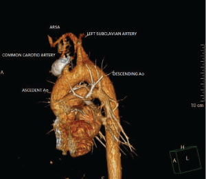 Figure 2: CT angiography of the aortic arch - 3D reconstruction.
Abberant right subclavian artery (ARSA) originates form aortic arch as the most distal branch. View Figure 2
Congenital aortic arch anomalies are mostly diagnosed sporadically, mostly during routine radiological diagnostics, due to its asymptomaticity [2,3]. If during coronary angiography, guiding wires repeatedly enter directly into the descending aorta when using right transradial access, one needs to think about ARSA and it should be excluded. According to the literature, transfemoral approach is used in 69%, and transradial in 31%, out of which left slightly more the right (83% vs. 17%) with conversion rate of 10% to transfemoral approach in settings of acute ST-elevation myocardial infarction [10]. Performing PCI in patients with ARSA is complicated and often requires multiple wires and catheters. In this settings the success rate of transradial approach is low (60%), so switching to transfemoral access is recommended [4]. Also, during maneuvering with guidewires and catheters there is certain risk of aortic dissection or rupture due to unfavourable angle between ARSA and aortic arch [11]. Treatment of such complication, as well as Kommerell diverticulum, often require hybrid procedures [12]. Limited data suggest that these procedures are relatively safe and effective with good short-term outcome [12].
In the reported case patient had no palpable left radial artery after performing primary PCI using left transradial access and clinical evidence of lower limb peripheral artery disease which was expected due to long-time type 2 diabetes mellitus. Due to mentioned, despite procedure success being lower and complication rate being greater [4,11], we continued the procedure without switching to transfemoral access. This was performed by the experienced interventional cardiologist in the high-volume PCI centre dedicated to transradial access. Usage of radiographic contrast was greater, however nearly as reported in the PCI of bifurcation lesions [9,13]. There are only two cases of complex PCIs using transradial access through ARSA described in the literature, one including primary PCI of right internal mammary artery graft originating from ARSA [9,13]. However, to the authors best knowledge, this is the first case of PCI of the bifurcation stenosis (Medina 1,0,1) using specific and technically complex culotte stenting via the ARSA.
In conclusion, the existence of ARSA makes coronary angiography and PCI using right transradial access more difficult and demanding procedure with success being lower and complication rate being greater. Although PCI of the bifurcation stenosis using culotte stenting technique is complex, the possibility of performing it through ARSA could be considered in high-volume PCI centres dedicated to transradial access.
The authors report no conflict of interest. There was no grant support.