

International Journal of Clinical Cardiology
Origin of Right Coronary Artery from the Ascending Aorta: An Extremely Rare Anomaly
Abdulrahman M Abdulbaki*, and Shivang Shah
Department of Cardiology, Louisiana State University and Health Science Center, USA
*Corresponding author:
Abdulrahman M Abdulbaki, Department of Cardiology, Louisiana State University and Health Science Center, 1501 Kings hwy, Shreveport. LA. 71103, USA, Tel: 318-675-5941, E-mail: aabdul@lsuhsc.edu
Int J Clin Cardiol, IJCC-2-041, (Volume 2, Issue 4), Case Report; ISSN: 2378-2951
Received: May 05, 2015 | Accepted: July 21, 2015 | Published: July 24, 2015
Citation: Abdulbaki AM, Shah S (2015) Origin of Right Coronary Artery from the Ascending Aorta: An Extremely Rare Anomaly. Int J Clin Cardiol 2:041. 10.23937/2378-2951/1410041
Copyright: © 2015 Abdulbaki AM, et al. This is an open-access article distributed under the terms of the Creative Commons Attribution License, which permits unrestricted use, distribution, and reproduction in any medium, provided the original author and source are credited.
Introduction
The incidence of coronary anomalies in patients undergoing coronary angiography varies from 0.27% to 1.66%1. Many of these anomalies are clinically benign; while, others are associated with serious morbidity. We describe the case of a patient undergoing coronary angiogram to evaluate her cardiomyopathy revealing an anomalous right coronary artery arising from the ascending aorta above the left sintubular junction plane. We discuss the rarity of this anomaly along with its clinical importance.
Case Report
A 54 year old female with a history of benign resected ovarian tumor and poorly controlled hypertension, who presented to the emergency department with three days of worsening lower extremity edema and exertional dyspnea. She denied having any chest pain, orthopnea, paroxysmal nocturnal dyspnea, palpitations, lightheadedness, or prior syncopal episodes. Patient was afebrile on presentation to the emergency department with a blood pressure of 189/143, pulse rate of 136 and respiratory rate of 18. On examination patient was found to have irregularly irregular heart sounds, tachycardia, jugular venous distention of 10cm H2O, lungs were clear to auscultation bilaterally, and 1+ pitting low extremity edema. EKG revealed atrial fibrillation with rapid ventricular rate in 130s. The transthoracic echocardiogram showed severely reduced Left ventricular ejection fraction of 20%, Left posterior wall thickness of 1.5cm, Intraventricular septal wall thickness of 1.5cm, and severely dilated left atrium suggestive of hypertensive cardiomyopathy.
During the hospital stay the patient was treated with AV nodal blocking agents and diuresis. She underwent coronary angiography to evaluate for ischemia as the etiology of her cardiomyopathy.
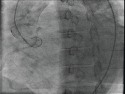
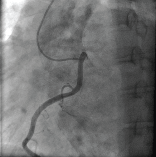
During the left heart catheterization procedure, the left main coronary artery was easily engaged using a Judkins left catheter. The angiogram of the left system revealed minimal non-obstructive disease. However, despite multiple attempts with the Judkins right and Williams right catheters, we were unable to cannulate or even visualize the right coronary artery (RCA) ostium. Hence an aortic root shot in LAO projection, 30 degrees angulation was performed, which revealed the location of an anomalous RCA arising from the ascending aorta above the left sinotubular junction (Figure 1). The RCA was then engaged successfully using an Amplatz left-1 (AL-1) catheter and an angiogram revealed a dominant, large RCA with mild luminal irregularities (Figure 2).
Discussion
Our patient's poor LV function couldn't be explained by the level of atherosclerosis she had, her cardiomyopathy appears to be secondary to her long standing history of poorly controlled hypertension, atrial fibrillation causing tachycardia induced CMP or a combination of both. Anomalous origin of a coronary artery poses a technical challenge during coronary angiograms. It increases the amount of radiation exposure as well as the contrast load utilized to patients. With an increase in the amount of time and the number of catheters, the likelihood of complications also increases, hence it is very important to be aware of the possibilities and have knowledge of them, when it is not possible to cannulate or visualize the coronary by the standard techniques. The most frequent anomalies of coronary origins are associated with the circumflex artery. Anomalies of the origin of RCA are more rare comparatively. Among the anomalous origins of the RCA, the origin from the left sinus of Valsalva was more common in one study 6-27% [1]. Another study found the RCA originating from the LAD or CX to be 12.5% [2]. However, there are only few reported cases of the RCA originating from ascending aorta above the sinotubular junction plane [2-6]. A study by Serkan et al. reported 0.006% prevalence of this rare anomaly, which constitutes only 2.1% of all the anomalously originating coronary arteries [2,3]. There are reports of typical angina pectoris, myocardial infarction, dyspnea, syncope, ventricular tachycardia, and sudden cardiac death related to anomalously arising RCA [7-11]. The origin of the vessel from the contralateral sinus of Valsalva or artery with subsequent passage between the aorta and right ventricular outflow tract has been clearly shown to be a dangerous anatomy [9,10]. Congenital coronary artery anomalies are frequently found to be responsible for sudden death in young athletes, in approximately 20% of cases, and represent the second most frequent disease responsible for athletic field deaths [11,12]. Since the standard catheters are shaped specifically in order to easily cannulate the normally originating coronaries, the success to engage the anomalously arising ostium using the same catheters is limited. In addition to being extremely rare, the anomalous RCA arising from the aorta above the sinotubular junction plane, poses more challenge in being engaged by the standard catheters, as compared to other anomalously arising coronaries. Literature review revealed that these anomalous RCA were very successfully and easily cannulated using Forward take off Judkins left guide (FL) 3.0 and/or Femoral Curve left (FCL) 3.0 and 3.5 catheters [3]. We had our success with using an AL-1 catheter. Coronary CT Angiography and Cardiac MRI play an important role in evaluating the exact anatomy without risk of any extra contrast dye or radiation or when it is not feasible to characterize the coronary anatomy by conventional coronary angiogram due to the anomalous origin. In patients undergoing diagnostic coronary angiograms or percutaneous coronary interventions the angiographic recognition of these anomalous coronary origins is vital to the success of the procedure, specifically to the choice of the diagnostic or guiding catheter used to engage the ostium of the anomalous artery.
References
-
Yamanaka O, Hobbs RE (1990) Coronary artery anomalies in 126,595 patients undergoing coronary arteriography. Catheter Cardiovasc Diagn 21: 28-40.
-
Yuksel S, Meric M, Soylu K, Gulel O, Zengin H, et al. (2013) The primary anomalies of coronary artery origin and course: A coronary angiographic analysis of 16,573 patients. Exp Clin Cardiol 18: 121-123.
-
Sarkar K, Sharma SK, Kini AS (2009) Catheter selection for coronary angiography and intervention in anomalous right coronary arteries. J Interv Cardiol 22: 234-239.
-
Yeoh JK, Ling LH, Maurice C (1994) Percutaneous transluminal angioplasty of anomalous right coronary artery arising from the ascending thoracic aorta. Catheter Cardiovas Diagn 32: 254-256.
-
Motamedi MH, Hemmat A, Kalani P, Rezaee MR, Safarnezhad S (2009) High take-off of right coronary artery: an extremely rare case of RCA anomaly. J Card Surg 24: 343-345.
-
Kadakia J, Gupta M, Budoff MJ (2013) Anomalous "High Take-Off" of the right coronary artery evaluated by coronary CT angiography. Catheter Cardiovasc Interv 82: E765-768.
-
Kimbiris D, Iskandrian AS, Segal BL, Bemis CE (1978) Anomalous aortic origin of coronary arteries. Circulation 58: 606-615.
-
Roberts WC, Siegel RJ, Zipes DP (1982) Origin of the right coronary artery from the left sinus of Valsalva and its functional consequences: analysis of 10 necropsy patients. Am J Cardiol 49: 863-868.
-
Kragel AH, Roberts WC (1988) Anomalous origin of either the right or left main coronary artery from the aorta with subsequent coursing between aorta and pulmonary trunk: analysis of 32 necropsy cases. Am J Cardiol 62: 771-777.
-
Frescura C, Basso C, Thiene G, Corrado D, Pennelli T, et al. (1998) Anomalous origin of coronary arteries and risk of sudden death: a study based on an autopsy population of congenital heart disease. Hum Pathol 29: 689-695.
-
Maron BJ, Shirani J, Poliac LC, Mathenge R, Roberts WC, et al. (1996) Sudden death in young competitive athletes. Clinical, demographic, and pathological profiles. JAMA 276: 199-204.
-
Angelini P, Velasco JA, Flamm S (2002) Coronary anomalies: incidence, pathophysiology, and clinical relevance. Circulation 105: 2449-2454.
