

International Journal of Critical Care and Emergency Medicine
High Flow Conditioned Oxygen Therapy for Prevention of Reintubation in Critically Ill Patients: A Preliminary Cohort Study
Gonzalo Hernandez1*, Concepción Vaquero Collado1, Susana García Plaza1, Ana Villasclaras Pacheco1, Candido Pardo Rey1, Eugenia de la Fuente O'Connor1, Rafael Cuena2, Paloma González Arenas1 and Rafael Fernandez3
1Intensive Care Unit, Hospital Universitario Infanta Sofía, Spain
2Research Unit, Medical Council, Toledo, Spain
3Intensive Care Unit, Hospital Sant Joan de Deu-Fundació Althaia, Spain
*Corresponding author:
Gonzalo Hernández Martínez, Critical Care Medicine, Hospital Universitario Infanta Sofía, Mezquite n°12, 6°A, 28045 Madrid, Spain, Tel: 003-491-527-5808, E-mail: ghernandezm@telefonica.net
Int J Crit Care Emerg Med, IJCCEM-1-009, (Volume 1, Issue 2), Original Research; ISSN: 2474-3674
Received: November 27, 2015 | Accepted: December 29, 2015 | Published: December 31, 2015
Citation: Hernandez G, Collado CV, Plaza SG, Pacheco AV, Rey CP, et al. (2015) High Flow Conditioned Oxygen Therapy for Prevention of Reintubation in Critically Ill Patients: A Preliminary Cohort Study. Int J Crit Care Emerg Med 1:009. 10.23937/2474-3674/1510009
Copyright: © 2015 Hernandez G, et al. This is an open-access article distributed under the terms of the Creative Commons Attribution License, which permits unrestricted use, distribution, and reproduction in any medium, provided the original author and source are credited.
Abstract
Objective: To determine the impact of delivering high flow conditioned oxygen therapy (HFO) through nasal cannula on prevention of reintubation in mechanically ventilated (MV) critically ill patients.
Design: Prospective cohort sturdy.
Setting: General ICU of a university hospital.
Patients: All patients under MV > 12-h and after scheduled extubation.
Exclusion criteria: Hypercapnia at extubation, non-scheduled extubation or with do-not-resuscitate orders. Patients were also divided between low and high reintubation risk.
Interventions: From September 2011 to September 2012, all patients received HFO after extubation for a fixed 24-h period and were compared with a historical cohort (2008-2011) treated with conventional oxygen therapy and matched for risk of reintubation.
Measurements and Main Results: The primary endpoint was reintubation rate within the 72-h following extubation. Statistical analyses included logistic multivariate model.
Main Results: Each cohort included 111 patients with similar clinical characteristics. The HFO group showed a non-significant lower reintubation rate (8.1% vs. 15.3%, p = 0.09). Variables independently related to reintubation rate in the multivariate analysis were HFO (OR 0.31 [0.10-0.95] p = 0.04), hypercapnia after extubation (OR 51.20 [11.55-226.63] p < 0.01), APACHE II > 12 at extubation (OR 1.06 [1.01-1.12] p = 0.04) and length of MV (OR 1.16 [1.03-1.30] p = 0.01). The area under the ROC curve for the model was 0.89 (0.77-0.94).
Conclusions: Routine HFO after planned extubation appears to be associated with lower reintubation rate.
Keywords
Mechanical ventilation, Weaning, Post-extubation respiratory failure, High flow conditioned oxygen therapy, Outcome
Key Messages
High flow conditioned oxygen therapy applied immediately after scheduled extubation reduces reintubation rate in a non-selected population of mechanically ventilated patients.
Introduction
Oxygen delivery after extubation is the cornerstone treatment to maintain adequate oxygenation and avoid reintubation. Oxygen is usually delivered through low-flow nasal prongs; when necessary, flow is increased or patients are switched to a high-flow face mask.
Some other interventions after extubation focus on specific causes of reintubation; for example, high risk patients are administered prophylactic corticosteroids before planned extubation to prevent laryngeal edema [1,2] and patients with hypercapnia at extubation are administered noninvasive mechanical ventilation [3]. However, to our knowledge, no other intervention has been proven to reduce reintubation rate in a general population of critically ill patients.
High flow conditioned oxygen therapy (HFO) is a novel therapy that delivers optimal oxygen through a nasal cannula. First, it generates low level positive pressure, ranging from 2.7 to 7.1 cm H2O, depending on the gas flow and with higher values when patients are breathing with their mouths closed [4-7]. This can attenuate the inspiratory resistance associated with the nasopharynx, thereby reducing the related work of breathing. Second, conditioning the gas mixture can improve conductance and pulmonary compliance with an increase in end-expiratory lung volume [8], probably decreasing irritation of the tracheal mucosa and increasing patient comfort. Technological improvements have increased the absolute humidity of the gas delivered up to 35 mg/l at 40 l/min and 30°C [9]. Finally, high flow ensures a constant FiO2 during inspiratory effort and washout of the nasopharyngeal dead space during expiration, theoretically improving oxygenation and carbon dioxide clearance. Nevertheless, clinical studies failed to demonstrate improvements in PaCO2 [10], and have even found mild to moderate increases in PaCO2 [11]. Studies of HFO after extubation in general and postsugical population [10,12,13], found HFO was more comfortable than conventional oxygen in terms of mouth and throat dryness [10,14]. Patients with HFO had fewer desaturation episodes and interface displacements [10]; however, the results for other outcomes like improvements in respiratory rate and oxygenation were inconsistent [10,12,14].
Extubation failure remains a clinical challenge, appearing in as much as 20% of critically ill patients. The main reasons for extubation failure are increased work of breathing, secretion retention, heart failure and loss of lung aeration, most of which might be alleviated by HFO's low-level airway pressurization and gas conditioning.
We hypothesized that delivering HFO through nasal prongs immediately after planned extubation would reduce the rate of reintubation.
Materials and Methods
Patients and Study design
In September 2011, we incorporated HFO after planned extubation into the routine clinical protocol routine for mechanically ventilated (MV) patients in our 8-bed closed medical-surgical ICU in a 300-bed university hospital. Then, we prospectively collected clinical data during the 12-month period from September 2011 through September 2012.
In this study, we included all patients in this period who underwent planned extubation after at least 12 hours MV except those with do-not-resuscitate orders, hypercapnia before extubation, or accidental or self extubation. We compared these patients treated with HFO after extubation with a historical cohort of consecutive patients treated with conventional oxygen therapy selected by matching criteria from medical records dating from February 2008 through July 2011. The institutional review board approved the study.
Endpoints
The primary outcome was extubation failure, defined as the need for reintubation within 72 hours after extubation. Patients with any of the following were immediately reintubated: respiratory or cardiac arrest, respiratory pauses with loss of consciousness or gasping for air, psychomotor agitation inadequately controlled by sedation, massive aspiration, persistent inability to remove respiratory secretions, heart rate below 50 min-1 with loss of alertness, or severe hemodynamic instability without response to fluids and vasoactive drugs. We also reintubated patients with postextubation respiratory failure defined as the presence and persistence of any of the following within 72 h of extubation: respiratory acidosis with arterial pH < 7.35 with PaCO2 > 45 mmHg; SpO2 < 90% or PaO2 < 60 mmHg at FiO2 ≥ 0.5; respiratory frequency > 35 min-1; agitation, diaphoresis, or decreased consciousness; or clinical signs suggestive of respiratory muscle fatigue and/or increased work of breathing, such as the use of accessory respiratory muscles, paradoxical motion of the abdomen, or retraction of the intercostal spaces [15].
In addition to analyzing reintubations due to all causes, we separately analyzed reintubations due to respiratory causes. We also analyzed predictors of extubation failure.
Study cohort
Patients from the two groups were matched according to stratification for high risk of reintubation based on the following criteria: > 65 years; MV for congestive heart failure; moderate-to-severe COPD; APACHE II > 12 at extubation; body mass index > 30; difficult or prolonged weaning; ≥ 2 comorbidities; airway patency problems, including inability to deal with respiratory secretions (inadequate cough reflex or > 2 suctioning during the 8 hours previous to extubation); and MV > 7 days.
Protocol
Our weaning protocol included a daily screening of weaning readiness according to the following criteria [16]: recovery from the precipitating illness; respiratory criteria: PaO2/FiO2 > 150 mmHg with positive end-expiratory pressure (PEEP) < 8 cmH2O and arterial pH > 7.32; and clinical criteria: absence of electrocardiographic signs of myocardial ischemia, no need for vasoactive drugs or dopamine (≤ 5 μg/kg/min), heart rate < 140 beats/min, hemoglobin > 8 g/dL, temperature < 38°C, no need for sedatives, presence of respiratory stimulus, and appropriate spontaneous cough. When patients fulfilled these criteria a spontaneous breathing trial (SBT) either with T-tube or 7 cm H2O pressure support for 30 minutes was performed. SBT failure criteria were: agitation, anxiety, depressed mental status, diaphoresis, cyanosis, evidence of increasing respiratory effort, increased accessory muscle activity, facial signs of distress, dyspnoea, PaO2 ≤ 60 mmHg or SaO2 < 90% on FiO2 ≥ 0.5, PaCO2 > 50 mmHg or > 8 mmHg increase, arterial pH < 7.32 or ≥ 0.07 decrease, respiratory rate > 35 breaths/min or ≥ 50% increase, heart rate > 140 beats/min or ≥ 20% increase, systolic arterial pressure >180 mmHg or ≥ 20% increase, systolic arterial pressure < 90 mmHg or cardiac arrhythmias.
Patients who tolerated the SBT were reconnected without changes to ventilator settings for rest and evaluation of airway patency. These patients were then extubated; those in the historic cohort received conventional supplementary oxygen and those in the contemporary group received HFO.
The three weaning categories (simple, difficult and prolonged weaning) were defined with standard criteria [16].
High flow oxygen therapy (Optiflow®, Fisher & Paykel Healthcare, Auckland, New Zealand) was applied immediately before extubation through a specific nasal cannula. Flow was initially set at 10 L/min and titrated upwards in 5 L/min steps until patients felt uncomfortable. The temperature of the administered gas was initially set to 37°C and maintained unless patients reported it was too hot, and FiO2 was regularly adjusted to target SpO2 > 92%. After 24 hours, HFO was stopped and switched to conventional oxygen therapy.
Data sources
The historical cohort population was defined with the use of Care Suite Critical Care Manager 8.0© Picis Inc™, and included all patients admitted to our ICU from February 2008 to July 2011. Historical cohort patients were selected adding the matching criteria simultaneously to the admission date to assure the exact combination of the high risk criteria and the consecutive selection. When an exact matching patient did not existed, we tolerated a mismatch in one criterion. When we found two patients fulfilling the same matching criteria we selected the one with similar primary diagnosis (medical vs surgical).
We recorded gender, APACHE II at ICU admission, primary diagnosis, gasometric variables, corticosteroids at extubation, risk factors for reintubation (including suctioning frequency), and lengths of ICU and hospital stays. We also recorded extubation-related complications and adverse events (mainly laryngeal edema and criteria for reintubation).
Statistical analysis
Comparison between the two groups: The univariate analysis to compare homogeneity of two cohorts was performed with Fisher's exact test, Student's t-test, the Mann-Whitney U test, or the chi-square test, as appropriate. The level of significance was set at 0.05.
Risks factors associated to reintubation: Raw relationships between reintubation and its risk factors were analyzed with contingency tables and X2 after tercile categorization. A logistic regression model was designed to assess the probability of reintubation, and the results were expressed as odds ratios (OR) with 95% confidence interval. The independent variables included in the model were HFO and variables that were statistically significant in the univariate analysis at p < 0.1.
Kaplan-Meier curves were plotted to assess the time from extubation to reintubation in the whole population and after classifying patients according to the tolerated flow and compared by means of the log-rank test.
Power estimation: because our historical reintubation rate was 13% in low risk patients and 17% in high risk patients, our 111 patient sample size may be able to detect a reduction in reintubation rate of 9%, for a power of 80% and an α-error of 0.05.
Analyses were performed using SPSS statistical software version 13.0 (SPSS Inc.; Chicago, IL).
Results
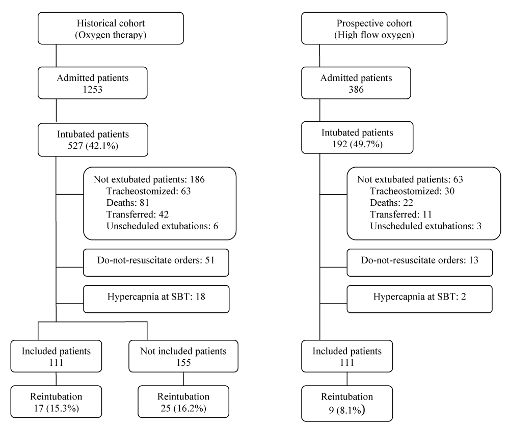
Both the prospective cohort of HFO patients and the historical cohort of conventional oxygen therapy included 111 patients. A flowchart of the study is presented in Figure 1. Perfect matching was not possible in 12 patients. The mean values of high risk criteria per patient in the HFO and control group were 4.2 vs 4.1, respectively (p = 0.8).
The univariable analysis comparing the baseline and outcome variables of the two cohorts are presented in table 1 and table 2 respectively. Baseline characteristics of both groups were similar.
![]()
Table 1: Baseline patients' characteristics. Data are expressed as mean ± SD, median (interquartile range), or number and percentage (%).
View Table 1
![]()
Table 2: Outcome variables. Data are expressed as mean ± SD, median (interquartile range), or number and percentage (%).
View Table 2
Reintubation rate showed a non-significant reduction in the HFO cohort (8.1% vs. 15.3%, p = 0.09). Interestingly, 2 out 9 (22%) reintubated patients in the HFO cohort and 6 out 17 (35%) in the conventional oxygen cohort did not have any risk factors for reintubation.
Unexpectedly, the low risk group showed a greater reduction in the reintubation rate (4% vs. 13%, absolute reduction 9%, relative reduction 67%) as compared to high risk group (11% vs. 17%, absolute reduction 6%, relative reduction 35%), with an even greater difference after excluding patients reintubated for a non-respiratory cause, mainly unscheduled surgery and neurologic complications (2.2% vs. 11.1%, absolute reduction 8.9%, relative reduction 80% in low risk and 7.5% vs. 12.1%, absolute reduction 4.6%, relative reduction 37.5% in high risk).
In the conventional oxygen group, 4 patients developed laryngeal edema after extubation and 3 (75%) needed reintubation, whereas no patient in the HFO group showed laryngeal edema.
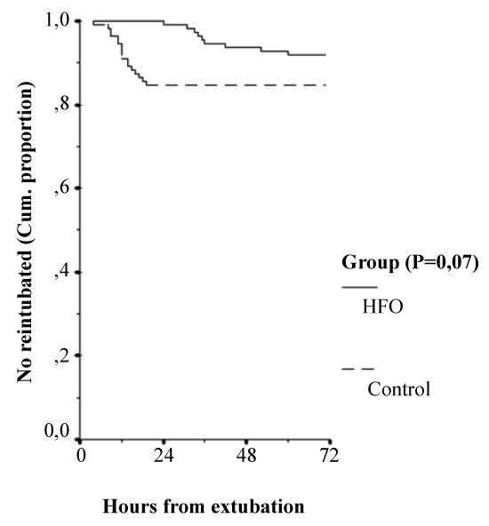
Time for reintubation requirement (Figure 2) was significantly longer in the HFO cohort (38 ± 9 vs. 11 ± 4 hours, p = 0.04), with a non-significant difference in ICU mortality rate (3.6% vs. 6.3%, p = 0.08). The ICU length of stay was shorter in the HFO group (11 vs. 14 days, p = .05), with similar hospital length of stay (17 vs. 18 days, p = 0.09).
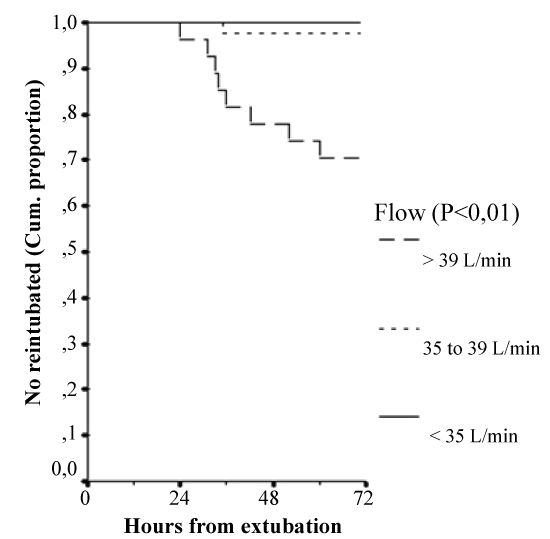
In the HFO group, FiO2 > 0.35 at 12-h after extubation was highly associated with reintubation (24 (21%) vs. 2 (2%), p < 0.01). Also gas-flow > 35 L/min at 12-h after extubation was highly associated with reintubation (9 (13%) vs. 0 (0%) patients, p < 0.01). Additionally, gas-flow showed a near-linear relationship with reintubation rate: 0/43 (0%) at flow < 35 L/min, 1/41 (2.4%) at flow 35-39 L/min and 8/27 (30%) with flow > 39 L/min, p < 0.01 (Figure 3). Tolerance to higher gas-flow was highly related to COPD diagnosis (p < 0.01) and APACHE II at ICU admission (p < 0.01).
The univariable analysis of variables associated with reintubation is shown in table 3, the multivariable analysis for all-cause reintubation in table 4 and multivariable analysis for respiratory-cause reintubation in table 5. The HFO appeared as a protector for reintubation (OR 0.31, 95% CI 0.10-0.95, p = 0.04). Among high risk criteria for reintubation, only APACHE II at extubation (OR 1.06, 95% CI 1.01-1.12, p = 0.04) and length of MV (OR 1.16, 95% CI 1.03-1.30, p = 0.01) appeared significant in the multivariate analysis.
![]()
Table 3: Univariable analysis of factors associated with all-cause reintubation. Data are expressed as mean±SD, median (interquartile range), or number and percentage (%).
View Table 3
![]()
Table 4: Multivariate analysis of factors associated with all-cause reintubation.
View Table 4
Development of hypercapnia after extubation was the clinical variable most strongly related to reintubation (OR 51.20, 95% CI 11.55-226.63, p < 0.01). Interestingly, we observed a trend toward a reduced reintubation rate in hypercapnic patients when using HFO: 50% (4/8) vs. 85% (6/7), p = 0.18. The model showed an area under the ROC curve of 0.89 (95% CI 0.77-0.94).
![]()
Table 5: Multivariate analysis of factors associated with respiratory-cause reintubation.
View Table 5
Discussion
The major finding of this study is that HFO is independently associated with reintubation reduction in our multivariate analysis.
The variable most strongly related to an increased risk for reintubation was development of hypercapnia after extubation. In the subgroup of patients who developed hypercapnia after extubation, we found a trend towards a lower reintubation rate in patients receiving HFO (50% vs 85% in controls, p = 0.2); however, HFO did not prevent hypercapnia (7% vs 6%, p = 0.9), as suggested by Maggiore et al. [10]. These findings are difficult to interpret in light of previous evidence. The evidence supporting the use of higher pressurization devices like noninvasive ventilation to prevent post-extubation respiratory failure in selected patients who develop hypercapnia during the spontaneous breathing trial is strong [3,15], but the evidence for using these devices to treat post-extubation hypercapnic respiratory failure is weaker [17]. Furthermore, in two other studies [18,19], post hoc analysis of hypercapnic patients failed to demonstrate significant reductions in the reintubation rate for noninvasive ventilation used to treat even mild (pH < 7.35) post-extubation hypercapnic respiratory failure. This complex scenario precludes any conclusion for this specific subgroup of patients until more information is available.
Not all the pre-defined factors for high risk of reintubation used to match the cohorts were confirmed in our study. The stratification of the risk for reintubation is complex. Although numerous factors have been described [20-22], subsequent studies have failed to confirm many of them, and no definitive model has yet to be established. Our study included no neurocritical patients [23], so we aimed to eliminate potential confounders by matching patients on a wide spectrum of risk factors (mainly those used by Ferrer [15] and Nava [24]). Risk factors selected by our univariate analysis were hypercapnia, airway patency problems (including laryngeal edema), MV longer than 7 days, heart disease, and APACHE II > 12 points on the day of extubation; however, only APACHE II on the day of extubation and MV length remained significant in the multivariate analysis, suggesting that not all the high risk factors selected are equally associated with a real increase in the risk of reintubation. In addition, in the multivariate analysis centered on reintubation due to respiratory causes, the APACHE II score was not significant, thus underlining the unpredictable etiology of reintubation due to non-respiratory causes.
The linear relationship between gas-flow and reintubation suggests that the higher the gas-flow tolerated by the patient, the greater the respiratory failure and the greater the risk of reintubation. Setting flow according to patient tolerance leads to higher flows than setting by oxygenation, even when HFO is indicated for acute hypoxemic respiratory failure [25]. In addition, tolerance to higher gas-flow was significantly associated with other variables associated with reintubation in the univariate analysis (COPD diagnosis, APACHE II at ICU admission and FiO2 at 12-h after extubation), supporting this hypothesis.
The apparent greater improvement observed in the low risk group merits some consideration. At first glance, the classification criteria used may have a low discriminating value, as suggested by the small difference in the reintubation rates between the subgroups within the control group (17% in high risk and 13% in low risk). An alternative explanation is that the beneficial effect of HFO is small and only able to reverse low-intensity complications.
Whether HFO can prevent laryngeal edema after extubation could not be determined due to our low incidence (4% in the control group vs. 0% in the HFO group), but both low level PEEP and gas conditioning can reduce inspiratory effort and dryness of the upper airways, so this is a very interesting possibility.
One concern related to using HFO as a preventive measure is the possibility of delaying reintubation, with its associated morbidity and mortality [26], as reported previously for NIMV [19]. The time to reintubation was significantly longer in the HFO group (35 h vs. 12 h in the control group), suggesting that the withdrawal of HFO unmasked respiratory insufficiency. Nevertheless, ICU mortality tended to be lower in HFO patients and was very similar among patients requiring reintubation in the two groups. However, there are some data suggesting that increasing time under high-flow therapy up to 48 hours, could improve outcomes in critically ill patients after extubation [10]. Additionally, our observation that hypercapnia after extubation, and that high FiO2 and high gas-flow al 12-h after extubation, are markers of failure may help to reduce the risk of delaying reintubation in the future.
Limitations of the Study
Some specific points of our protocol could limit the extrapolation of our conclusions. First, in any study with historical controls, the comparability between groups is always debatable. Although we used a wide number of reported risk factors for extubation failure to select historical controls and the resulting data was comparable, unrecorded variables could affect extubation outcome. Another issue is whether a temporary trend may reduce comparability. In our study, both groups were consecutive, without any time-lag in between them, and our reintubation rate was fairly stable in the three years comprising the period from which controls were selected (13/70 (18.6%), 16/107 (14.9%), and 15/89 (16.8%), p = 0.1). Moreover, co-interventions other than HFO remained unchanged in our ICU during the two study periods.
Second, we cannot identify which component of the HFO system is most important for its beneficial effect. The CPAP effect is higher at higher gas flow, but in our patients higher gas flow was associated with higher reintubation rates. Hence, despite the higher CPAP effect, HFO was unable to reverse respiratory insufficiency in patients asking for more flow, which is probably a marker of much greater severity. The dead-space washout may reduce ventilatory needs, but our design did not allow for comparisons of minute ventilation, respiratory rate, or dyspnea between groups. Improved humidification can also play a beneficial role, but bedside bronchial ciliary movement or mucus viscosity measurements were not available.
We conclude that routine clinical use of HFO was associated with a reduction in the reintubation rate. The multicenter randomized controlled trial necessary to confirm these results is currently recruiting patients (ClinicalTrials.gov Identifier: NCT01191489).
Acknowledgments
We thank all the patients and medical and nursing staff for their cooperation.
Author contributions
Dr. Hernandez: contributed to the conception, coordination, design, and interpretation of the study, as well as to drafting, critical revision, reading, and approval of the manuscript; Dr. Pardo, Dra. Gonzalez, Dra. Villasclaras, Dra. García and Dra. De la Fuente: contributed to coordination and interpretation of the study, as well as to critical revision, reading, and approval of the manuscript; Dr. Cuena: contributed to statistical analyses and interpretation of the study, as well as to critical revision, reading, and approval the manuscript; Dra. Vaquero, and Dr. Fernandez: contributed to interpretation of the study, and to drafting, critical revision, reading, and approval of the manuscript.
References
-
Khemani RG, Randolph A, Markovitz B (2009) Corticosteroids for the prevention and treatment of postextubation stridor in neonates, children and adults. Cochrane Database Syst Rev.
-
Fan T, Wang G, Mao B, Zeyu Xiong, Yu Zhang, et al. (2008) Prophylactic administration of parenteral steroids for preventing airway complications after extubation in adults: meta-analysis of randomised placebo controlled trials. BMJ.
-
Ferrer M, Sellares J, Valencia M, Andres Carrillo, Gumersindo Gonzalez, et al. (2009) Non-invasive ventilation after extubation in hypercapnic patients with chronic respiratory disorders: randomised controlled trial. Lancet 374: 1082-1088.
-
Dysart K, Miller TL, Wolfson MR, Shaffer TH (2009) Research in high flow therapy: mechanisms of action. Respir Med 103: 1400-1405.
-
Parke R, McGuinness S, Eccleston M (2009) Nasal high-flow therapy delivers low level positive airway pressure. Br J Anaesth 103: 886-890.
-
Parke RL, McGuinness SP (2013) Pressures delivered by nasal high flow oxygen during all phases of the respiratory cycle. Respir Care 58: 1621-1624.
-
Ritchie JE, Williams AB, Gerard C, Hockey H (2011) Evaluation of a humidified nasal high-flow oxygen system, using oxygraphy, capnography and measurement of upper airway pressures. Anaesth Intensive Care 39: 1103-1110.
-
Riera J, Perez P, Cortes J, Roca O, Masclans JR, et al (2013) Effect of high-flow nasal cannula and body position on end-expiratory lung volume: a cohort study using electrical impedance tomography. Respir Care 58: 589-596.
-
Bouchard PA, Bojmehrani A, Bouchard C, Francois Lellouche (2012) Hygrometry of gas delivered with high flow oxygen humidifiers and comfort in healthy subjects. Am J Respir Crit Care Med.
-
Maggiore SM, Idone FA, Vaschetto R, Festa R, Cataldo A, et al. (2014) Nasal high-flow versus venture mask oxygen therapy after extubation. Effects on oxygenation, comfort, and clinical outcome. Am J Respir Crit Care Med 190: 282-288.
-
Sztrymf B, Messika J, Bertrand F, Hurel D, Leon R, et al. (2011) Beneficial effects of humidified high flow nasal oxygen in critical care patients: a prospective pilot study. Intensive Care Med 37: 1780-1786.
-
Parke RL, McGuinness SP, Dixon R, Jull A (2012) Protocol for a randomized controlled trial of nasal high flow oxygen therapy compared to standard care in patients following cardiac surgery: the HOT-AS study. Int J Nurs Stud 49: 338-344.
-
Stephan F, Barrucand B, Petit P, Saida Rezaiguia-Delclaux, Anne Medard, et al.(2015) High-flow nasal oxygen vs noninvasive positive airway pressure in hypoxemic patients after cardiothoracic surgery. A randomized clinical trial. JAMA 313: 2331-2339.
-
Tiruvoipati R, Lewis D, Haji K, Botha J (2010) High-flow nasal oxygen vs high-flow face mask: a randomized crossover trial in extubated patients. J Crit Care 25: 463-468.
-
Ferrer M, Valencia M, Nicolas JM, Bernadich O, Badia JR, et al (2006) Early Noninvasive Ventilation Averts Extubation Failure in Patients at Risk. A Randomized Trial. Am J Respir Crit Care Med 173: 164-170.
-
Boles JM, Bion J, Connors A, Herridge M, Marsh B, et al. (2007) Weaning from mechanical ventilation. Eur Respir J 29: 1033-1056.
-
Hilbert G, Gruson D, Portel L, Gbikpi-Benissan G, Cardinaud JP (1998) Noninvasive pressure support ventilation in COPD patients with postextubation hypercapnic respiratory insufficiency. Eur Respir J 11: 1349-1353.
-
Keenan SP, Powers C, McCormack DG, Block G (2002) Noninvasive positive-pressure ventilation for postextubation respiratory distress. A randomized controlled trial. JAMA 287: 3238-3244.
-
Esteban A, Frutos-Vivar F, Ferguson ND, Yaseen Arabi, Carlos Apezteguía, et al. (2004) Noninvasive positive-pressure ventilation for respiratory filure after extubation. N Engl J Med 350: 2452-2460.
-
Knaus WA, Draper EA, Wagner DP, Zimmerman JE (1985) APACHE II: a severity of disease classification system. Crit Care Med 13: 818-829.
-
Menon N, Joffe AM, Deem S, Yanez ND, Grabinsky A, et al. (2012) Occurrence and complications of tracheal reintubation in critically ill adults. Respir Care 57: 1555-1563.
-
Saugel B, Rakette P, Hapfelmeier A, Schultheiss C, Phillip V, et al. (2012) Prediction of extubation failure in medical intensive care unit patients. J Crit Care 27: 571-577.
-
Brown CV, Daigle JB, Foulkrod KH, Brouillette B, Clark A, et al. (2011) Risk factors associated with early reintubation in trauma patients: a prospective observational study. J Trauma 71: 37-41.
-
Nava S, Gregoretti C, Fanfulla F, Squadrone E, Grassi M, et al. (2005) Noninvasive ventilation to prevent respiratory failure after extubation in high-risk patients. Crit Care Med 33: 2465-2470.
-
Roca O, Riera J, Torres F, Masclans JR (2010) High-flow oxygen therapy in acute respiratory failure. Respir Care 55: 408-413.
-
Kang BJ, Koh Y, Lim CM, Huh JW, Baek S, et al. (2015) Failure of high-flow nasal cannula therapy may delay intubation and increase mortality. Intensive Care Med 41: 623-632.
