Human Epidermal growth factor Receptor type 2 (HER2) is over expressed in 20.0-30.0% of breast cancers and is currently evaluated histopathologically. Immunohistochemistry and fluorescence in situ hybridization require invasive enucleation of the tumor tissue and may be affected by heterogeneity. Serum marker tests are more objective because of the uniformity of the study material. Serum HER2 levels are important for breast cancer care. However, the clinical utility of serum HER2 testing is unclear. We evaluated serum HER2 as a marker of therapeutic response in breast cancer.
Serum HER2 levels were measured in 64 tissue HER2-positive breast cancer patients during routine care. Relationships between serum HER2 levels, clinical stage, tumor diameter, therapeutic response, and Distant Metastasis (DM) were analyzed.
Serum HER2 levels correlated with therapeutic response. An association was observed between serum HER2 levels and tumor diameter. Serum HER2 levels were significantly higher in patients with DM than in those without DM.
Serum HER2 levels are associated with therapeutic response, tumor diameter, and DM. We confirm the clinical utility of serum HER2 testing for monitoring therapeutic response and disease progression in patients with breast cancer.
Breast cancer, Distant metastasis, Human epidermal growth factor receptor type 2, Serum marker, Therapeutic response
HER2: Human Epidermal Growth Factor Receptor type 2; ECD: Extracellular Ligand-Binding Domain; IHC: Immunohistochemistry; FISH: Fluorescence In Situ Hybridization; DM: Distant Metastasis .
Human Epidermal growth factor Receptor type 2 (HER2) is a glycoprotein of 185 kDa that comprises an Extracellular ligand-binding Domain (ECD), a transmembrane domain, and an intracellular domain [1,2]. HER2 functions as a transmembrane receptor and tyrosine kinase that is activated by the binding of its ligand to the ECD and autophosphorylation of its tyrosine residue [3], leading to the nuclear transmission of intracellular signals. HER2 is expressed at low levels in normal cells, and is involved in cell proliferation, differentiation, and growth. HER2 overexpression leads to neoplastic transformation and malignancy [4].
HER2 overexpression occurs in 20.0-30.0% of breast cancers, usually as a result of gene amplification [4,5], and is associated with a poor prognosis. Therefore, an accurate diagnosis is crucial for breast cancer patients [4].
In Japan, therapeutic agents that molecularly target HER2 (e.g., trastuzumab, lapatinib, pertuzumab, and trastuzumab emtansine) have been developed in rapid succession since 2001 [6]. Therefore, a need has arisen for a test that can accurately diagnose HER2-positive breast cancer patients [7]. HER2-positive breast cancer is detected based on HER2 protein overexpression using Immunohistochemistry (IHC) or HER2 gene amplification using Fluorescence In Situ Hybridization (FISH) [8]. IHC and FISH require invasive enucleation of the tumor tissue and may be affected by heterogeneity [9]; there is also the potential for mismatches to occur between technicians [10].
HER2 testing using the ADVIA Centaur system was approved by the United States Food and Drug Administration in 2003. The HER2 ECD that is released into the serum can now be quantitatively measured by chemiluminescent immunoassays using blood samples [11]. Evaluation of serum HER2 levels is more advantageous than IHC or FISH, because it is less invasive and does not require tumor tissue sampling [12]. Additionally, high serum HER2 levels in tissue HER2-negative breast cancer patients can predict the development of metastatic tumors. Even with HER2-negative primary cancer, high serum HER2 levels after distant metastasis could suggest the possibility of positive tissue HER2 status in the metastatic cancer site. Therefore, this method may be useful for reevaluating HER2 status in metastatic tissue [12,13]. Moreover, needle biopsy is not always possible for all metastatic lesions. Serum HER2 testing using blood samples is advantageous in that it can be conducted more frequently than tissue HER2 testing. However, the clinical utility of serum HER2 testing is unclear. Therefore, we evaluated serum HER2 as a marker of therapeutic response in tissue HER2-positive breast cancer patients from Japan.
This work is an extension of our previous reports [14,15] on the study of drug-induced cardiotoxicity in breast cancer, the study of serum HER2 in tissue HER2-negative breast cancer patients. We measured serum HER2 levels in 220 breast cancer patients from Japan and analyzed the relationships between serum HER2 levels, clinical stage, tumor diameter, therapeutic response, and Distant Metastasis (DM). The study design was approved by the Institutional Review Board for Clinical Research at Tokai University School of Medicine (Kanagawa, Japan). All participants provided informed written consent. Research was conducted in accordance with the Helsinki Declaration.
Tissue and serum samples were obtained from 64 female patients with tissue HER2-positive breast cancer (median age, 59.5 [range, 30.0 - 82.0] years), who were examined in the Department of Breast and Endocrine Surgery (Tokai University School of Medicine, Kanagawa, Japan) between February 2010 and January 2014 (Table 1). Blood samples were collected routinely from the start of treatment during outpatient treatment course. Subjects were excluded if they had ovarian, lung, prostate, or gastric cancer. Serum samples were separated and cryopreserved at -80.0 °C.
Table 1: Patient characteristics. View Table 1
IHC staining was performed on target tissue sections using the HER2 antibody (Ventana I-VIEW Pathway HER2 [4B5]; Roche Diagnostics K.K., Tokyo, Japan). The HER2 protein was detected through an antigen-antibody reaction [16,17].
The copy number of the HER2 gene was detected in interphase nuclei of tumor cellsusing a fluorescently labeled DNA probe (PathVysion HER-2 DNA Probe Kit; Abbott Co., Ltd., Tokyo, Japan) [17,18] in formalin-fixed paraffin-embedded tissue sections.
Tissue HER2 status was considered positive with an IHC score of 3+ (positive cells: > 30.0%)/2+ (positive cells: 10.0-30.0%) and a HER2/centromere 17 gene ratio of ≥ 2.2 on FISH [16].
Serum HER2 levels were measured during routine hospital visits using the ADVIA Centaur XP System and ADVIA Centaur HER2/neu assay (Siemens Healthcare Diagnostics K.K., Tokyo, Japan). Briefly, serum HER2 forms an immune complex after reacting with the acridinium-ester-labeled anti-HER2 antibody among the labeling reagents and the fluorescein-labeled anti-HER2 antibody among the fluorescein-conjugated reagents, as well as, the anti-fluorescein antibody binding to magnetic particles among the solid-phase reagents [11]. The immune complex is washed after bound/free separation and serum HER2 levels are measured based on the quantity of chemiluminescence that is generated by the addition of an oxidant or supplementary oxidant. Serum HER2 levels of ≥ 15.3 ng/mL were considered positive [19].
Tumor diameter was measured as the maximum diameter of the tissues demonstrating invasion within the excised tumor.
An anthracycline regimen, followed by taxane regimen, is the standard neoadjuvant and adjuvant chemotherapy treatment. Taxane, combined with 1-year administration of trastuzumab, was used in the tissue HER2-positive patients. The chemotherapy regimens chosen by the physicians included: Bevacizumab + paclitaxel, paclitaxel, docetaxel, eribulin, vinorelbine, gemcitabine, or oral 5-fluorouracil. In addition, anti-HER2 agents were used concomitantly with chemotherapy in tissue HER2-positive breast cancer patients with disease progression or recurrence. Chemotherapy drugs were also administered to the patients with triple negative breast cancer or lymph node metastasis who were in Stage I–III.
Therapeutic response was evaluated in accordance with the General Rules for Clinical and Pathological Recording of Breast Cancer (17th edition) [20]: Complete response (all target lesions, including secondary tumor-related changes, are eliminated), partial response (the sum of the diameters of the target lesion is reduced by ≥ 30.0% in comparison to the sum of the diameters before initiating treatment), stable disease (neither a reduction in tumor size constituting a partial response nor an increase in tumor size constituting progressive disease), and progressive disease (the sum of the diameters of the target lesion is increased by ≥ 20.0% in comparison to the time when the sum of the diameters was at its smallest and/or the sum of the diameters is increased by an absolute value of ≥ 5.0 mm).
Mann-Whitney U tests were used to compare the non-normally distributed groups. Relationships were analyzed using Spearman's rank-order correlation coefficients. Statistical analyses were conducted using Statistical Package for the Social Sciences (software version 23.0; IBM Corp., Armonk, NY, USA). A P < 0.05 was considered significant.
Relationships between serum HER2 levels and clinical stage were analyzed, with no significant findings (Figure 1). Significant differences in serum HER2 levels and therapeutic response were observed between tissue HER2-positive patients with a complete response and those with stable or progressive disease. No other significant findings were detected (Figure 2).
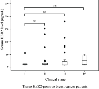 Figure 1: Relationships between serum Human Epidermal Growth Factor Receptor type 2 (HER2) levels and clinical stage in tissue HER2-positive breast cancer patients. NS: Not Significant.
View Figure 1
Figure 1: Relationships between serum Human Epidermal Growth Factor Receptor type 2 (HER2) levels and clinical stage in tissue HER2-positive breast cancer patients. NS: Not Significant.
View Figure 1
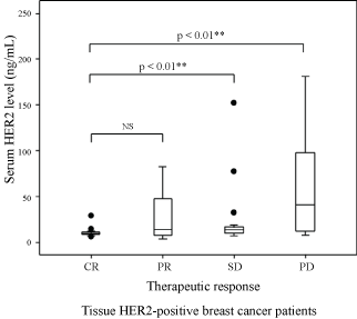 Figure 2: Relationships between serum Human Epidermal Growth Factor Receptor type 2 (HER2) levels and therapeutic response in tissue HER2-positive breast cancer patients. CR: Complete Response; PD: Progressive Disease; PR: Partial Response; SD: Stable Disease; NS: Not Significant.
View Figure 2
Figure 2: Relationships between serum Human Epidermal Growth Factor Receptor type 2 (HER2) levels and therapeutic response in tissue HER2-positive breast cancer patients. CR: Complete Response; PD: Progressive Disease; PR: Partial Response; SD: Stable Disease; NS: Not Significant.
View Figure 2
Positive associations between serum HER2 levels and tumor diameter were observed in tissue HER2-positive patients (Figure 3). Serum HER2 levels were significantly higher in patients with DM than those without DM (Figure 4).
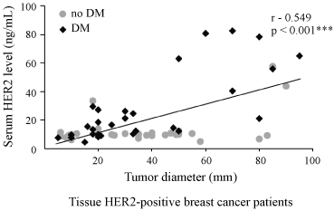 Figure 3: Relationships between serum Human Epidermal Growth Factor Receptor type 2 (HER2) levels and tumor diameter in tissue HER2-positive breast cancer patients. DM: Distant Metastasis.
View Figure 3
Figure 3: Relationships between serum Human Epidermal Growth Factor Receptor type 2 (HER2) levels and tumor diameter in tissue HER2-positive breast cancer patients. DM: Distant Metastasis.
View Figure 3
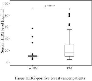 Figure 4: Relationships between serum Human Epidermal Growth Factor Receptor type 2 (HER2) levels and Distant Metastasis (DM) in tissue HER2-positive breast cancer patients.
View Figure 4
Figure 4: Relationships between serum Human Epidermal Growth Factor Receptor type 2 (HER2) levels and Distant Metastasis (DM) in tissue HER2-positive breast cancer patients.
View Figure 4
Currently, IHC, in which the HER2 ECD is immunostained, and FISH, in which amplified HER2 DNA is fluorescently labeled [16,21], are the two most frequently used techniques to evaluate HER2 status in breast cancer patients. HER2 testing is an important tool for administering personalized treatment [16]. However, IHC and FISH require invasive enucleation of the tumor tissue and may be affected by heterogeneity [9]. There is also the potential for mismatches to occur between technicians [10,22]. Metastatic breast cancer sites have an increased potential for malignancy and therefore cancer at the distant site could be HER2 positive, even if the primary lesion is HER2 negative. However, needle biopsy cannot always be performed for all metastatic lesions; for these cases, measuring serum HER2 levels may indicate the tissue HER2 status in metastatic lesions. The HER2 ECD is released into the blood from cell surfaces through protease-accelerated shedding [23]. It is detected in the serum by enzyme or chemiluminescent immunoassays [24,25]. In this study, serum HER2 measurements were performed using an assay-based system that is not affected by trastuzumab, lapatinib, pertuzumab, or trastuzumab emtansine. Anti-HER2 antibodies are detected at positions 78 - 242 on the amino acid chain of the ECD. Since this is separated from positions 529 - 625, where trastuzumab binds, there is no interference from the administration of trastuzumab [26].
Whilst serum HER2 may be present at low levels in healthy individuals, it is frequently detected at high levels in breast cancer patients [19]. Serum HER2 is also detected in epithelial tumors other than breast cancer [27,28]. A consensus regarding the serum HER2 cutoff value has not been established [29]. In Western countries, a cutoff value of 15.0 ng/mL has been widely adopted from the United States Food and Drug Administration [19], while 15.2 ng/mL is commonly used in Japan.
We reported in previous reports that high serum HER2 levels in tissue HER2-negative breast cancer patients is associated with clinical condition [15].
Since tumor size is related to Stage I-III disease, but not Stage IV disease, it has been suggested that Stage I-III disease may correlate with serum HER2 levels [30]. However, this was not the case in our study.
A significant association was observed between serum HER2 levels and therapeutic response, with high serum HER2 levels correlating with a poor therapeutic response. We postulated that serum HER2 could be a marker of therapeutic response in breast cancer patients. This study did not a specific time-period to measure serum HER2 levels. Future studies are needed to identify the ideal time-period of serum HER2 testing for more accurate define of therapeutic response.
Serum HER2 levels have also been reported to correlate with tissue HER2 status [29,31]. However, this has not always been the case [32]. Differences in serum and tissue HER2 status were observed in this study. Therefore, serum HER2 levels could not be used to predict tissue HER2 status. False evaluation of the tissue HER2 status of the primary tumor (e.g., due to testing inaccuracies or the intratumoral heterogeneity of HER2 expression) could explain this discrepancy [23]. Several studies [33-35] have also suggested that HER2 status may differ between primary and metastatic cancer. HER2-negative primary cancer patients may have HER2-positive DMs, and HER2 overexpression may be lost during metastatic progression [23,26].
We also observed a positive association between serum HER2 levels and tumor diameter in tissue HER2-positive breast cancer patients. Serum HER2 levels were significantly higher in patients with DM than in those without DM. Thus, serum HER2 levels were related to tumor diameter and tumor cell metastasis in tissue HER2-positive patients, with high serum HER2 levels correlating with poor clinical condition. Since increased serum HER2 levels are related to tumor diameter and distance metastasis, serum HER2 levels are suggested to be a potential index of metastatic lesions [37]. However, given the wide variety of chemotherapy drugs, administration methods, mechanisms of action, as well as the unclear effect of chemotherapy-induced tumor lysis on serum HER2 levels, further studies are needed.
Currently, trastuzumab is not administered to serum HER2-positive/tissue HER2-negative patients because its effects remain unclear. Since the use of anti-HER2 therapies (e.g., trastuzumab) improves the clinical course of tissue HER2-positive patients, an accurate evaluation of HER2 status is crucial [38]. Whilst the evaluation of tissue HER2 status may be useful for selecting those patients who are mostly likely to benefit from anti-HER2 therapy, such evaluation by IHC or FISH may produce inconsistent results, due to differences in technique among institutions and/or the intratumoral heterogeneity of HER2 expression [9]. Some studies have reported that tissue HER2 status are routinely detected in primary breast cancers, although the positivity was not consistent, having been noted in 10.0-30.0% of recurrent tumors [29,40]. The American Society of Clinical Oncology guidelines suggest that approximately 20.0% of findings from current HER2 testing methods are inaccurate [10]. Therefore, it is essential to improve the testing techniques for HER2-positive breast cancer patients to provide optimal therapy.
The evaluation of serum HER2 status is less invasive than tissue HER2 status as it does not require tumor tissue sampling. Thus, when tumor tissue sampling is problematic, it may be possible to measure HER2 levels through serum HER2 testing [12]. Since serum HER2 levels reflect the clinical condition of the patient better than carcinoembryonic antigen or cancer antigen 15-3 levels [13], it can be useful to compare serum and tissue HER2 status. Serum HER2 is not a predictive marker of anti-HER2 therapy. However, serum HER2 testing may be beneficial for assessing clinical condition [12,13].
Serum HER2 is associated with therapeutic response, tumor diameter, and DM in Japanese breast cancer patients. Our findings support the clinical utility of serum HER2 testing for monitoring therapeutic response and disease progression in breast cancer patients. However, since the sample size is relatively small, future large-scale studies are needed to contribute to breast cancer treatment.
We wish to thank all the patients who participated in this study.
None.
None declared.