The incidence of malignant melanoma is increasing rapidly. Following clinical diagnosis, excisional biopsy is performed to confirm the diagnosis. Wide local excision down to the deep fascia must be performed in order to completely remove the melanoma. Large plantar soft tissue defects are often a consequence of resection for oncological purposes. In order to reconstruct the weight bearing sole of the foot, the tissue used must be resistant to weight, shear stress and pressure. Previous cases report the use of free and pedicled flaps to cover soft tissue defects. Other cases have involved transmetatarsal amputations and amputations of toes to include metatarsal heads in order to fully excise melanoma and achieve skin closure, while avoiding below knee amputation. We present two cases, which have successfully excised malignant melanoma while maintaining the biomechanics of the foot and avoiding below knee amputation.
Free flap, Limb salvage surgery, Melanoma, Pedicled flap
Melanoma is a malignant tumour, which arises from melanocytes [1]. The prevalence of melanoma is increasing. It is the most serious form of skin cancer and is the sixth most common cancer in North America [2]. It is estimated that there will be 87,110 newly diagnosed cases of melanoma and 9,730 deaths due to melanoma in the United States in 2017 [2].
Risk factors for melanoma include having blue or green eyes, fair hair, fair complexion, history of intense sun and ultraviolet exposure or sun burn, personal or family history of melanoma, p16 mutation, more than 100 typical nevi or any atypical nevi [3].
There are four subtypes of melanoma, invasive cutaneous melanoma, nodular melanoma, lentigo maligna melanoma and acral lentiginous melanoma, all of which can occur on the foot, with the exception of lentigo maligna melanoma which arises mainly on the face [4,5].
Acral lentiginous melanoma (ALM) accounts for approximately 2-8% of melanomas in Caucasians, however, affects 60-72% of African Americans and 29-46% of Asians [3]. The incidence of ALM is similar in all ethnicities, accounting for less than 5% of all melanomas [3,5]. The median age of onset of ALM is 65, with the most common sites being the sole, palm and subungual locations [3].
Nodular melanoma (NM) is the second most common subtype of melanoma, accounting for approximately 15-30% of all melanomas. NM rapidly evolves, arising over several weeks to months, more commonly de novo, rather than in a pre-existing nevus. It typically presents as a dark blue-black or blue-red raised lesion, however 5% are amelanotic. Amelanotic lesions can be mistaken for pyogenic granulomas, basal cell carcinomas or haemangiomas, while pigmented lesions have been mistaken for pigmented basal cell carcinomas or blue nevi [3].
Up to fifteen percent of cutaneous melanomas arise in the foot and ankle region [1]. These are associated with poor prognosis. Diagnosis is often delayed due to lesions being misdiagnosed as haematomas or plantar warts, resulting in lesions being more advanced at the time of final diagnosis [3].
Definitive management of melanoma is wide local excision down to the deep fascia with margins of 2 cm [6]. Below knee amputation has previously been employed for treatment of foot and ankle melanomas in order to achieve disease free margins and adequate skin closure. While this reduces recurrence rate, it results in both psychological and financial burden on the patient [1].
Extended plantar soft tissue defects due to oncological resection, result in the loss of shock-absorbing and friction resistant tissue. This leads to changed walking patterns and pain [7].
Therapeutic options, which ensure complete resection of the tumour, while maintaining the biomechanical function of the foot and ankle, are necessary to avoid below knee amputation. Replacement of plantar tissue requires soft tissue resistant to weight, pressure and shear stress [7].
When reconstructing tissue defects in the foot, the reconstruction must be durable enough to withstand the weight of the body. These reconstructions should also be capable of healing quickly in order to allow adjuvant chemotherapy in melanoma patients [8].
Previous cases have reported the use of innervated and non-innervated free flaps and pedicled flaps for foot reconstruction with varied degrees of success [1,7,8,9-14], however, these do not fully recreate the tissue of the plantar surface of the foot and have a risk of failure on weight bearing surfaces. Other cases have reported use of ray amputations and forefoot amputations in order to fully excise melanoma of the foot, however these amputations have been aggressive and left the patients at risk of loosing function of the foot [15].
We present two cases, which have successfully excised malignant melanoma while maintaining the biomechanics of the foot and avoiding below knee amputation.
A 62-year-old man with a background history of hypertension and benign prostatic hypertrophy was referred to the orthopaedic service by the general surgeons. He presented with a two-month history of plantar surface nodules on his right foot overlying the first and second metatarsal heads. He denied any previous trauma to the area. His symptoms were progressive in that the pain from these lesions resulted in him only being able mobilise when he had padding applied to the area. On examination these nodules were discharging and malodourous, however, there was no obvious surrounding infection or cellulitis.
Radiographs of the right foot were obtained and there was no bony involvement (Figure 1 and Figure 2). He underwent right plantar biopsy. Histology revealed amelanotic malignant melanoma of the nodular subtype. The Breslow thickness was > 4 mm, ulceration was present, all margins were involved, however, there was no lymphovascular or perineural invasion. The lesion was staged as pT4b.
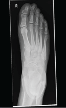 Figure 1: Case 1: Pre-operative anteroposterior foot radiograph-No evidence of bony involvement. View Figure 1
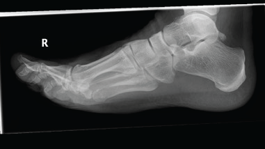 Figure 2: Case 1: Pre-operative lateral foot radiograph-No evidence of bony involvement. View Figure 2
PET scan showed no evidence of distant disease. He subsequently underwent wide local excision such that there was no residual invasive melanoma and resection margins were free of involvement by a minimum of 15 mm. Sentinel node biopsy was negative for metastatic melanoma. The final pathological stage was pT4a N0.
In order to cover the large remaining defect following wide local excision, the second toe was degloved and the distal one third of the metatarsal bone was excised (Figure 3, Figure 4, Figure 5 and Figure 6). This provided full thickness sensate skin cover and a narrowed foot to allow partial primary closure of the defect. Total surgery time for this procedure was less than thirty minutes.
 Figure 3: Case 1: Post skin incision to demarcate area - demonstrates size of the lesion and the size of the wide local excision needed in order to achieve clear margins. View Figure 3
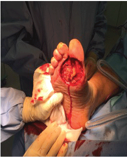 Figure 4: Case 1: Post excision of lesion - remaining defect. View Figure 4
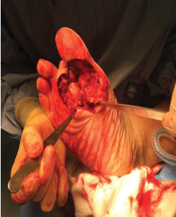 Figure 5: Case 1: The second toe was degloved and the distal one third of the metatarsal bone excised providing both full thickness sensate skin cover and a narrowed foot to allow partial primary closure. View Figure 5
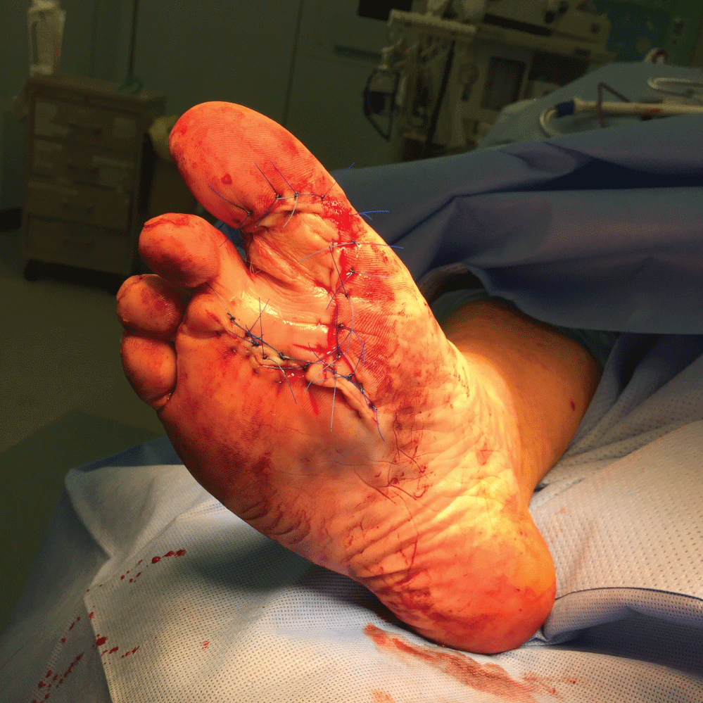 Figure 6: Case 1: Post closure. View Figure 6
The patient was referred to the oncology service and opted for active surveillance. He has no signs of recurrence on his most recent CT-TAP, two years post diagnosis.
He was prescribed an accommodative foot orthotic post-operatively and can fully weight bear.
A 67-year-old man with a background history of angina, gastritis, dyslipidaemia and gastrointestinal bleeding was referred by his GP to the dermatology service with a one-year history of a "blue scratch" on his right heel. This lesion had rapidly increased in size in the six weeks prior to referral. The lesion was sometimes painful and occasionally bled. The patient had no personal or family history of skin cancer or melanoma.
On examination there was a 3 cm exophytic amelanotic lesion, which was non-tender (Figure 7). The patient had bilateral groin nodes, however, it was thought that these were physiological in nature. The differential diagnosis was amelanotic melanoma or pyogenic granuloma, given the rapid increase in size of the lesion in the weeks prior to presentation.
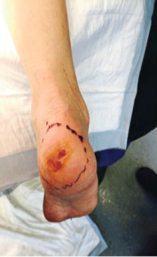 Figure 7: Case 2: Preoperative wide local excision. View Figure 7
Radiographs of the right calcaneous revealed a 2.8 cm soft tissue lesion arising from the skin of the heel, but no bony involvement (Figure 8). Incisional biopsy was performed. Histology revealed malignant melanoma of the right heel with a Breslow thickness of 9 mm. Ulceration was present, however there was no lymphovascular invasion or perineural invasion. Adjacent papules were present, and these were thought to be likely intransit metastases. PET scan and MRI brain did not reveal any distant disease.
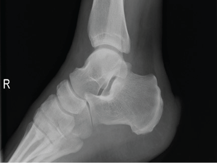 Figure 8: Case 2: Preoperative lateral radiograph of right calcaneus showing a 2.8 cm soft tissue lesion, which arises from the skin of the heel. View Figure 8
He subsequently underwent wide local excision (Figure 9). Calcaneal excision was performed in order to allow adequate skin closure. A bone anchor was used to reattach the Achilles tendon to the residual calcaneus (Figure 10 and Figure11). The calcaneal tuberosity, the origin of plantar fascia, and the subtalar joints were retained (Figure12, Figure 13, Figure 14 and Figure15) The patient was counseled on the side effect of reduced Achilles tendon power, however, the alternative to achieve margins and get skin closure was a below knee amputation. Total surgery time for this procedure was less than thirty minutes.
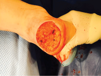 Figure 9: Case 2: Post excision of lesion - remaining defect. View Figure 9
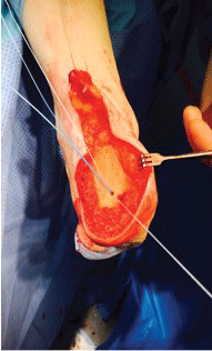 Figure 10: Case 2: Calcaneal excision was performed to allow adequate skin closure. A bone anchor was used to reattach Achilles tendon. View Figure 10
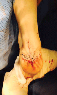 Figure 11: Case 2: Post Closure. View Figure 11
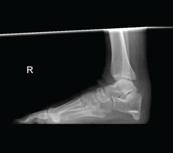 Figure 12: Case 2: Three-month post-operative lateral foot radiograph. The calcaneal tuberosity, origin of the plantar fascia, was retained, as was the subtalar joint; The Achilles tendon was reattached to the residual calcaneous. View Figure 12
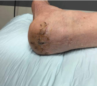 Figure 13: Case 2: Post-operative image. View Figure 13
 Figure 14: Case 2: Post-operative image. View Figure 14
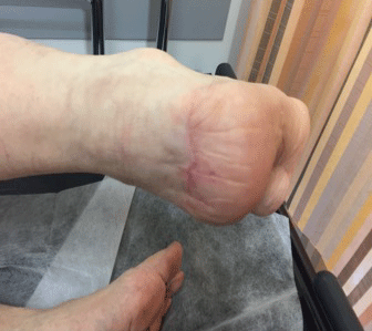 Figure 15: Case 2: Post-operative image. View Figure 15
The patient was referred to the oncology service and opted for active surveillance. At a recent outpatient’s appointment, one-year post diagnosis, he was found to have palpable right groin nodes. Fine needle aspirate cytology of these groin nodes was positive for melanoma. PET scan and CT-TAP have since revealed metastatic melanoma with groin nodes and a pulmonary nodule. The patient is currently being treated with immunotherapy under the oncology service.
Two cases of melanoma affecting the foot were successfully excised with negative margins by the orthopaedic service, achieving skin closure, while avoiding below knee amputation and maintaining the biomechanics of the foot.
Risks of failure of free and pedicled skin flaps were avoided and operating time was much reduced.
One patient has remained recurrence free, while the second patient has developed a recurrence and is currently being treated with immunotherapy under the oncology service.
With the rising incidence of melanoma, treatment must evolve in order to provide adequate resection of margins and achieve adequate skin coverage, all while providing the greatest functional outcome to the patient, in the quickest possible time. With rising difficulties in obtaining theatre time due to staff shortages and increasing demands on the health service, time efficient operations need to be developed such that more can be treated, without compromising their quality of care.
Struckmann, et al. [7] described a retrospective study of 21 patients who were treated for tissue defects in the weight-bearing plantar area due to trauma, peripheral arterial occlusive disease or decubitus ulcers using free or pedicled flaps over a 9-year period. They did not observe any flap loss, however one of the flaps required revision due to haematoma. Operation time in the free flap group (n = 12) was significantly longer (371.6 ± 68.4 minutes) than in the pedicled flap group (n = 9) (158.9 ± 77.7 minutes), however in-patient stay time did not differ significantly between the two groups. Functional outcome was not statistically significant between the two groups, however they found that myofasciocutaneous flaps yielded the best functional results, but those with myofasciocutaneous flaps also reported higher rates of pain on activity. Of the 21 patients only 7 had the ability to distinguish between warm and cold probes. Gait analysis also revealed that there was a tendency for prolonged contact time in the reconstructed plantar areas. 17 of the 21 patients had started full weight bearing after 12 weeks, 2 further patients were full weight bearing after 6 months while one patient was not full weight bearing until after 6 months.
Kang, et al. [8] reported 13 cases of pedicled flaps for hindfoot and lateral arch reconstruction following wide excision of malignant melanoma between 2005 and 2009. A distal-based island sural flap was used in 10 cases of hindfoot reconstruction, while lateral supramalleolar fasciocutaneous flap was used in 3 cases of lateral arch reconstruction after wide local excision of malignant melanomas. Four of these cases, two of each type of flap, were complicated by partial necrosis of the flaps. All patients were allowed to start partial weight bearing after six weeks, while full weight bearing commenced at twelve weeks.
Liu, et al. [1] reported on a retrospective review of 21 patients who either underwent cutaneous flaps following limb salvage surgery (n = 11) or amputation (n = 10) for resection of melanoma. All flaps survived and provided adequate defect coverage, however only 9 of the 11 patients were able to weight bear fully with no pain. Patients who underwent reverse sural island flaps reported loss of sensation on the lateral aspect of the foot. Two patients who underwent cutaneous flap reconstruction developed infection at the incision site, one developed oedema, while a fourth developed partial necrosis. A fifth patient experienced limitation of plantar flexion. Those who underwent limb amputation, including below knee, tarsometatarsal and toe amputation did not demonstrate differing oncological outcomes in comparison to those who underwent limb salvage surgery.
Cowles, et al. [15] reported several different techniques to adequately resect and provide skin coverage for melanomas occurring on the foot. They reported cases of lateral ray amputations with removal of the metatarsal heads to allow adequate skin closure, wide local excision with secondary wound healing and delayed split thickness skin grafting, transmetatarsal forefoot amputation following which the patient was able to ambulate with a customised shoe. All cases resulted in the patients being able to ambulate post-operatively, however due to the extensive nature of these amputations there was risk of defunctioning the foot.
Wan, et al. [9] report on the use of a medial plantar flap for heel reconstruction harvested with a branch of the medial plantar nerve in order to preserve innervation. Two of the cases were for skin closure following excision of malignant melanoma. Both patients were able to fully weight bear after six months. There was good preservation of sensation in both cases, however ability to sense cold temperature, static and dynamic two-point discrimination and light pressure was reduced.
While it has been reported that both free and pedicled, sensate and insensate flaps [1,7-15], serve well to provide adequate tissue coverage for plantar defects, both operation time and recovery for these flaps is lengthy, with the minimum time to full weight bearing being 12 weeks, as reported by Struckmann, et al. [7]. They are also at risk of failure, necrosis and ulceration [1,7,8] and indeed injury due to the reported inability to distinguish between hot and cold temperatures [7,9]. Performing free and pedicled flaps is also complicated by the need to replace the defects with tissue, which can appropriately meet the functional demands of the foot. Tissue used to replace defects on the foot should have durable skin, be capable of maintaining both sensation and limb function.
We present two cases of melanoma affecting the foot. These were successfully excised with negative margins by the orthopaedic service, achieving skin closure, while avoiding below knee amputation and maintaining the biomechanics of the foot.
These cases maintained the biomechanical function of the foot, adequately resected melanoma providing appropriate skin coverage, were less taxing on operating theatre time, and indeed patient recovery allowing immediate weight bearing as tolerated.