

International Journal of Ophthalmology and Clinical Research
Macular Multifocal Granular Hyperfluorescence on Fluorescein Angiography
Taiichi Hikichi*
Ohtsuka Eye Hospital, Sapporo, Japan
*Corresponding author: Dr. Taiichi Hikichi, Ohtsuka Eye Hospital, Kita-16 Nishi-4, Kita-ku, Sapporo 001-0016, Japan, Tel: +81-11-747-5211, Fax; +81-11-757-5223, E-mail: taiichi-hikichi@hokkaido.med.or.jp
Int J Ophthalmol Clin Res, IJOCR-2-010, (Volume 2, Issue 1), Research Article; ISSN: 2378-346X
Received: December 24, 2014 | Accepted: January 28, 2015 | Published: January 30, 2015
Citation: Hikichi T (2015) Macular Multifocal Granular Hyperfluorescence on Fluorescein Angiography. Int J Ophthalmol Clin Res 2:010. 10.23937/2378-346X/1410010
Copyright: © 2015 Hikichi T. This is an open-access article distributed under the terms of the Creative Commons Attribution License, which permits unrestricted use, distribution, and reproduction in any medium, provided the original author and source are credited.
Abstract
Purpose: To investigate the causes of multifocal granular hyperfluorescence at the macula on fluorescein angiography (FA) in Japanese patients.
Methods: I retrospectively studied 1,457 consecutive Japanese patients (1,457 eyes) who underwent digital simultaneous FA and indocyanine green angiography (ICGA).
Results: One hundred forty-seven of the 1,457 eyes had multifocal granular hyperfluorescence on FA images. Of the 147 eyes, 94 (64%) eyes had polypoidal choroidal vasculopathy (PCV); 25 (17%) eyes had either recurrent or chronic central serous chorioretinopathy (CSC); 19 (13%) eyes had occult choroidal neovascularization (CNV) associated with age-related macular degeneration (AMD); and three (2%) eyes had multiple evanescent white-dot syndrome. No diagnosis was established in 6 (4%) eyes because of the difficulty differentiating chronic CSC, PCV, and occult CNV.
Conclusion: The current results showed that PCV, occult CNV, and chronic CSC are the main causes of multifocal granular hyperfluorescence on FA. Thus, clinicians first should consider such disorders when examining patients with scattered lesions with pigment epithelial degeneration on ophthalmoscopy and multifocal granular hyperfluorescence on FA.
Keywords
Age-related macular degeneration; Central serous chorioretinopathy; Fluorescein angiography; Multifocal granular hyperfluorescence; Polypoidal choroidal vasculopathy
Introduction
Fluorescein angiography (FA) is an imaging examination that highlights the retinal and choroidal circulation and useful in the diagnosis of fundus disorders. Hyperfluorescence is defined as an abnormal presence of fluorescence or increase in normal fluorescence in fluorescein angiogram. Frequently hyperfluorescence is seen with window defects and with abnormal blood vessels. A window defect refers to the choroidal fluorescence produced by a relative decrease or absence of pigment in the retinal pigment epithelium (RPE). Multifocal granular hyperfluorescence seen on FA indicates diffuse RPE damage [1]. For example, in eyes with longstanding or recurrent central serous chorioretinopathy (CSC), the clinical picture is more complex with the possible emergence of a myriad of secondary changes, including patchy RPE atrophy or atrophic RPE tracks [2-5]. Multiple areas of diffuse RPE decompensation can evolve, resulting in gradual leakage through the posterior blood-retinal barrier and an indistinct zone of staining or oozing seen on FA images [1]. In contrast, in eyes with occult choroidal neovascularization (CNV) secondary to (age-related macular degeneration) AMD, although RPE decompensation can develop with window defects seen on FA images, the multifocal granular hyperfluorescent lesions are not as common [6,7]. Polypoidal choroidal vasculopathy (PCV) is a distinct exudative macular disorder and a highly prevalent and clinically relevant disease in Asian patients [8-12]. While its pathogenesis is unclear, PCV generally is considered to be a primary abnormality of the inner choroid characterized by a network of vessels with two distinct components: a complex of branching vessels and multiple, terminal, reddish-orange aneurysms or polypoidal lesions. Because PCV has a chronic natural history, diffuse retinal pigment epithelium (RPE) damage may develop in these eyes [8-12]. Multifocal granular hyperfluorescence occasionally is seen on FA images in eyes with PCV. In this study, the causes of multifocal granular hyperfluorescence on FA at the macula of Japanese patients were investigated and the clinical findings among the causes were compared to evaluate the essential elements in a differential diagnosis.
Patients and Methods
The institutional review board of Ohtsuka Eye Hospital approved the study. All patients provided informed consent. This study adhered to the tenets of the Declaration of Helsinki. I retrospectively studied 1,457 consecutive Japanese patients who had undergone digital simultaneous FA and indocyanine green angiography (ICGA) examinations at Ohtsuka Eye Hospital, Sapporo, Japan, between August 2011 and January 2013. If the patients had bilateral disease, the eye with the more recent symptom onset was included in this study. The patients had more than one angiogram performed in my institute; the angiogram performed firstly was chose in this study. All patients underwent a complete ophthalmic examination including measurement of the visual acuity (VA) using a Landolt ring chart, digital simultaneous FA and ICGA using a confocal laser scanning system (Heidelberg Retina Autograph 2, Heidelberg Engineering Inc., Heidelberg, Germany) [13], and spectral-domain optical coherence tomography (OCT) (Spectralis, Heidelberg Engineering Inc.). A mixture of 25 mg of indocyanine green (Ophthagreen, Daiichi-Seiyaku Co., Tokyo, Japan) and 500 mg of sodium fluorescein (Fluorescite, Japan Alcon, Tokyo, Japan) was injected intravenously. Eyes with multifocal granular hyperfluorescence on FA were identified and the causes of this finding were studied. Multifocal granular hyperfluorescence (Figure 1) was defined as the presence of multiple regions of hyperfluorescence seen early during choroidal filling with the greatest intensity at peak choroidal filling with or without late leakage. PCV was defined as the presence of one or multiple focal areas of hyperfluorescence arising from the choroidal circulation within the first 6 minutes after injection of ICGA, with or without an associated branching vascular network [14,15]. The polypoidal lesions could have been solitary (arbitrarily defined as one or two polyps) or multiple; if multiple polypoidal lesions were present, they could have been arranged in a ring or cluster. Occult CNV secondary to AMD was diagnosed when FA showed increasing subretinal hyperfluorescence, no clear delineation, and irregular staining, and ICGA showed early vascular hyperfluorescence and late staining of abnormal vessels [16-18]. Cases with a choroidal branching vascular network and no polypoidal lesions were diagnosed with occult CNV. CSC was defined as an idiopathic serous detachment of the neurosensory retina in the macular area with or without pigment epithelial changes and one or more focal leaks at the level of the RPE [19]. Chronic or recurrent CSC, referred to as diffuse retinal pigment epitheliopathy, was defined as a neurosensory detachment associated with an area of RPE atrophy and pigment mottling, granular hyperfluorescence with subtle leaks on FA, and no characteristics of PCV or occult CNV [2-5].
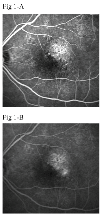
Figure 1: Multifocal granular hyperfluorescence on (A) early- and (B) latephase
fluorescein angiography images. Multiple regions of hyperfluorescence
are seen early during choroidal filling with the greatest intensity at the peak
choroidal filling in the absence of late leakage.
View Figure 1
Results
Of the 1,457 eyes, 147 eyes had multifocal granular hyperfluorescence at the macula on FA. Of the 147 eyes, 94 (64%) eyes had PCV; 25 (17%) eyes had either recurrent or chronic CSC; 19 (13%) eyes had occult CNV associated with AMD; and three (2%) eyes had multiple evanescent white-dot syndrome. No diagnosis was established in 6 (4%) eyes because of the difficulty differentiating chronic CSC, PCV, and occult CNV. Although multifocal hyperfluorescence on FA can be shown in Stargardt�s disease and Harada syndrome, these disorders were not found in the147 eyes reviewed in this study. The 94 eyes with PCV had characteristic polypoidal lesions on ICGA (Figure 2). The 19 eyes with occult CNV had granular hyperfluorescence on FA and CNV that was confirmed by ICGA (Figure 3). In 25 eyes with chronic or recurrent CSC, diffuse retinal pigment epitheliopathy was seen on FA, and no characteristics of PCV or occult CNV were seen on ICGA (Figure 4). Choroidal vascular hyperpermeability was observed in 36 (38%) eyes with PCV, two (11%) eyes with occult CNV, and 19 (76%) eyes with CSC. Although six eyes not established definite diagnosis had multifocal granular hyperfluorescence at the macula on FA and small branching vascular network as observed in PCV that was confirmed by ICGA, characteristic polypoidal lesions could not be observed on ICGA? Thus, these six eyes were not coincided with the diagnostic criteria of PCV. The mean � standard deviation (SD) ages of patients with PCV, occult CNV associated with AMD, and CSC, respectively, were 75 � 9, 77 � 8, and 52 � 8 years. The mean age of patients with CSC was significantly (P< 0.001 for both comparisons by the unpaired Student�s t-test) younger than those of patients with PCV and occult CNV associated with AMD. Fifty-five (59%) eyes with PCV, 12 (63%) eyes with occult CNV, and 18 (72%) eyes with CSC were those of male patients; the percentages did not reach significance.
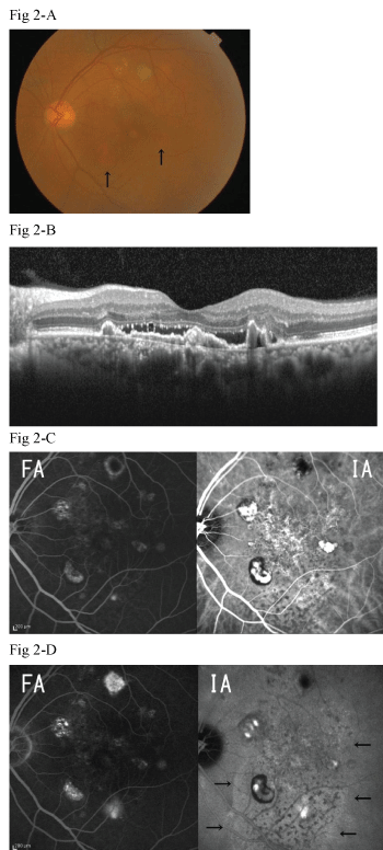 Figure 2: The left eye of a 68-year-old man with polypoidal choroidal
vasculopathy. A: A fundus photo shows some lesions with pigment epithelial
degeneration and orange-red lesions (arrows). B: A horizontal optical
coherence tomography scan shows a serous sensory detachment; the elevated
retinal pigment epithelium line separating from Bruch�s membrane, which was
irregular and thickened due to deposition; and the thickened choroid. C: The
early phase of simultaneous fluorescein angiography (FA) and indocyanine
green angiography (ICGA) scans shows multifocal granular hyperfluorescence
on FA and three relatively large polypoidal lesions and a choroidal branching
vascular network on ICGA. D: The late phase of simultaneous FA and ICGA
scans shows late geographic hyperfluorescence (arrows) on ICGA.
View Figure 2
Figure 2: The left eye of a 68-year-old man with polypoidal choroidal
vasculopathy. A: A fundus photo shows some lesions with pigment epithelial
degeneration and orange-red lesions (arrows). B: A horizontal optical
coherence tomography scan shows a serous sensory detachment; the elevated
retinal pigment epithelium line separating from Bruch�s membrane, which was
irregular and thickened due to deposition; and the thickened choroid. C: The
early phase of simultaneous fluorescein angiography (FA) and indocyanine
green angiography (ICGA) scans shows multifocal granular hyperfluorescence
on FA and three relatively large polypoidal lesions and a choroidal branching
vascular network on ICGA. D: The late phase of simultaneous FA and ICGA
scans shows late geographic hyperfluorescence (arrows) on ICGA.
View Figure 2
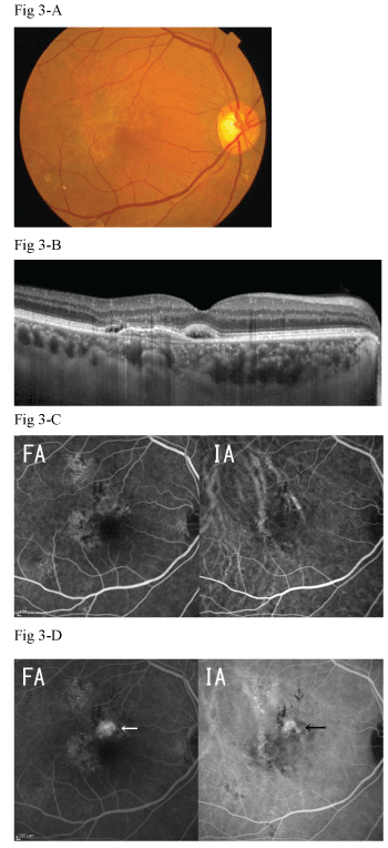 Figure 3: The right eye of a 73-year-old man with occult choroidal
neovascularization associated with age-related macular degeneration. A: A
fundus photo shows a pigment epithelial degeneration. B: A horizontal optical
coherence tomography scan shows a focal serous sensory detachment,
hyperreflective layers beneath the flat elevation of the retinal pigment
epithelium, and the thickened choroid. C: The early phase of simultaneous
fluorescein angiography (FA) and indocyanine green angiography (ICGA)
scans shows several granular hyperfluorescence lesions on FA. D: The late
phase of simultaneous FA and ICGA scans shows a lesion with increasing
subretinal hyperfluorescence, lack of clear delineation, irregular staining
(white arrow) on FA, and a plaque lesion (black arrow) on ICGA.
View Figure 3
Figure 3: The right eye of a 73-year-old man with occult choroidal
neovascularization associated with age-related macular degeneration. A: A
fundus photo shows a pigment epithelial degeneration. B: A horizontal optical
coherence tomography scan shows a focal serous sensory detachment,
hyperreflective layers beneath the flat elevation of the retinal pigment
epithelium, and the thickened choroid. C: The early phase of simultaneous
fluorescein angiography (FA) and indocyanine green angiography (ICGA)
scans shows several granular hyperfluorescence lesions on FA. D: The late
phase of simultaneous FA and ICGA scans shows a lesion with increasing
subretinal hyperfluorescence, lack of clear delineation, irregular staining
(white arrow) on FA, and a plaque lesion (black arrow) on ICGA.
View Figure 3
In 126 (86%) of the 147 eyes with multifocal granular hyperfluorescence on FA, OCT showed the presence of a serious retinal detachment (SRD). In eyes with PCV, the RPE line was elevated and separated from Bruch�s membrane, the vascular pigment epithelial detachment (PED) corresponded to the branching vascular network, which was irregular and thickened due to deposition, and the choroid was thickened (Figure 2). The OCT images of eyes with occult CNV showed a flat elevation of RPE, beneath which hyperreflective layers were observed that corresponded to fibrovascular infiltration (Figure 3). In eyes with CSC, OCT showed an undulating RPE line, elongation of the photoreceptor outer segment, and a thicken choroid (Figure 4). Small serous PEDs were observed in the area of the SRD in 13 (52%) eyes.
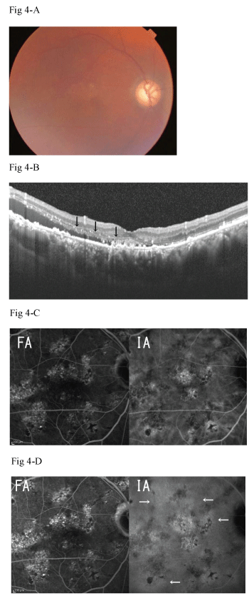 Figure 4: The right eye of a 73-year-old man with chronic central serous
chorioretinopathy. A: A fundus photo shows some lesions with pigment
epithelial degeneration. B: A vertical optical coherence tomography scans
shows a serous sensory detachment, an undulating retinal pigment epithelium
line, elongation of the photoreceptor outer segment (arrows), and a thickened
choroid. The early (C) and late phases (D) of simultaneous fluorescein
angiography (FA) and indocyanine green angiography (ICGA) scans show
multiple window defects on FA and choroidal vascular hyperpermeability
(arrows) without polypoidal lesions or choroidal neovascularization on ICGA.
View Figure 4
Figure 4: The right eye of a 73-year-old man with chronic central serous
chorioretinopathy. A: A fundus photo shows some lesions with pigment
epithelial degeneration. B: A vertical optical coherence tomography scans
shows a serous sensory detachment, an undulating retinal pigment epithelium
line, elongation of the photoreceptor outer segment (arrows), and a thickened
choroid. The early (C) and late phases (D) of simultaneous fluorescein
angiography (FA) and indocyanine green angiography (ICGA) scans show
multiple window defects on FA and choroidal vascular hyperpermeability
(arrows) without polypoidal lesions or choroidal neovascularization on ICGA.
View Figure 4
Discussion
The current results showed that PCV, occult CNV, and chronic CSC are the main causes of multifocal granular hyperfluorescence on FA. Thus, clinicians first should consider such disorders when examining patients with scattered lesions with pigment epithelial degeneration on ophthalmoscopy and multifocal granular hyperfluorescence on FA. Clinical factors such as age and the findings on ICGA and OCT images should be considered in the differential diagnosis. In some cases of CSC with a persistent or recurrent neurosensory detachment, a myriad of secondary effects may develop at the level of the RPE, and fluorescein leakage may no longer be focal or discrete as in eyes with typical CSC. A localized or diffuse area of RPE decompensation instead may evolve, resulting in granular leakage through the posterior blood-retinal barrier and an indistinct zone of staining or so-called �ooze� [20-22]. PCV is associated with multiple, recurrent serosanguineous detachments of the RPE and neurosensory retina secondary to leakage and bleeding from choroidal vascular lesions [8-12,14,23]. However, in some cases, a small-caliber choroidal vascular branching network with a neurosensory detachment at the macula without hemorrhage is a major manifestation associated with RPE atrophy. Because FA shows multifocal areas of granular hyperfluorescence, it is often difficult to distinguish PCV from persistent or recurrent CSC [12,24]. Such cases of PCV were reported to masquerade as CSC [12]. PCV was reported to have a particularly strong association with a history of CSC and be part of a sequence after chronic CSC [25,26]. PCV sometimes had choroidal vascular hyperfluorescence seen on ICGA images, which has been interpreted as regional vascular hyperpermeability of the choriocapillaris [24]. Choroidal vascular hyperpermeability also has been reported in eyes with CSC, and these findings may be involved in the pathogenesis of CSC [4,27,28]. Thus, those authors speculated that chronic CSC or subclinical CSC might contribute to development of PCV. Furthermore, increased choroidal thickness has been seen on OCT images obtained from eyes with both disorders [29-34]. The pathogenesis is similar between the two disorders and some eyes with PCV had a medical history of CSC [24,25], raising the possibility that the two disorders belong to the same category. The recent advent of OCT, especially spectral-domain OCT with high-resolution images, provides an opportunity to observe new findings and contributes to the differential diagnosis among disorders characterized by deterioration around the outer retina and subretinal space, such as PCV, occult CNV, and chronic CSC [34-36]. Although the incidence rates of the characteristic findings on OCT images vary among the diseases, such as the so-called double-layer sign, which is comprised of two highly reflective layers, i.e., an undulating RPE line and a hyperreflective straight line representing Bruch�s membrane, the findings are not pathognomonic for only one disease but also are found in other diseases [34-36]. Thus, although OCT can provide essential information about the outer retina and subretinal space, establishing a diagnosis seems to be difficult using only OCT findings.
As PCV is a relatively new entity, it had been regarded as occult CNV in many patients and studies previously [8-12]. In the present study, PCV was defined as focal areas of hyperfluorescence after injection of ICGA with or without an associated branching vascular network and cases with a choroidal branching vascular network and no polypoidal lesions were not diagnosed with PCV. As the focal hyperfluorescence in PCV may not always show up on ICGA because it was obscured by blood or fluid or it has shrunken after treatment or bleeding or did not develop polypoidal lesions yet, only branching vascular network may be seen as occult CNV [37]. Thus, the stage of the disease may have associated with the incidences of the diseases in the current study. PCV is also suggested to be an underlying cause of CSC in some patients. If the angiogram showed PCV and OCT showed serous detachment, were the patients regarded as PCV in the current study. Therefore, the number of patients reported in the two conditions may be misleading if this study distinguished them based on the presence of PCV on ICGA alone.
PCV, occult CNV, and chronic CSC are primary causes of multifocal granular hyperfluorescence on FA images obtained from Japanese patients. In eyes with multifocal granular hyperfluorescence on FA images, multi-modality findings such as fundus ophthalmoscopy, OCT, and ICGA are necessary to establish a definitive diagnosis.
However, since relevance among PCV, occult CNV, and chronic CSC is suggested, only fragmentary examinations may be difficult in the definite differential diagnosis in some cases. In such cases, follow-up examinations are also necessary for the exact diagnosis.
Acknowledgements
Publication of this article was not supported by grants. Dr. Hikichi has received lecture fees from Novartis Pharma Japan, Bayer Japan, Santen, Kowa Souyaku, Alcon Japan, and Senjyu Pharmaceutical. All processes of the study from conception and design of study; analysis and interpretation; writing of the article; critical revision of the article; final approval of the article; to data collection, were performed by the author (T.H.). The author declares that there is no conflict of interests regarding the publication of this paper.
Ethical Statement
The current study was conducted with the approval of the Institutional Review Board of Ohtsuka Eye Hospital, and informed consent was received from all enrolled patients.
References
-
Jalkh AE, Jabbour N, Avila MP, Trempe CL, Schepens CL (1984) Retinal pigment epithelium decompensation. I. Clinical features and natural course. Ophthalmology 91: 1544-1548.
-
Castro-Correia J, Coutinho MF, Rosas V, Maia J (1992) Long-term follow-up of central serous retinopathy in 150 patients. Doc Ophthalmol 81: 379-386.
-
Gilbert CM, Owens SL, Smith PD, Fine SL (1984) Long-term follow-up of central serous chorioretinopathy. Br J Ophthalmol 68: 815-820.
-
Iida T, Kishi S, Hagimura N, Shimizu K (1999) Persistent and bilateral choroidal vascular abnormalities in central serous chorioretinopathy. Retina 19: 508-512.
-
Schatz H, Madeira D, Johnson RN, McDonald HR (1992) Central serous chorioretinopathy occurring in patients 60 years of age and older. Ophthalmology 99: 63-67.
-
Bressler NM, Finklestein D, Sunness JS, Maguire AM, Yarian D (1990) Retinal pigment epithelial tears through the fovea with preservation of good visual acuity. Arch Ophthalmol 108: 1694-1697.
-
Malamos P, Sacu S, Georgopoulos M, Kiss C, Pruente C, et al. (2009) Correlation of high-definition optical coherence tomography and fluorescein angiography imaging in neovascular macular degeneration. Invest Ophthalmol Vis Sci 50: 4926-4933.
-
Sho K, Takahashi K, Yamada H, Wada M, Nagai Y, et al. (2003) Polypoidal choroidal vasculopathy: incidence, demographic features, and clinical characteristics. Arch Ophthalmol 121: 1392-1396.
-
Uyama M, Matsubara T, Fukushima I, Matsunaga H, Iwashita K, et al. (1999) Idiopathic polypoidal choroidal vasculopathy in Japanese patients. Arch Ophthalmol 117: 1035-1042.
-
Uyama M, Wada M, Nagai Y, Matsubara T, Matsunaga H, et al. (2002) Polypoidal choroidal vasculopathy: natural history. Am J Ophthalmol 133: 639-648.
-
Yannuzzi LA, Ciardella A, Spaide RF, Rabb M, Freund KB, et al. (1997) The expanding clinical spectrum of idiopathic polypoidal choroidal vasculopathy. Arch Ophthalmol 115: 478-485.
-
Yannuzzi LA, Freund KB, Goldbaum M, Scassellati-Sforzolini B, Guyer DR, et al. (2000) Polypoidal choroidal vasculopathy masquerading as central serous chorioretinopathy. Ophthalmology 107: 767-777.
-
Freeman WR, Bartsch DU, Mueller AJ, Banker AS, Weinreb RN (1998) Simultaneous indocyanine green and fluorescein angiography using a confocal scanning laser ophthalmoscope. Arch Ophthalmol 116: 455-463.
-
Koh AH; Expert PCV Panel, Chen LJ, Chen SJ, Chen Y, Giridhar A, et al. (2013) Polypoidal choroidal vasculopathy: evidence-based guidelines for clinical diagnosis and treatment. Retina 33: 686-716.
-
Lim TH, Laude A, Tan CS (2010) Polypoidal choroidal vasculopathy: an angiographic discussion. Eye (Lond) 24: 483-490.
-
Bressler NM, Frost LA, Bressler SB, Murphy RP, Fine SL (1988) Natural course of poorly defined choroidal neovascularization associated with macular degeneration. Arch Ophthalmol 106: 1537-1542.
-
(1982) Argon laser photocoagulation for senile macular degeneration: results of a randomized clinical trial. Arch Ophthalmol 100: 912-918.
-
(1996) Occult choroidal neovascularization. Influence on visual outcome in patients with age-related macular degeneration. Macular Photocoagulation Study Group .Arch Ophthalmol 114: 400-412.
-
Berger AR, Olk RJ, Burgess D (1991) Central serous choroidopathy in patients over 50 years of age. Ophthalmic Surg 22: 583-590.
-
Iida T, Spaide RF, Haas A, Yannuzzi LA, Jampol LM, et al. (2002) Leopard-spot pattern of yellowish subretinal deposits in central serous chorioretinopathy. Arch Ophthalmol 120: 37-42.
-
Spaide RF, Campeas L, Haas A, Yannuzzi LA, Fisher YL, et al. (1996) Central serous chorioretinopathy in younger and older adults. Ophthalmology 103: 2070-2079.
-
Spaide RF, Hall L, Haas A, Campeas L, Yannuzzi LA, et al. (1996) Indocyanine green videoangiography of older patients with central serous chorioretinopathy. Retina 16: 203-213.
-
Hatz K, Pr�nte C (2014) Polypoidal choroidal vasculopathy in Caucasian patients with presumed neovascular age-related macular degeneration and poor ranibizumab response. Br J Ophthalmol 98: 188-194.
-
Sasahara M, Tsujikawa A, Musashi K, Gotoh N, Otani A, et al. (2006) Polypoidal choroidal vasculopathy with choroidal vascular hyperpermeability. Am J Ophthalmol 142: 601-607.
-
Ueta T, Obata R, Inoue Y, Iriyama A, Takahashi H, et al. (2009) Background comparison of typical age-related macular degeneration and polypoidal choroidal vasculopathy in Japanese patients. Ophthalmology 116: 2400-2406.
-
Ahuja RM, Downes SM, Stanga PE, Koh AH, Vingerling JR, et al. (2001) Polypoidal choroidal vasculopathy and central serous chorioretinopathy. Ophthalmology 108: 1009-1010.
-
Kitaya N, Nagaoka T, Hikichi T, Sugawara R, Fukui K, et al. (2003) Features of abnormal choroidal circulation in central serous chorioretinopathy. Br J Ophthalmol 87: 709-712.
-
Pr�nte C, Flammer J (1996) Choroidal capillary and venous congestion in central serous chorioretinopathy. Am J Ophthalmol 121: 26-34.
-
Chung SE, Kang SW, Lee JH, Kim YT (2011) Choroidal thickness in polypoidal choroidal vasculopathy and exudative age-related macular degeneration. Ophthalmology 118: 840-845.
-
Imamura Y, Fujiwara T, Margolis R, Spaide RF (2009) Enhanced depth imaging optical coherence tomography of the choroid in central serous chorioretinopathy. Retina 29: 1469-1473.
-
Jirarattanasopa P, Ooto S, Nakata I, Tsujikawa A, Yamashiro K, et al. (2012) Choroidal thickness, vascular hyperpermeability, and complement factor H in age-related macular degeneration and polypoidal choroidal vasculopathy. Invest Ophthalmol Vis Sci 53: 3663-3672.
-
Koizumi H, Yamagishi T, Yamazaki T, Kinoshita S (2013) Relationship between clinical characteristics of polypoidal choroidal vasculopathy and choroidal vascular hyperpermeability. Am J Ophthalmol 155: 305-313.
-
Maruko I, Iida T, Sugano Y, Ojima A, Ogasawara M, et al. (2010) Subfoveal choroidal thickness after treatment of central serous chorioretinopathy. Ophthalmology 117: 1792-1799.
-
Ojima Y, Hangai M, Sakamoto A, Tsujikawa A, Otani A, et al. (2009) Improved visualization of polypoidal choroidal vasculopathy lesions using spectral-domain optical coherence tomography. Retina 29: 52-59.
-
Sato T, Kishi S, Watanabe G, Matsumoto H, Mukai R (2007) Tomographic features of branching vascular networks in polypoidal choroidal vasculopathy. Retina 27: 589-594.
-
Yang L, Jonas JB, Wei W (2013) Optical coherence tomography-assisted enhanced depth imaging of central serous chorioretinopathy. Invest Ophthalmol Vis Sci 54: 4659-4665.
-
Squirrell DM, Bacon JF, Brand CS (2009) To investigate the prevalence of polypoidal choroidal vasculopathy in presumed age-related peripapillary subretinal neovascular membranes. Clin Experiment Ophthalmol 37: 368-372.
