

International Journal of Ophthalmology and Clinical Research
Can OCT Scan Averaging Induce a Loss of Information?
Desmettre T*, Menard M, Colas E and Tadayoni R
Ophthalmology Department, Hopital Lariboisiere, AP-HP, Université Paris 7-Sorbonne Paris Cite, Paris, France
*Corresponding author: Desmettre T, Ophthalmology Department, Hôpital Lariboisière, AP-HP, Universite Paris 7-Sorbonne Paris Cité, Paris, France, E-mail: thomas@desmettre.org
Int J Ophthalmol Clin Res, IJOCR-4-069, (Volume 4, Issue 1), Research Article; ISSN: 2378-346X
Received: September 16, 2016 | Accepted: March 23, 2017 | Published: March 25, 2017
Citation: Desmettre T, Menard M, Colas E, Tadayoni R (2017) Can OCT Scan Averaging Induce a Loss of Information? Int J Ophthalmol Clin Res 4:069. 10.23937/2378-346X/1410069
Copyright: © Desmettre T, et al. This is an open-access article distributed under the terms of the Creative Commons Attribution License, which permits unrestricted use, distribution, and reproduction in any medium, provided the original author and source are credited.
Abstract
Purpose: An averaging of retina scans is integrated in the algorithm of most Optical Coherence Tomography (OCT) devices to improve final image quality. This study aimed to evaluate image modification or alteration induced by this averaging.
Methods: Thirty-three consecutive patients examined with the Opko®, SLO/OCT OTI were included in this retrospective study. Final OCT-B images were built with an averaging of 20 OCT-B Scans. Fifteen of these patients were followed for the monitoring of antimalarials while the 18 others were followed up for various retina conditions. Final OCT-B images were decomposed into their Scan 1, Scan 5 and Scan 10. Based on the Scan 1 image, we evaluated changes on 4 items (i) Choroid, (ii) Retinal Pigment Epithelium (RPE) and or Ellipsoid zone (EZ), (iii) Neuroretina, and (iv) Anterior profile line. The items were noted 0 or 1 (absence or presence of subtle alteration). We then compared the percentages of alterations for Scan 5, Scan 10 and Scan 20. For the 15 patients treated with antimalarials, based on clinical evaluation, visual field, multifocal electroretinogram, fundus autofluorescence and on the presence of alteration of the outer retinal layers on OCT the patients were separated into group (A) : no sign of antiamarials intoxication (n = 7) and group (B) : presence of antiamarials intoxication (n = 8).
Results: All the images of the 33 patients included in this series were used for the analysis. Subtle alterations with loss of some details (retina vessels and their shadows, exudates, choroid lobules) were observed along with increased averaging. For the whole series, with 4 items × 33 patients (132 items) we noticed 24 changes on Scan 5 (18.18%), 44 changes on Scan10 (33.33%), and 47 changes (35%) on Scan 20. For the 15 patients with antiamarials, on Scan 10 and Scan 20, image modification was more frequent in the group with intoxication (75%) than in the group without intoxication (42.8%).
Conclusion: For OCT devices with technical characteristics similar to the Opko® and for patients with fixation characteristics similar to those of our series, it would be worth discussing the possibility of image alteration induced by scan averaging if Scan 10 or above images are used. For current OCT devices, as the number of scans averaged has now increased, our data suggest that acquisition speed, better co-localization of repeated OCT Scans and other technical improvements could influence the benefit/loss ratio of averaging.
Introduction
Optical coherence tomography (OCT) is a well-established imaging modality for the diagnosis of retinal conditions. Final images arise from computed scans derived from the backscatter of short coherence light. An averaging of retina scans is regularly integrated in the algorithm of most OCT devices: several scans are averaged to build the final image. Depending on the technology and on the availability of an eye-tracking system the averaging process can even be on real time. On the Spectralis® the Automatic Real-Time Averaging (Art) even computes the images during the acquisition (Art 1: one scan, Art 2: two scans etc…). The speckle noise is indeed reduced by averaging the images and this process has been used for years for improvement of image quality in computed tomography and magnetic resonance images [1-3].
However, for OCT B-Scans one can wonder if this averaging process is always a benefit, especially for pathologic conditions with subtle details to control over time or for patients with visual defects. Firstly, an increase of the acquisition time is logically induced with the number of B-Scan averaged. Secondly, while the averaging increases the apparent quality of the final OCT image, it also may be a source of image modification. Primary fixation is frequently interrupted by slow drifts, micro saccades and saccadic intrusions [4]. These micro-movements, even contra-balanced by an eye-tracking system could be a source of image modification with a slight blurring of the final image and a certain thickening of the outer retina layers.
In the literature, few authors have inquired about the optimal averaging, i.e. the optimal number of scans to add to meet up the quality of the images and the accuracy of the medical information. Pappuru, et al. [5] and Sakamoto, et al. [6] have shown that image quality improved with averaging of up to 16 or 20 B-Scan. In the same way, Shirasawa, et al. recently showed that although the image quality of OCT images of the retina improved with an increase in the number of images averaged, it didn't improve significantly by averaging more than 20 Scans [7]. For their study they used 9 healthy eyes and a phantom eye model.
The speed of acquisition of the OCT, the presence or absence of an eye-tracker and the possibility of a loss of fixation, were not addressed in these preclinical studies. We therefore thought that some clinical information would help to evidence the influence of scan averaging on image quality.
In this study, we compared different averaging of the same image acquired among patients mainly treated with antiamarials (chloroquine or hydroxychloroquine) monitored with a spectral domain OCT (Opko®, OTI, Toronto, ONT, Canada). Retinal toxicity of antimalarials first affects retinal ganglion cells and photoreceptors, especially in the perifoveal region [8,9]. The drugs also have affinity for pigmented cells, including the Retinal Pigmented Epithelium (RPE). For these reasons, our study was mainly focused on image alteration on the outer retinal layers, the Ellipsoid Zone (EZ) and the RPE. Some patients followed in our department for another specific condition (Age related Macular Degeneration, Central Serous Chorioretinopathy…) were examined on the same OCT device and were also included in the study.
For averaging up to 5 Scans the modification of the final image was minimal except on the edges of the images. For 10 Scans and above we noticed more frequent and obvious image modifications induced by averaging. Then, for OCT devices with technical characteristics similar to the Opko® (acquisition speed 27,000 A-scans/s, no eye-tracking) and for patients with fixation characteristics similar to those of our series, it would be worth discussing the possibility of image alteration induced by scan averaging if Scan 10 or above images are used. For current OCT devices, with higher acquisition speed and eye-tracking system, because the number of scans averaged has now increased, our data suggest that the benefit/loss ratio of averaging can still be discussed.
Patients and Methods
This exploratory retrospective study was conducted in accordance with the principles of the Declaration of Helsinki and in compliance with the ethical committee of the French Society of Ophthalmology.
Thirty three patients (mean 56.3 year old; from 27 to 79) were included in this consecutive series of patients.
Fifteen of them, followed for Lupus, Sarcoidosis, Rheumatoid Arthritis or other inflammatory diseases were treated with antimalarials. Based on clinical evaluation, visual field, multifocal electroretinogram, fundus autofluorescence and on the presence of alteration of the outer retinal layers on OCT images the patients were separated into 2 groups: (A) No sign of antimalarials intoxication (n = 7), (B) presence of antimalarials intoxication (n = 8).
Another group labeled (C) Other condition (n = 18) included patients not treated with antimalarials but followed up on the same OCT device. These patients had various retinal conditions (Diabetes, Age Macular Degeneration, Venous Occlusion, and Central Serous Chorioretinopathy).
The images used for this monitoring were acquired on the Opko®, SLO/OCT OTI. The device displayed OCT images built with a computation of 20 B-Scan.
All the images were blindly saved on a computer dedicated to clinical research. The Scan 20 images were decomposed into Scan 1, Scan 5, and Scan 10 for the use of the study. Three readers (TD, MM, RT) had access to the images and their decomputations. On the basis of the Scan 1 image, they evaluated the changes (loss or appearance) of details on Scan 5, Scan 10 and Scan 20 images. As this study was only observational and retrospective, no modification of the initial diagnosis was attempted by the readers.
Among different OCT devices available in our department (Avanti®, Optovue, Fremont USA; Cirrus 4000®, Zeiss USA; Spectralis®, Heidelberg Germany; Opko®, OTI Canada), the OTI Opko® OCT/SLO was chosen because it allows a retrospective analysis of the images with a decomposition of the averaged images.
The readers rated gross modifications induced by scan averaging (flawed overcrossing of the edges of scans, significant blurring of the image) and sought for alterations of more subtle details (modification of choroid lobules, shading of the RPE, thickening or interruption of ellipsoid zone, loss of retinal vessel, loss of edema cyst walls, alteration of retina anterior profile line, modification of retina/vitreous interface).
Based on the Scan 1 image, we reported changes on 4 items (i) Choroid, (ii) RPE and or EZ, (iii) Neuroretina, and (iv) Anterior profile line. The items were noted 0 or 1 according the absence or presence of any subtle alteration. Such a notation was reported for Scan 5, Scan 10 and Scan 20. We then compared the percentages of alterations for the different Scan #.
For the patients followed under antimalarials, because of the location of drug toxicity we also did a simpler subgroup evaluation focused on the changes noted 0 or 1 on the RPE and the Ellipsoid zone.
Since scan averaging is widely used for more recent OCT devices we also examined a few patients with images acquired on the Cirrus 4000®, on the Spectralis® and on the Avanti® to seek out the same kind of alteration than observed with the Opko®. A case imaged with the Cirrus is displayed as an example.
Results
All the images of the 33 patients included in this series were used for the analysis. We first noticed that for images built with good quality scans, successive additions appeared generally as a benefit. Conversely, images built with poor quality scans seemed to suffer the most from the averaging process. Some obvious alterations associated with scan averaging were almost always noticeable on the edges of the images. More subtle alterations including loss of details (edema cyst walls, retina vessels and their shadows, exudates, choroid lobules) were harder to disclose and could in some cases interfere with a diagnosis.
General evaluation
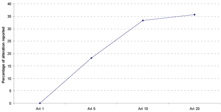
For the 33 patients, on the total of 4 × 33 = 132 items we noted 24 changes on Scan 5 (18.18%), 44 changes on Scan 10 (33.33%), and 47 changes (35%) on Scan 20. Figure 1 displays the progression of these percentages (Figure 1).
.
Figure 1: Grading of image modification (%) for the 33 patients. Four items were analyzed on each eye (4 × 33 = 132 items). We quoted 24 changes on Scan 5 (18.18%), 44 changes on Scan 10 (33.33%), and 47 changes (35%) on Scan 20. The slow rise of the curve after Scan 10 is explained by the fact that the changes observed on Scan 5 are sustained on Scan 10; changes quoted on Scan 5 and Scan 10 are sustained on Scan 20.
View Figure 1
Subgroup evaluation: Antimalarials
For groups A and B, we also achieved a simpler evaluation based on the presence of any loss of details on the RPE or EZ (Table 1 and Table 2). The modifications noticed on retina vessels and their shadows, on exudates, and on choroid lobules were not reported in this subgroup analysis.
![]()
Table 1: Group A (no sign of antimalarials intoxication): comparison of changes on RPE or Ellipsoid Zone or ONL on Scan # compared to Scan 1. (*): see details of changes on Figure 2.
View Table 1
![]()
Table 2: Group B (presence of antimalarials intoxication-4 with onset alterations, 4 with advanced alterations): comparison of changes on RPE or Ellipsoid Zone or ONL on Scan # compared to Scan 1.
View Table 2
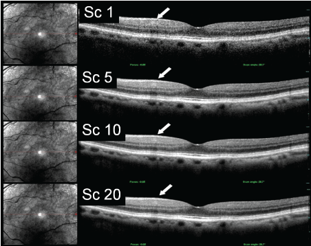
No sign of antimalarials intoxication (n = 7): These 7 patients had no sign of antimalarials intoxication. The percentages of image alterations for Scan 5, Scan 10 and Scan 20 were respectively 28.5%, 42.8%, and 42.8% (Table 1). Once observed at Scan 5 or Scan 10 the false positive changes on the RPE or EZ persisted or increased with further averaging. Figure 2 displays some of these image modifications associated with scan averaging (Figure 2).
.
Figure 2: Patient of group A (no sign of antimalarials intoxication). Although the general image quality is rather improved from Scan 1 to Scan 20, gross alteration related to summation appear on the edges of images from Scan 5. Moreover, nasal to the foveola a vessel with his slight shadow (arrow) on Scan 1 progressively disappears from Scan 5 to Scan 20. Conversely his shadow is progressively deepened and enlarged. On Scan 20 projection of this shadow on the underlying layers could mimic an alteration of the Ellipsoid Zone.
View Figure 2
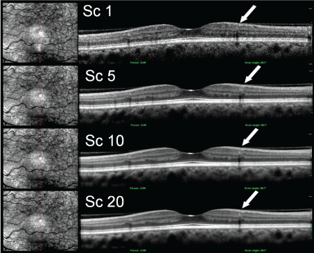
Signs of antimalarials intoxication (n = 8): These 8 patients had slight (n = 4) to advanced (n = 4) signs of antimalarials intoxication. The percentages of image alterations for Scan 5, Scan 10 and Scan 20 were respectively 12.8%, 75%, and 75% (Table 2). Figure 3 displays some example of these image alterations (Figure 3).
.
Figure 3: Patient of group B with onset antimalarials intoxication. Temporal to the foveola, 2 vessels and their shadows appear separated on Scan 1 (arrow). The signals corresponding to those vessels are progressively merged from Scan 5 to Scan 20.
View Figure 3
Subgroup evaluation: "other conditions"
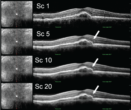
For this group C (n = 18), as in the general evaluation we sought out changes on 4 items ((i) Choroid, (ii) RPE and or EZ, (iii) Neuroretina, and (iv) Anterior profile line). For each of these 18 patients, a total of 72 items (4 × 18) were examined on different Scans #. The percentages of image alterations for Scan 5, Scan 10 and Scan 20 were respectively 18.7%, 26.4% and 29.2%. Figure 4 shows an example of a modification with changes of the shape of the elevation of the RPE corresponding to a choroidal neovascularization (Figure 4).
.
Figure 4: Patient of group C (other condition) with an irregular detachment of the RPE corresponding to a type I choroidal neovascularization. The size of the neovascularization is enlarged on Scan 5 as compared to Scan 1. On Scan 5, a faint line of separation appears between the neovascularization imaged on Scan 1 and what seems to be an added lobule. On Scan 10 and Scan 20 this line is no longer visible.
View Figure 4
Patients examined on Spectralis®, on Cirrus 4000® and on Avanti®
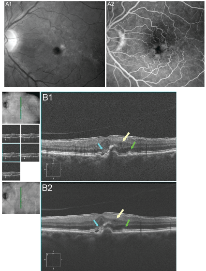
On these 3 devices the number of scans used for an image acquisition has to be determined in advance in a prospective way and one cannot decompose the final image to retrospectively analyse the effect of scans averaging. We nevertheless analysed the images acquired on 3 patients to explore whether the results obtained with the Opko® were not solely relied on this OCT device. We observed the same type of loss of details with a slight blurring with increasing number of scans very much alike the changes observed with the Opko®. On Figure 5 the blurring with withdrawing of some details characterizes the Scan 20 image compared with Scan 5 images (Figure 5). According to the small number of images no statistical analysis was performed.
.
Figure 5: 76-year-old patients with pigment clumps, material deposit and an Epiretinal Membrane (ERM). The blue channel image (A1) shows retina folds induced by the ERM while the middle phases of the angiography (A2) shows progressive staining of the material. On the OCT (Cirrus 4000, Zeiss) some details are better delineated on the Scan 5 images (B1) than on the Scan 20 image (B2): the yellow arrow points a little retina cyst that is hardly seen on Scan 20, the green arrow points a shallow elevation of RPE hardly seen on Scan 20, the blue arrow points the EZ no longer visible on Scan 20. On the contrary the nice smothering of the Scan 20 image make it easier to see the different layers of the inner retina, the ERM.
View Figure 5
Discussion
Conversely to fundus photographs and fluorescein angiograms, OCT images result of an artificial "constructed" production. The algorithms of image elaboration differ from one OCT device to another. The magnitude of scans averaging also differs between devices. The aim of this study is not to try to discuss the complex process of image construction but to draw the attention of clinicians and of clinical studies designers on the potential diagnosis drawback of an excess of scans averaging.
Averaged OCT images often display a slight blurring and a certain thickening of outer retina layers, in particular among patients with fixation instability. On the other hand, these averaged images display a better quality. In our clinical practice the choice can be somewhat tricky between a beautiful image that can include some flaws and a raw image that can be more accurate but less attractive. This study used the Opko® as a surrogate to evaluate image changes induced by OCT Scan averaging. This OCT device is not commercialized anymore and it can be labelled as a first generation SD-OCT. Nevertheless it allowed retrieving the different scans that compose the final averaged image. Though the number of scan averaged is low (20) it matches the technical characteristics of the device (acquisition speed 27,000 A-scans/s, no eye-tracking). It allowed us to have some information about averaging for current OCT devices.
On this 33 patient's series, with the Scan 1 image as a reference, we quoted 18.18% the image alteration on Scan 5, 33.33% on Scan 10 and 35% on Scan 20. These percentages were based on the presence or absence of alteration on 4 items from the choroid to the anterior profile line. Since they depend on the number of items analyzed the figures of the percentages are not of great interest. The ratio of progression of these percentages better point our issue (Figure 1). The loss of details, mainly by blurring is noticeable on Scan 5 but more obvious on Scan 10 and 20. The slow rise of the curve after Scan 10 is explained by the fact that the changes observed on Scan 10 are sustained on Scan 20. Overall scans averaging up to Scan 5 improved the quality of the final image without too much modification. On the contrary for Scan 10 and further averaging the modifications were further increased.
Some authors have recently shown limitations to scan averaging [7] but to the best of our knowledge this is the first clinical study showing image alterations induced by scan averaging. Because this study was only exploratory, we did not address the fact that image modification may influence the diagnosis.
The subgroup analysis of patients treated with antimalarials brings some further information. In group A (no sign of antimalarials intoxication) the percentages of modifications linearly increase with the number of Scan. On the contrary in group B (signs of antimalarials intoxication), the increase of the percentages is almost exponential. This could be related to a greater proportion of fixation instability. Given the small number of patients we have not sought to detail the degree of intoxication (onset or advanced). For the patients of group C (other condition), the percentages of the grading doesn't seem too critical. However the image on figure 4 displays an example of an enlargement of the size of choroidal neovascularization.
Subjective image quality is logically meant to relate to the diagnostic utility of these images. The results of this small series may raise some questions as OCT Scan averaging meant to improve the image quality may also lead to some image alteration. For more recent OCT devices, the use of an eye-tracking system as well as a high acquisition speed aims to balance the eye micro-movements. However, the few images from the Spectralis® the Cirrus® and the Avanti® that we analysed showed the same tendency of image modification when an increasing number of scans composes the final image. The speed of acquisition of the OCT devices has dramatically increased during the last 10 years, evolving from 600 scans/s on the Stratus® to 25,000 scans/s on the Cirrus® and 40,000 scans/s on the Spectralis® [10] and even more recently 100,000 to 400,000 scans/s with the Atlantis® Swept Source OCT [11]. Nonetheless, the ratio between micro saccades (30 to 50 ms) or macro saccades (≥ 150 ms) and the speed of acquisition (around 3 seconds to get 20 Scan) still put the averaging process into perspective. Interestingly the Spectralis® uses an eye tracking while the Cirrus 4000® doesn't. The speed of acquisition of the Avanti® (70,000 scans/s) brings some reduction of the blurring that goes along with averaging but this blurring is not completely avoided on the images. On this device, the improvement of acquisition speed and of the eye tracking has prompted the designers to raise the number of scans that are averaged: for a single line the default scan is 80.
In everyday practice as for clinical studies the size of the details that we analyze is closely related on the pathologic conditions. The slight blurring with scans averaging observed in our study would not be equally significant for different diseases. For diabetic macular edema, the enlargement of macular cysts may be related to a progressive loss of retinal tissue, possibly due to Müller cells degeneration. Then the evaluation of the size of cysts is logically of some importance to assess a visual prognosis before treatment. On the contrary, during the follow up the decrease of macular thickness is the main outcome to be followed. In our study, the anterior profile line as the RPE line were not much altered by scan averaging allowing a fair evaluation of retinal thickness.
Our study obviously shows a number of limitations. A greater number of patients, a greater number of OCT devices analysed would have better pointed the drawbacks of too many scans averaging. The Opko® used here can be labelled as a "first generation of SD-OCT" with its 6 scans/s acquisition speed. However, as surrogate, it was the perfect tool for this retrospective study because it allows a decomposition of the final image into its different Scans. Besides, as discussed an increase of the modification of the final image with the number of averaged scans can be shown with any OCT device. Figure 5 shows loss of some details on a Scan 20 image acquired with a Cirrus4000® compared to a Scan 5 image.
The possible consequences of image modification induced by averaging on a diagnosis were not addressed in this study mainly because it was only designed to show that image averaging can induce some image alteration.
In conclusion our study showed that an excess of scan averaging can alter the OCT-B images. This demonstration was performed using a first generation OCT that is no more available. However, for current OCT devices, as the number of scans averaged has now increased, our results suggest that acquisition speed, better co-localization of repeated OCT Scans and other technical improvements could influence the benefit/loss ratio of averaging.
References
-
Seitz J, Strotzer M, Volk M, Held P, Djavidani B, et al. (2000) Reduction of motion artifacts in magnetic resonance imaging of the neck and cervical spine by long-term averaging. Invest Radiol 35: 380-384.
-
Swindell W, Mosleh-Shirazi MA (1995) Noise reduction by frame averaging: a numerical simulation for portal imaging systems. Med Phys 22: 1405-1411.
-
Sprawls P (1992) AAPM tutorial. CT image detail and noise. Radiographics 12: 1041-1046.
-
Abadi RV, Gowen E (2004) Characteristics of saccadic intrusions. Vision Res 44: 2675-2690.
-
Pappuru RR, Briceno C, Ouyang Y, Walsh AC, Sadda SR (2012) Clinical significance of B-scan averaging with SD-OCT. Ophthalmic Surg Lasers Imaging 43: 63-68.
-
Sakamoto A, Hangai M, Yoshimura N (2008) Spectral-domain optical coherence tomography with multiple B-scan averaging for enhanced imaging of retinal diseases. Ophthalmology 115: 1071-1078.
-
Shirasawa M, Sakamoto T, Terasaki H, Yamashita T, Uchino E, et al. (2014) Objective determination of optimal number of spectral-domain optical coherence tomographic images of retina to average. PLoS One 9: e110550.
-
Stepien KE, Han DP, Schell J, Godara P, Rha J, et al. (2009) Spectral-domain optical coherence tomography and adaptive optics may detect hydroxychloroquine retinal toxicity before symptomatic vision loss. Trans Am Ophthalmol Soc 107: 28-33.
-
Rodriguez-Padilla JA, Hedges TR 3rd, Monson B, Srinivasan V, Wojtkowski M, et al. (2007) High-speed ultra-high-resolution optical coherence tomography findings in hydroxychloroquine retinopathy. Arch Ophthalmol 125: 775-780.
-
Wolf-Schnurrbusch UE, Ceklic L, Brinkmann CK, Iliev ME, Frey M, et al. (2009) Macular thickness measurements in healthy eyes using six different optical coherence tomography instruments. Invest Ophthalmol Vis Sci 50: 3432-3437.
-
Potsaid B, Baumann B, Huang D, Barry S, Cable AE, et al. (2010) Ultrahigh speed 1050nm swept source/Fourier domain OCT retinal and anterior segment imaging at 100,000 to 400,000 axial scans per second. Opt Express 18: 20029-20048.
