The piriformis syndrome is an entrapment neuropathy emerging with the compression of sciatic nerve by a muscle due to its close proximity with piriformis muscle. However its pathophysiology hasn't been understood clearly, it is thought that it arises from the oedema and inflammation in the piriformis muscle. This syndrome, which we meet with sciatica table clinically, is often likened to lumbar disc herniation (LDH). Multidisciplinary, the treatment of piriformis syndrome may include the administration of nonsteroidal anti-inflammatory drugs (NSAID), muscle relaxants, physical therapy and interventional procedures. Treatment with diagnosis of LDH was applied to each three cases we shared (intradiscal intervention for two cases and micro discectomy by microsurgery for one case). Three patients, whose pain scores hadn't been improved in the surveillance period, were evaluated in pain clinics. In comply with ultrasonography (USG) injection with 10 ml 0.25% bupivacaine and 40 mg methylprednisolone was administered to three patients who were diagnosed with piriformis through physical examination and imaging methods. Pre-treatment, post-treatment and post-treatment 1st and 3rd months VAS scores of the patients were recorded. 3 months VAS scores were found as pre-treatment (PT) VAS:6, after treatment (AT) VAS:3, AT 1st month VAS:3, AT 3rd month VAS:2 for 1st case, PT VAS:8, AT VAS:2, İS 1st month VAS:3, AT 3rd month VAS:2 for 2nd case, PT VAS:6, AT VAS:2, AT 1st month VAS:3, AT 3rd month VAS:2 for 3rd case.
Diagnosis of piriformis syndrome is often likened to LDH. Injection administration in company with USG can be used for both diagnosis and treatment. We aimed to share 3 cases who were diagnosed with LDH before, to whom we administered injection in company with USG by diagnosing piriformis syndrome with examination and monitoring findings and VAS scores of whom were decreasing.
Piriformis syndrome, Ultrasound-guided corticosteroid injection, Sciatica, Pain
Piriformis syndrome, which was defined as trapping of sciatic nerve by piriformis muscle, was defined by Yeoman [1]. While leaving pelvis from sciatic notch, sciatic nerve progresses under piriformis muscle and above obturator internus muscle. This is the area where sciatic nerve is compressed most [2]. In the piriformis syndrome, which is classified as premier and seconder, premier reasons are arisen from intrinsic pathologies (myofascial pain, myositis ossificans etc.) of the muscle. Seconder reasons are due to pathologies of sciatic notch adjacent structures [1,3,4].
Formation of the syndrome is an entrapment and neuropathy table formed as a result of compression by piriformis muscle to sciatic nerve. As a result of entrapment, it causes leg pain along sciatic nerve apart from hip pain. As a result of the entrapment of the motor fibers, weakness at foot and gait disorder can be seen. This pain is almost as ache or cramping and may cause sensorial disorder as paresthesia in the area of sciatic nerve [2].
In the diagnosis, physical examination, special tests to piriformis syndrome (Freiberg Test: hip pain due to stretching of piriformis muscle by forced internal rotation of femoral in extension, FADIR Test: occurrence of the pain due to flexion, adduction and internal rotation of hip, Pace Symptom: External rotation and abduction of hip made against resistance in sitting position causes the spasm of piriformis muscle and it creates pain and loss of strength), imaging methods as computed tomography (CT), Magnetic Resonance Imaging (MRI) and Magnetic resonance neurography (MRN) can be used [3-5]. In electroneuromyography (ENMG), lack of denervation (fibrillation and positive waves) in paraspinal muscles may suggest piriformis syndrome [2,4].
In piriformis syndrome, treatment is multidisciplinary. It includes relaxing and stretching exercises for the strain of piriformis muscle with non-steroid anti-inflammatory drugs (NSAID), muscle relaxants, drugs for neuropathic pain (gabapentin, carbamazepine etc.) and physical therapy and rehabilitation (PTR) applications as ultrasound (US) and transcutaneous electrical nerve stimulation(TENS) [2]. In cases, which don't respond to conventional treatments, injection can be done [4,6]. Local anesthetics-steroid mixture and botulinum toxin can be used as option for injection [7,8].
With LDH diagnosis, minimal invasive procedure (intradiscal radiofrequency treatment) and invasive (micro discectomy) surgical procedures were administered to 3 patients to whom we made diagnosis as piriformis syndrome through implemented physical examination and imaging methods. We aimed to share the 3 months follow-up of 3 patients to whom we administered 10 ml bupivacaine + 40 mg methylprednisolone injection at 0.25% concentration in company with USG.
36-year old female patient admitted to the pain clinic because of pain moving from hip to right leg for 2 years. She had PTR program 2 times before but she couldn't benefit. Six months before she admitted to the pain clinic, intradiscal procedure (radiofrequency thermo coagulation) had been administered to her by neurosurgery for L4-L5 disc range but reduction in pain hadn't been observed. Pain complaint of the patient, who was taking pregabalin 150 mg 2x1 for two months with neuropathic pain diagnosis, was still ongoing. There weren't any feature in the history of the patient and she wasn't describing a trauma for her hip area. In her examination, her Visual Analog Scale (VAS) score was 6. In physical examination lumbar ROMs range was painful at the end. Paravertebral spasm was present. Right leg Straight Leg Raising (SLR) test was positive in 60°. Her hip ROMs' were clear but range-end was painful. There was sciatic nerve walleix sensitivity at right. Freiberg Test, FADIR Test and Pace Test were positive. Her neurological examination was normal. Lumbar MRI as imaging method of the patient: there was bulging which doesn't create L4-L5 nerve root compression. Right piriformis muscle hypertrophy was detected in the pelvic MRI (Figure 1).
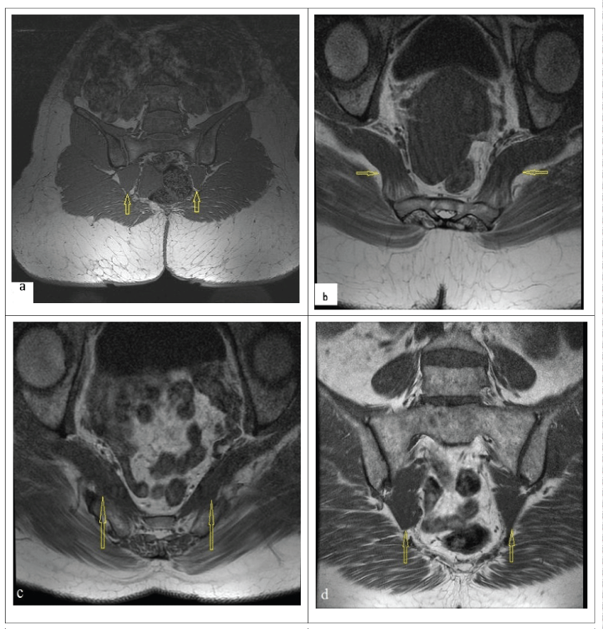 Figure 1: Imaging findings. a) Coronal T1 Pelvic MRI right piriformis muscle hypertrophy (case 1); b) Normal pelvic MRI (case 2); c) Axial T1 pelvic MRI, piriformis muscle hypertrophy/irregularity (case 3); d) Coronal T1 pelvis MRI (case 3);
Yellow arrows: piriformis muscle.
View Figure 1
Figure 1: Imaging findings. a) Coronal T1 Pelvic MRI right piriformis muscle hypertrophy (case 1); b) Normal pelvic MRI (case 2); c) Axial T1 pelvic MRI, piriformis muscle hypertrophy/irregularity (case 3); d) Coronal T1 pelvis MRI (case 3);
Yellow arrows: piriformis muscle.
View Figure 1
65-year old female patient admitted to our clinic because low back pain and left leg pain for 10 years. She had PTR program 7 months ago but she couldn't benefit. Then she had intradiscal procedure (radiofrequency thermo coagulation) by neurosurgery for L4-L5 disc range but reduction in pain hadn't been observed. There wasn't left hip trauma in the history of the patient. In her examination, her VAS score was 8. There was sensitivity at left gluteal region with palpation. Lumbar extension and flexion were clear but painful at the end of the range. There was L4-L5 paravertebral spasms. Left Straight Leg Raising (SLR) test was positive in 70°. Freiberg Test, FADIR Test and Pace Test were positive. Lumbar MRI as imaging method of the patient: protrusion and facet joint hypertrophy which don't create nerve root compression at L4-L5 and L5-S1 level. Pelvic MRI was normal (Figure 1).
54-year old female patient admitted to our clinic because of pain moving from right hip to down for 1, 5 years. She had PTR program 2 times with LDH diagnosis but she couldn't benefit. Three months before she admitted to the pain clinic, surgical intervention (laminectomy micro discectomy) had been administered to her by neurosurgery for L5-S1 disc range but reduction in pain hadn't been observed. There wasn't hip trauma in the history of the patient. In her examination, her VAS score was 8. All lumbar ROMs' were limited at the end of the range. There were paravertebral spasms. Right Straight Leg Raising (SLR) test was positive in 400. There was sensitivity at right gluteal region with palpation. Freiberg Test, FADIR Test and Pace Test were positive. Lumbar MRI of the patient: there was bulging which doesn't create L4-L5 nerve root compression and laminectomy defect and soft tissue increment, facet joint hypertrophy at L5-S1 were detected. In pelvic MRI, piriformis muscle hypertrophy and irregularity were detected (Figure 1).
Injection was recommended to patients with piriformis syndrome. By taking their approvals, interventional procedures were administered.
In operating room environment, position, in which upper hip was at flexion from the knee at lateral decubitus, position was given to each three patients. After proper area cleaning, sciatic nerve and piriformis muscle were monitored through 2-8 MHz convex USG probe after 5 cm caudal was signed from the midpoint of line connecting trochanter major with posterior superior iliac crest. Intervention was made by 100 mm, 22 gauge USG visible block needle (Stimuplex, B: Braun Melgusen AG; Germany). When appropriate motor response (contradictions in hamstring, calf, foot and thumb) was taken at 0.2-0.3 mA, 10 ml 0.25% bupivacaine and 40 mg methylprednisolone mixture was injected intramuscularly and around the sciatic nerve. Any neurologic deficit and complication hadn't been observed during the follow-up of patients.
Values of patients, who were followed by pre-treatment (PT), after treatment (AT), AT 1st and 3rd month VAS scores, are presented in table 1.
Table 1: VAS scores. View Table 1
Prevalence of piriformis syndrome in literature varies from 0.5% to 17%. Most of these patients administer to clinic because of chronic low back and leg pain [4,6,9]. A definitive diagnosis can be made by physical examination with the exclusion of other pathologies. Imaging methods as MR can detect the pathological changes in piriformis muscle [3,4]. In addition, electroneuromyography (ENMG) can be used for the evaluation of diagnosis and response to treatment. In EMG, during FADIR test, patients with extension piriformis syndrome in H reflex can be detected [10].
26 of 3550 patients examined by Jawish et al. were diagnosed with piriformis syndrome and Dorsolumbar MRI was used in all of them as a method adjunct to diagnosis. While sacral MRI was needed for 7 patients, in 7 of 13 patients, H reflex was detected in ENMG. However, they stated that anatomical variations may give inaccurate results in ENMG [10].
In all 3 patients, Freiberg, FADIR and Pace Tests were positive. Lumbar and pelvic MRI was used as imaging method. In each 3 cases, there were bulging not forming nerve compression and protrusions in lumbar MRI. In pelvic MRI, there was hypertrophic piriformis muscle disorder in case 2 and there was disorder in piriformis muscle in cases 1 and 3.
There isn't golden standard method among current diagnostic methods. Due to fact that there wasn't golden standard for diagnosis, Jankovic et al. shared the opinion as physical examination and tests were main coadjutors in diagnosis [9].
In piriformis syndrome treatment, there are treatment options as PTR methods, medical treatment, interventional procedures and surgical treatments [7,11,12]. Success rate is at 79% level with conservative treatment [13].
Injections used as interventional procedure can be applied intramuscularly or to sciatic nerve. Often steroid and local anesthetic mixture or botulinum toxin is used [8,12,13]. Injections can be applied in company with MRI, Fluoroscopy, CT and USG [6,14]. In two different studies aiming to compare reliability of USG use with other methods; Fowler et al. used USG and fluoroscopy for injection with 10 ml 80 mg triamcinolone, 1% lidocaine mixture. After both of these 2 applications, statistically significant difference wasn't detected during the 3 month follow-up [6]. In other study, through MRI, they confirmed the success of injection administered to piriformis muscle in company with USG. It was detected that a successful intervention was applied in 9 of 10 patients to whom injection was administered [14]. We benefited from USG and peripheral nerve stimulator while we were applying injection to these three patients. In this way, we aimed to reduce the possibility of sciatic nerve damage and increase the success of the operation.
Medication selection seems disputable for piriformis syndrome. Although there are popular approaches as local anesthesia and local anesthetic steroid mix, BTX use increases day by day [9,12,13]. Due to fact that similar results were obtained in studies carried out by Fishman and Lanb despite BTX was used at two different doses, it is though that further dose studies are needed for BTX-B use [12,13]. Therefore, we preferred to use local anesthesia and steroid mixture for injection. After injection with 10 ml 0.25% bupivacaine and 40 mg methylprednisolone in similar amounts with literature, we observed reduction in 3 months VAS scores and improvement in symptoms of these three patients. We think that injection use in company with USG is more useful and convenient for diagnosis and treatment in comparison with other methods.
However it is not required to think piriformis syndrome in each patient coming with sciatica table, they are often treated with LDH diagnosis as seen in 3 cases submitted by us. However piriformis syndrome is kept in mind in patients who have examination inconsistency and clinic findings, it is possible to reach correct diagnosis with detailed physical examination and diagnostic injection.