A 74-year-old with burnt out autoimmune hepatitis, portal hypertension with thrombocytopenia, oesophageal varices and previous hepatic encephalopathy has presented with a recurrence of a left sided pleural effusion. She was very breathless at rest and therapeutic aspiration of 1.1 litres of straw coloured fluid has good symptomatic benefit. However, this is her sixth presentation in as many months and on top of four previous therapeutic aspirations, she has had one 12 French Seldinger drain inserted for fluid removal accompanied by an attempt at medical pleurodesis using 4 g of talc.
She is very frail and has a low body mass index (18.6) and doses of diuretics beyond 80 mg of furosemide and 25 mg of spironolactone cause systemic hypotension with dizziness on standing and renal dysfunction. Even removing more than 1 L from her pleural space causes hemodynamic compromise and needs concomitant albumin replacement.
The pleural fluid has always been a transudate with an average fluid protein of 9 g/l. The ascitic fluid is also a transudate with a fluid protein of 7 g/l. The patient thus has a Hepatic Hydrothorax (HH), which is a transudative pleural effusion which occurs in patients with liver cirrhosis in the absence of cardiac or pleural disease [1]. Repeated cytological examinations of both ascitic and pleural fluid have been negative. She has had a normal echocardiogram and has never smoked. She has had previous hypo-albuminaemia but this is now normal. A Magnetic Resonance (MR) scan of her liver has confirmed extensive cirrhosis, large amount of ascites and no hepatocellular carcinoma.
This patient is well known to the gastroenterological services. She has had autoimmune hepatitis for many years now and has become very frail recently with development of ascites and significant weight loss. She has been admitted on a few occasions with worsening confusion and ammonia levels have been raised suggesting hepatic encephalopathy. No obvious cause was found for this and she was treated conservatively. She has also had repeated abdominal paracenteses to try control her ascites.
There are many reasons postulated as to why HHs develop; namely leakage of ascites via diaphragmatic defects, hypo-albuminaemia, and lymphatic leakage from thoracic ducts. Portal hypertension and splanchic vasodilatation also play an important role. HHs are also predominantly right sided, as the diaphragmatic defects tend to be there, but 17.5% of effusions on the left and 3% bilateral [2]. Negative intrathoracic pressure and piston-like effect of the diaphragm cause unidirectionalmovement of fluid into the pleural cavity.
Treatment options beyond drainage and diuretics are limited. The patient has previously been declined for a liver transplant on grounds of frailty and is not fit for a Transjugular Intrahepatic Port Systemic Shunt (TIPS) given her previous encephalopathy. The latter procedure enables the creation of an intra-hepatic artificial channel between the inflow portal vein and the outflow hepatic vein but carries up to 25% risk of encephalopathy and has a very important application as a bridge to transplant which is not applicable here.
She is on a salt restricted diet and fluid restriction, but up to a quarter of hepatic hydro thoraces are resistant to such therapies [1].
Her symptoms have been deemed terminal on a few occasions but her survivability has impressed us all. The patient has agreed to have a permanent Do Not Attempt Resuscitation decision in her notes and for her not to be treated in level 2 or 3 care.
A medical pleurodesis has been attempted before when her hemi thorax was drained to dryness. There was no trapped lung but the pleurodesis was unsuccessful. This is not surprising as the rapid rate of fluid accumulation would have prevented pleural apposition and small amounts of fluid would not have been visible on a chest x-ray. Various studies have had pleurodesis success rates from 47% to 100% but they have not been adequately powered randomized controlled trials [2]. A second opinion was sought as to how to control this fluid.
HHs is usually amenable to diuretic treatment. She is frail, has mild thrombocytopenia and her clotting is deranged with a varying prothrombin time between 16 and 22 seconds. Various options were discussed with her and her family and are listed below:
1. Repeated therapeutic aspirations - These carry a risk of infection, of bleeding and pneumothorax [3] which would not be inconsequential in her. They also require her to come to clinic fortnightly to monthly. This was not recommended.
2. Gastroenterological options - They seemed to have been exhausted. There are case reports of octreotide and terlipressin use in HH [2] but they are not used routinely. Nor are vesico-peritoneal shunts. Repeated paracenteses have not helped.
3. Pleurodesis - Talc medical slurry has been tried. There are studies of video-assisted thoracoscopy or medical thoracoscopy with or without repair of diaphragmatic defects that have quotes success rates between 35%-91% [2]. There are significant problems with any of these approaches in terms of morality, some quoted as high as 50%, as well as bleeding risks and this do not recommend them.
4. An Indwelling Pleural Catheter (IPC) - IPCs have an established role in the management of malignant pleural effusions and their use in non-malignant effusions is rising. A recent review of IPCs such a setting revealed the commonest indications to be HH. Rates of pleurodesis in those patients were less, as expected and infection rates were just under 4%. We provide a safe and streamlined pleural service in our trust and think that IPCs provide an excellent patient centered approach to management of symptoms due to effusions [4].
Thus, we opted for option 4 and a Rocket IPC was inserted under fresh frozen plasma cover (Figure 1). There were no complications. 6 months later, the patient has been managing well at home and 500 mls are removed two to three times a week depending on symptoms. However, recently, increasing ascites has required paracenteses and the district nurses wondered whether it was possible to than 500 mls of fluid at every visit. One of the limitations of the Rocket vacuum bottle is that they come only in 500 mls. There are 2 companies providing such bottles and the other one, PleurX enables drainage of 1 L of fluid at a time. However, the 2 systems are not inter-changeable. A viable option was to connect a Rocket ascitic drainage bag to the IPC and allow more fluid to be drained, but one needs to be very careful of inducing significant fluid shifts and having albumin replaced at the same time pleural fluid is removed [5].
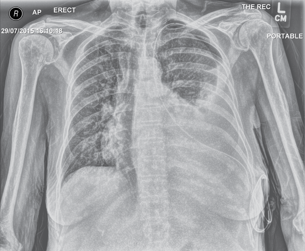 Figure 1: Showing IPC inserted and left effusion. View Figure 1
However, before the above could be actioned, the patient was admitted with a suspected abscess at the IPC site. There was a small red lump just next to the distal IPC insertion site. An ultrasound showed a small flocculated abscess, which was not amenable to drainage according to the radiologists. A swab of the site was negative and the pleural fluid seems was still a transudate and straw coloured. We were asked by the admitting team whether the IPC should be removed.
However, we knew that this is not an infected pleural space. The options for this patient were still limited and the IPC has allowed her to be out of hospital and for her symptoms to be controlled. We suggested that a 2 week course of antibiotics should help and indeed it did, enabling complete resolution of the abscess. Unfortunately for the patient, she has become frailer, has developed hypersplenism and has required transfusions. After a prolonged admission, she was discharged with palliative care input with still ongoing drainage every few days. Another 6 months later, the patient presented to clinic again and admitted that there has been no fluid drained from her IPC for the last 4 weeks. A chest radiograph confirmed a clear pleural space and it was our conclusion that she had auto-pleurodesed. This is described in the literature [2].
The above case was a difficult case that spanned many months. It required a lot of discussions with the various specialities concerned as well as with the patient to make sure she was on board with what we proposed to do to her.
We hope the foregoing provides a clear account. Reviewing each management option in detail is beyond the remit of this article. Hence the main learning points are of
1. The definition and the pathophysiology of HHs.
2. An introduction to the various management options in HHs.