High-resolution computed tomography (HRCT) is an essential technique for the diagnosis and follow-up of idiopathic pulmonary fibrosis (IPF), but it is not routinely used to evaluate severity. A semi-quantitative HRCT score was developed to determine its relation with physiologic variables, to assess severity and fibrosis progression, and to establish prognostic factors.
The HRCTs of 48 newly diagnosed IPF patients were assessed. Reticular pattern, ground-glass opacities, distortion and honeycomb patterns were scored by two independent radiologists to obtain these scores and the Total Score of Fibrosis. Its relation with pulmonary function tests, the 6-minute walking test (6MWT) and air blood gases was evaluated, and with the survival and identification of the worse prognosis factors. The lower HRCT level (everything below the highest point of the right diaphragm) was independently assessed to evaluate differences.
The Honeycomb Score (HS) correlated with DLCO% (r = -0.50, p < 0.001) and KCO% (r = -0.55, p < 0.001). The Total Score (TS) correlated with DLCO% (r = -0.55, p < 0.001) and KCO% (r = -0.50, p < 0.001). Final SatO2 in the 6MWT correlated with the TS (r = -0.45, p < 0.01), and desaturation correlated with both HS (r = -0.34, p < 0.05) and TS (r = -0.45, p < 0.01). Survival was 42 (37-48) months. DLCO < 40 (p = 0.005) and the TS of Fibrosis ≥ 20 (p = 0.03) were independent predictors of mortality. In the lower HRCT level assessment HS correlated with DLCO (r = -0.533; p < 0.001), KCO (r = -0.426; p < 0.01) and CPI (r = 0.505; p < 0.001), and also the TS correlated with both DLCO (r = -0.362; p < 0.01) and KCO (r = -0.481; p < 0.001); the HS was an independent mortality factor (p = 0.015) at this level.
A semi-quantitative HRCT score is useful for determining severity upon diagnosis and for predicting IPF progression, and it correlates well with physiologic variables. DLCO < 40 and the TS of Fibrosis ≥ 20 were independent mortality predictors. The honeycomb score at lower HRCT levels determined mortality.
HRCT, Semi-quantitative score, IPF, Severity, Progression, Mortality
Idiopathic Pulmonary Fibrosis (IPF) is a chronic progressive disease of unknown etiology, fatal prognosis and has no effective treatment, save lung transplant, accessible to very few patients, or new antifibrotic drugs, e.g., pirfenidone and nintedanib, which are used in a small group of patients defined functionally as being mild-to-moderate affected [1-4]. To date, diagnostic criteria are clearly established and high-resolution computed tomography (HRCT) plays a first-order role [1,2]. Functional criteria are very well-defined to evaluate the progression and stability of, or improvement in, the disease. The HRCT and pulmonary function tests (PFT) are essential tools to diagnose, monitor and make a prognosis of IPF. However, HRCT reports are usually limited to the description and location of radiological patterns and provide no information on the (quantified) extent of the described findings. Some studies have shown a correlation between PFT and the IPF extent on HRCT findings by identifying poor prognosis and early mortality factors with both [5,6].
Our hypothesis was that quantifying the extent of the HRCT findings of IPF would not only be important at the time a diagnosis was made but would also be essential to establish initial IPF severity, and to even monitor its progression. We aimed to evaluate the usefulness of a Semi-quantitative Score in the HRCT extent of IPF [7], which was redesigned to suit our workplace, and to establish the possible relation to lung function parameters, the 6-minute walking test (6MWT), arterial blood gases (ABG) and to bronchoalveolar lavage (BAL), all performed in a cohort of patients upon diagnosis. The secondary objectives were to identify poor prognosis factors in the radiological score findings, to assess whether quantifying IPF extension would be useful as a disease progression measurement, and to determine if a simplified scoring of the lower HRCT level would be enough to evaluate disease progression.
Consecutive IPF patients were selected according to the established clinical and radiological (usual interstitial pneumonia -UIP-) criteria [1,2]. Those patients who refused to participate, had severe comorbidity or could not perform all the required tests were excluded. The study was approved and authorized by the Ethics and Clinical Trials Committee at the Dr. Peset University Hospital of Valencia (Spain), and an informed consent was obtained.
A prospective study was performed for 5 consecutive years (2006-2010) and included newly diagnosed IPF patients. An analysis of the obtained findings was done at the time of the diagnosis. HRCT and PFT were done within 1 month.
Patients underwent spirometry with a bronchodilator test, static lung volumes and the single breath carbon monoxide transfer factor, 6 MWT and ABG on room air, which were always performed by the same respiratory nurse and followed proper guideline recommendations [8-11].
All the functional tests were done at the PFT Laboratory: spirometry and DLCO with V-MAX equipment Spectra 22®, static volumes with 20® Encore V.Max team in an Autobox cabin VS62J®; an ABG analysis was run with patients at rest for 10 minutes before puncture, analyzed in ABL 520 Radiometer equipment. A 6 MWT was performed in a flat 30-meter-long corridor.
All the flexible bronchoscopies (Pentax bronchoscope® EB-1570K 2.0) were performed by the same pulmonologist, who had over 10 years' experience. BAL was obtained at the middle lobe or lingula, and at least four transbronchial biopsies (TBB) were taken, preferably at lower lobes, in previously located areas in the HRCT with less honeycombing [12].
A General Electric Light Speed VCT 64-detector was used. Images were obtained with the patient lying in supine position and after maximal inspiration with collimation (1.25 mm) at 10 mm intervals, with 120 kV and 335 mAs. The reconstruction algorithm was the high spatial resolution (bone). The windows to display lung parenchyma were 1500 (width) and amplitude level (level) -600 HU [13]. The characteristics for each identifiable radiological pattern in IPF [14] have been previously defined to improve not only identifications by each radiologist, but to also improve the agreement among evaluations [15]:
a) Honeycombing: Cystic air spaces, from several millimeters to 1 cm diameter, with thick clearly defined walls, located mainly in subpleural areas.
b) Ground-glass: Increased lung attenuation (as mist) that allows to view the underlying vessels through it.
c) Reticular: Irregular intralobular crisscrossed lines, randomly arranged and separated by a few millimeters.
d) Distorted lung architecture: Abnormal displacement of bronchi, pulmonary vessels, interlobar fissures and interlobular septa.
HRCT was read by two independent core radiologists according to the following protocol: three levels were selected from each HRCT (Figure 1) to evaluate six different areas (three from the right lung and three from the left lung).
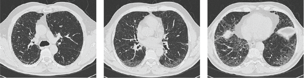 Figure 1: The different HRCT levels.
View Figure 1
Figure 1: The different HRCT levels.
View Figure 1
• Upper: Everything above the level of the carina;
• Lower: Everything below the highest point of the right diaphragm;
• Middle: Between the previous two, right at the midpoint.
Each level was divided into each lung by drawing a horizontal line and leaving a measurable area of 50%; a second line, which ran perpendicularly to the horizontal one, left a 25% assessable area of each lung. Each 25% was subdivided into five portions, which corresponded to an area of 5% each. In each slice and each lung, the presence or absence of previously described radiographic patterns was identified by assigning the following scores: 0 (none), 1 (≤ 10% affectation), 2 (11-20%), 3 (21-30%), 4 (31-40%), 5 (41-50%) and 6 (> 50%) (Figure 2). The final sum of the scores for each pattern in each HRCT slice defined it as the Pattern Score. Thus, the pattern with the highest score was defined as the predominant pattern. The sum of all the HRCT scores taken from the same patient was known as the Total Score (TS). The amount of emphysema in these patients was also taken into account by using the theoretical calculation of the "Composite Physiologic Index" (CPI), which results from the following equation: CPI = 91.0 - (0.65 × %DLCO) - (0.53 × % FVC) + (0.34 × % FEV1) [16]. CPI correlates well with disease extent in HRCT and eliminates the influence of emphysema on PFT, which is commonly found in current smokers, and occasionally in pulmonary fibrosis [1,2,17-19].
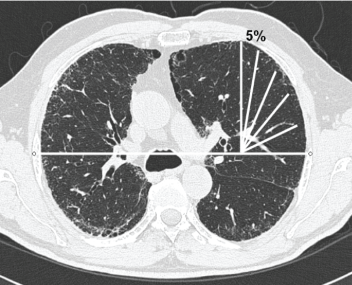 Figure 2: Quantification of the HRCT slices.
View Figure 2
Figure 2: Quantification of the HRCT slices.
View Figure 2
Data were tabulated and analyzed in a database designed for this purpose, which is found in the PASW Statistics 18 statistical program. After checking the normality of the variables, they were compared using an ANOVA or a quantitative test unpaired t-test according to the purpose of the analysis. Qualitative variables were studied with a Chi-square test. Non-parametric tests were used when variables did not follow a normal distribution, in which case data were expressed as the median and interquartile range. The relationship between HRCT scores and functional parameters was analyzed by Spearman's or Pearson's correlation test according to the distribution and type of variables included in this analysis. A Kaplan-Meier survival test and a COX proportional hazards regression analysis were performed to determine bad prognosis factors. The intraclass correlation coefficient was used to evaluate the concordance score between two core radiologist readers. The level of significance for all the statistical analyses was p < 0.05.
A separate analysis of the lower HRCT level was done within the same statistical tests.
Seventy-three IPF patients were initially studied and 48 were finally included in the study. The main reasons for exclusion were patients refused to participate, inability to perform any test (HRCT, bronchoscopy) or failure to meet the deadlines set out in the study design.
Demographics: The mean age was 71.6 ± 8 years, 68.7% of the participants were male; 66.7% were current or former smokers. The Charlson index was 2.77 (1.8-3.8). To date, 17 patients have died (35.4%; Table 1). The excluded patients had a mean age of 75.8 ± 6.8 years (46.7% males), eight of whom have died (32%). No significant differences were found for the study population (p > 0.05).
Table 1: Demographic characteristics of the study population. View Table 1
Bronchoscopy with TBB was performed in 21 cases (58.3%), whose results were consistent with usual interstitial pneumonia (UIP) in two cases. Surgical lung biopsy was performed in six patients (12.5%), all with typical UIP features.
Patients had mild restriction, FVC 87.5% (81-93) and FEV1/FVC% 79.7% (77-81), with a moderate drop in DLCO, 55.1% (49-60). The 6MWT had an average distance, 395 m (347-442), an initial SatO2 of 94.3% (93-95) and a final SatO2 of 87.2% (84-89), with an average desaturation (initial SatO2 minus final SatO2) of 7.2%. The average AaPO2 was 29.7 mmHg (26-32); 68.2% of the patients had hypoxemia (PaO2: 72.1 mmHg (66-77) and 12.5% displayed respiratory failure at the time of diagnosis (PaO2 = 49.8 ± 8 mmHg) (Table 2).
Table 2: Characteristics of PFT, 6MWT, ABG and BAL. View Table 2
The reticular pattern was predominant in 85.4% of the patients, with a honeycombing pattern in 14.6%. The intraclass correlation index among the radiologists who read the Honeycombing Score was 0.92 and the Total Score (TS) of fibrosis was 0.78.
A significant correlation was found between the HS and %DLCO (r = -0.50, p < 0.001), %KCO (r = -0.55, p < 0.001), and CPI (r = 0.47, p < 0.001), and also between the TS and %DLCO (r = -0.55 , p < 0.001), %KCO (r = -0.50, p < 0.001) and CPI (r = 0.51, p < 0.001) (Figure 3).
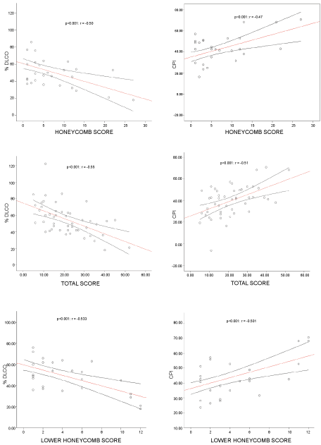 Figure 3: Correlations between the PFT and HRCT Scores.
View Figure 3
Figure 3: Correlations between the PFT and HRCT Scores.
View Figure 3
In 6MWT, a significant correlation was observed between the HS and desaturation (r = -0.34, p = 0.04), and also between the TS and final SaO2 (r = 0.45, p < 0.01). There was not any relation between the scores and air blood gases or with BAL cellularity.
Seventeen patients (35.4%) died during the study period (survival of 30 ± 4 months), seven of whom (53.8%) exhibited a reticular predominant pattern in HRCT and six (46.2%) displayed a honeycombing pattern. These patients displayed greater functional impairment at the time of diagnosis and obtained a higher HS, TS and CPI. A tendency for BAL neutrophilia among these patients was also found (Table 3 and Table 4).
Table 3: PFT differences between living and deceased. View Table 3
Table 4: HRCT and BAL differences between living and deceased. View Table 4
Global survival was 3.5 years (3.1-4). The patients with DLCO < 40 had a worse prognosis (121 vs. 201 days; p = 0.003) and were also those with a TS ≥ 20 (155 vs. 207; p = 0.012) (Figure 4 and Figure 5). The bad prognosis factors after the COX regression analysis were DLCO < 40 (Expβ = 3.985; 95CI: 1.503-10.561; p = 0.005) and a TS ≥ 20 (Expβ = 3.352; 95CI: 1.128-9.963; p = 0.03) (Table 5 and Table 6).
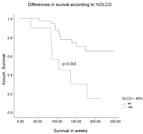 Figure 4: Kaplan-Meier survival curves depending on DLCO%.
View Figure 4
Figure 4: Kaplan-Meier survival curves depending on DLCO%.
View Figure 4
 Figure 5: Kaplan-Meier survival curves depending on the Total Score.
View Figure 5
Figure 5: Kaplan-Meier survival curves depending on the Total Score.
View Figure 5
Table 5: COX regression univariate analysis. View Table 5
Table 6: COX regression multivariate analysis. View Table 6
A good correlation was found between the HS and DLCO (r = -0.533; p < 0.001), KCO (r = -0.426; p < 0.01) and CPI (r = 0.505; p < 0.001), and also between the TS with both DLCO (r = -0.362; p < 0.01) and KCO (r = -0.481; p < 0.001) (Table 7). In the COX regression univariate analysis, the HS was a bad prognosis factor (Expβ = 1,149; 95 CI: 1.027-1.285; p = 0.015) (Table 8).
Table 7: Pearson correlation between the HRCT scores and PFT at the lower HRCT level. View Table 7
Table 8: COX regression univariate analysis with the lower HRCT level. View Table 8
This study aimed to evaluate IPF extent by a semi-quantitative HRCT score in IPF patients by using it to determine severity and its possible role in prognosis based on its correlation with PFT, which have been the only parameters considered to date to determine the stability, improvement or progression of IPF [2].
The agreement between our radiologist HRCT readers was good. Subjective reading is always a matter of discussion, especially in centers where radiologists are not experienced enough given the low prevalence of ILD. However, the pre agreement between radiologists to define not only HRCT disorders, but also scoring values, significantly improves their reading agreement, which makes it possible to implement it at any care level [6].
Similarly to other published studies [5,6,20-23], our results indicated that a semi-quantitative score was able to establish severity at diagnosis, especially due to the fact that the evaluated extent of IPF correlated well with DLCO, which has been considered to be the main parameter to date, together with FVC and AaPO2, in order to monitor disease severity and progression [24,25].
The good correlation found between the HS and DLCO has already been reported in other series [23,25,26], but neither its extension nor severity of patients has yet been quantified in the same way as PFT are currently used. What most concerns clinicians when evaluating patients with IPF is not only radiological changes, but also having better detailed information to allow them to obtain the extent and severity of pulmonary fibrosis. Xaubet, et al. [5] have shown the usefulness of a semi-quantitative HRCT score at the time of IPF diagnosis, how easy it is to implement and a good correlation with the functional results. However, these authors only defined two patterns: Ground-glass, which is barely relevant itself in IPF, and Honeycombing. We observed the same problems in other studies, which were limited to certain HRCT patterns and did not evaluate the real extent [26,27], or read too many different disorders, which could make both reading and scoring extremely difficult [28,29]. Conversely in our study, the sum of the four radiographic patterns found in IPF, and resulted in the TS, provided more complete information about the real extent of the disease, especially when it correlated with those parameters that best reflect IPF severity.
We also defend that our results provided more information as we considered emphysema a confounding factor by calculating the CPI, which also showed a good correlation with the TS. This parameter, which confers disease extent and severity by relating functional and morphological data, is valid to determine IPF severity, and has even been associated with mortality [14], as in our series.
Best, et al. [30] evaluated the utility of HRCT by quantitative scores using computerized parameters (skewness, kurtosis and mean lung attenuation) as a result of analyzing the curve shape that derives from the frequency histogram on HRCT cuts [31]. However, these repeated measurements over a 12-month follow-up of patients with IPF did not add any predictive ability to the visual score, which itself proved a strong independent predictor of mortality [24]. Computer-aided diagnosis (CAD), as a new system to quantify interstitial disorders, can be better than visual scoring in the near future, but as the radiologist has to determine the normal and abnormal structures, it will not mean less work [32]. Multidectector CT scans can also be more accurate than HRCT [33,34], but radiation exposure is higher, which is possibly a non-desired secondary effect that can be easily avoided [35].
As the average survival was 3.5 years, which is similar to the published IPF average, we noticed that having a TS of fibrosis over the average was the cut-off to independently determine mortality. These results were good enough to confirm that obtaining fibrosis extension was possible by this HRCT Score, and it could establish not only the diagnosis of IPF, but also its prognosis. In another series [36-40], a worse prognosis at diagnosis was made in patients with DLCO levels below 40% and also with higher HRCT scores of fibrosis. However, the main difference in our study came from the scoring data, as getting a cut-off score in the TS that equaled or exceed 20 points was the worse prognosis cut-off.
Finally, we analyzed the results of the lower HRCT level by assuming that the more disorders at the bottom, the more UIP we get. We observed a very good correlation between PFT, especially DLCO, KCO and CPI, with the HS and the TS. A clear relation was also noted between the HS and the worse prognosis and less survival in the univariate analysis, which was not confirmed in the multivariate analysis, probably due to the small number of patients recruited in the cohort. These data are clearly a new pathway to be studied in the future because of the vast amount of information obtained only at one reading level, including severity and prognosis, which could be a better way to simplify prognosis research into IPF.
Loss of a significant number of patients during recruitment was a limiting factor to obtain more conclusive results as many functional parameters showed a clear trend to statistical significance without actually achieving it. The few surgical biopsies performed could be another bias to the certainty of diagnosing IPF. However, all the patients who did not undergo surgical biopsy fulfilled the radiological criteria accepted for IPF diagnosis [2], with sensitivity above 90% when the HRCT showed a characteristic UIP pattern [41]. We consider that this should not significantly bias our results. We also consider not making a comparison with other prognosis factors, such as the biomarkers or other indices and scales, to be another limitation.
In conclusion, a semi-quantitative HRCT score is very useful for evaluating the extent and initial severity of IPF. In addition, it may be the only assessable evidence in advanced disease stages when the main functional restrictions already come into play. Having more amount of honeycombing at lower HRCT levels of lungs could be related with poorer survival. In any case, more studies with larger numbers of cases and long-term monitoring are necessary to ensure a reliable way to quantify the usefulness of extending IPF in the prognostic evaluation and evolution of these patients.
This study had no funding.
Dr. Ricardo Peris Sánchez has no conflict of interest. Dr. Estrella Fernández-Fabrellas, reports receiving advisory board fees from Roche, Boehringer Ingelheim. Dr. Gustavo Juan Samper reports receiving advisory board fees from Actelion, Bayer. Dr. María Luisa Domingo Montañana has no conflict of interest. Dr. Lidia Navarro Vilar has no conflicto of interest.
Number 9/09 Ethics and Clinical Trials Committee at the Dr. Peset University Hospital of Valencia), February the 2nd 2009.