Renal artery dissections (RADs) are lesions that disrupt vessels that primarily occur in patients with a known history of hypertension and caused by stenosis or enlargement of the renal artery typically due to underlying connective tissue disorders. However, RADs may occur spontaneously from trauma and no previous history of hypertension. Here, we report a rare case of bilateral isolated spontaneous RADs that characteristically occurs in healthy males. A 52-year-old male presented with left lower quadrant abdominal pain and renal insufficiency. Two years prior, he had experienced a similar episode of pain on the contralateral side, which was due to an infarct of the right kidney. On this admission, a computed tomography angiogram confirmed a new infarct of the left kidney, with dissection of a branch of the renal artery supplying the upper lobe. Work-up for cardiovascular, hematologic, radiographic or connective tissue causes was negative. We postulate that both RADs were potentially associated with the rapid twisting and turning of the abdominal area on a daily basis required for his occupation as an air traffic controller. The patient was treated with a renin angiotensin system inhibitor. After one year, both RADs had significantly improved and his renal function increased by ~23%. Isolated RAD may be associated with consistent or long-term activities that require rapid twisting and turning of the abdominal area. If left untreated, this may result in malignant hypertension, bilateral dissections, renal ischemia. To avoid misdiagnosis; we provide a comprehensive review of the typical presentation and necessary assessment and management.
Renal artery, Hypertension, Dissection, Connective tissue disorder, Spontaneous, Flank pain, Abdominal pain
In general, arterial dissections occur in small, medium and large-sized vessels. Dissections of the renal artery may be difficult to diagnose and manage largely due to the nature of its non-specific symptoms, moderate imaging sensitivity and low incidence. Typically, a patient will present with severe pain in a unilateral upper abdomen with radiation to the epigastric region, flank pain, acute or uncontrollable hypertension, and gross hematuria. Due to the similarity of the pain to renal colic, it is often assumed that the diagnosis is kidney stones. Some patients are assumed to have appendicitis, and undergo unnecessary appendectomy. In particular, isolated spontaneous dissections of the renal artery are rare and require an extensive work-up to establish the diagnosis. This review describes the salient features of the clinical presentation that should assist with the differential diagnosis and medical and surgical management of renal artery dissection (RAD).
A 52-year-old male presented to the emergency department with the sudden onset of sharp, left lower quadrant abdominal and flank pain while at work. In the emergency department, he was noted to be afebrile with a pulse of 58 beats per minute, an elevated blood pressure of 141/84 mmHg, and an otherwise unremarkable physical examination. Laboratory data revealed blood urea nitrogen of 14 mg/dl, creatinine of 1.4 mg/dl and normal urinalysis. There was no prior history of hypertension or clotting disorders, but he described his occupation as an air traffic controller as being very strenuous, requiring rapid twisting and turning of his abdominal area on a daily basis.
His medical history was significant for the parallel quality of abdominal pain that occurred two years previously on the right side. At that time, a non-contrast computed tomogram (CT) suggested acute appendicitis for which he underwent an appendectomy. However, the pathology report revealed a normal appendix. After two months of persistent pain, he obtained a second evaluation at a tertiary academic center, which showed a wedge-shaped infarct of the right kidney on contrast CT. In addition, a renal angiogram demonstrated multiple infarcts of the right kidney without evidence of a definite vascular etiology, and a normal left renal kidney. Eventually, his pain gradually subsided.
In order to diagnose the recent presentation of left-sided abdominal pain, a contrast-enhanced CT angiogram was performed, which confirmed a new left kidney infarct with a focal area of decreased perfusion at the lateral aspect. The left artery to the upper pole showed a focal area of narrowing near the hilum with distal dilation and possible dissection within it. The right kidney showed a small focal area of scarring in the mid pole and a focal area of cortical loss. These findings were consistent with a dissection in the renal artery branch supplying the upper lobe of the left kidney (Figure 1).
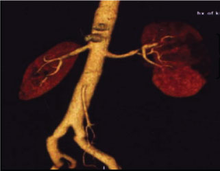 Figure 1: Spiral CT angiogram showing area of infarct in the left kidney. View Figure 1
In order to identify the underlying etiology of the RAD, the patient subsequently underwent a chest CT angiogram, electrocardiogram and echocardiogram, all of which were normal. In addition, evaluation for thrombotic diathesis was negative; review of radiological films uncovered no evidence of fibromuscular dysplasia, atherosclerosis or vasculitis and skin biopsy was unremarkable. The final diagnosis was bilateral isolated spontaneous RAD and he was started on an angiotensin converting enzyme inhibitor for persistently elevated blood pressure. However, no surgical intervention was deemed necessary and anticoagulation was not started. An iothalamate renal clearance showed glomerular filtration rate (GFR) of 57 ml/min.
Follow-up at our facility one year later showed an improvement in ambulatory blood pressure. The patient reported earlier symptoms of lightheadedness, which necessitated discontinuation of his antihypertensive medication. His GFR had improved to 73.7 ml/min and repeat CT scan noted normal bilateral renal arteries without evidence of dissection. The right kidney was noted to have 2-3 wedge-shaped areas of cortical loss, and the left kidney showed small wedge-shaped areas of cortical loss in the lateral aspect and mid-portion.
Renal artery dissection and/or subsequent renal infarction are a rare cause of abdominal pain. Arterial dissections involve dissection of the subintimal-medial or medial-adventitial area of the vessels accompanied by an intimal tear [1]. Common locations include large vessels such as the aorta, medium-sized vessels such as the renal artery, and small-sized vessels such as the coronary artery. Currently, there is no major classification system for RAD. A review of the medical literature notes that RAD is categorized by the onset: acute or chronic.
Acute RAD is further categorized into three major classes as a result of their etiologies: (1) Iatrogenic RAD occurs as a result of blunt trauma, angioplasty of the abdominal aorta and/or renal arteries; (2) Agonal RAD occurs as result of acute systemic illness such as sepsis, cirrhosis, and malignancy, typically diagnosed post-mortem [2]; and (3) Spontaneous RAD refers to all types of acute RAD that are neither iatrogenic nor agonal in nature. Spontaneous RAD can be a result of several underlying diseases such as atherosclerosis, malignant hypertension, fibromuscular dysplasia, and connective tissue disorders. Although atherosclerosis is attributed to the majority of spontaneous RAD cases, the actual etiology of most cases remains unknown.
A subset of spontaneous RAD cases is referred to as isolated spontaneous RAD, in which patients present without any predisposing factors. They are usually healthy males in their fourth and fifth decades with a male:female ratio of 10:1 [3]. The onset of symptoms is temporally related to unusual physical exertion or emotional stress. This is a very rare presentation with almost 200 published cases [4,5]. Unilateral isolated dissections are the more common presentation, however bilateral lesions are noted in 10-15% of cases [6]. Our patient had a sudden increase in intra-abdominal pressure via valsalva maneuver-like movements, potentially causing injury as a result of the rapid rate of rise of his blood pressure. Also, there may have been abnormal stretching of the renal artery given the vigorous activities associated with his occupation as an air traffic controller.
Chronic RAD is categorized as functional or silent. Functional chronic RAD may cause renovascular hypertension and may be detected in patients undergoing evaluation for new onset or uncontrolled hypertension. These patients are typically younger than patients with spontaneous RAD with male:female ratio of 1.7:1 versus 3:1 [7]. Similarly, etiologies of chronic RAD commonly include connective tissue disorders that lead to vascular endothelium fragility such as fibromuscular disease, Marfan's syndrome and Type IV vascular Ehlers-Danlos syndrome [4,8].
The diagnosis of RAD requires a high index of suspicion. The diagnosis of RAD of our patient was determined two months after he was found to have a right kidney infarct. Renal infarcts necessitate a thorough work-up to evaluate the underlying etiology in order to direct clinical management [9]. The work-up should begin with an electrocardiogram and holter monitor to evaluate for cardiac arrhythmias. An echocardiogram should be performed to identify valvular or ventricular dysfunction and a chest CT angiogram will rule out pulmonary embolism. Thrombo-embolism and arterial thrombosis may be identified by filling-defects on angiogram and testing for hypercoagulable disorders such as Factor Leiden V and G20210A mutations is necessary. Vascular abnormalities such as vasculitis or atherosclerosis may be evaluated by ultrasound, CT and/or angiogram of the renal arteries for exclusion of fibromuscular dysplasia. Skin biopsy and genetic testing may exclude the diagnosis of Ehler's Danlos or Marfan's syndrome.
A contrast-enhanced CT of the abdomen and pelvis can help establish the diagnosis of RAD. However, high-resolution CT scan and magnetic resonance angiogram will detect RAD with a sensitivity of approximately 90%, as small branch and end dissections are not easily detected [10]. Conventional renal angiogram remains the gold standard for diagnosis but recent studies suggest that CT angiogram of the abdomen and pelvis may be diagnostic.
Complications of RAD include new-onset, uncontrolled hypertension, and renal failure resulting from diminished or absent blood flow and infarction of the renal parenchyma with subsequent permanent loss of renal mass. It may account for a rare cause of renovascular hypertension although the exact mechanism remains uncertain; hypoperfusion and activation of the renin-angiotensin-aldosterone system is thought to play a role.
The management of RAD can be either conservative or operative. Non-operative management includes blood pressure and pain control, and serial radiographic imaging. An elevated blood pressure noted during the acute onset of the RAD may be attributed to a hyperreninemic state in response to parenchymal necrosis [11]. As a result of this pathophysiological response, angiotensin converting enzyme inhibitors are the first-line agent of choice [6]. Elevated blood pressure or decline in renal function noted during long-term follow-up may suggest stenosis or changes in arterial flow via Doppler ultrasonography [11].
Additional non-operative management of RAD includes long-term anticoagulation with coumadin, given the high thromboembolic risk at the point of entry [11]. Surgical treatment for RAD still remains debated, as prospective, randomized control trials are difficult to coordinate in a rare pathological finding such as RAD. Surgical treatments include endovascular procedures such as stenting of a segmental truncal lesion and coil embolization of the dissected branch [12]. Additional approaches include extracorporeal reconstruction and nephrectomy. Hemodynamically significant occlusions of the main or major segmental renal arteries, renovascular hypertension refractory to medical therapy and/or irreversible injury of the kidney [11] necessitate surgical intervention.
Isolated spontaneous RAD can be difficult to diagnose, but should be considered in the setting of new-onset hypertension or abdominal pain, particularly when renal function is affected. Once diagnosed, and categorized, the treatment option for this arterial dissection depends on the stability of the lesion, control of blood pressure and the level of renal function. The vigorous activities associated with our patient's occupation reiterate the importance of investigating a patient's daily lifestyle and social history to rule out causes of high suspicion. If there remains high suspicion, RAD should be considered differential diagnosis.
The authors have no ethical conflicts to declare.
The authors have no conflicts of interest to disclose.
This work was supported by NIH grants 1DP3 DK094311 and UL1TR000124 to SBN.