A 46-year-old man with a history of anal condyloma and HIV infection presented with four years of bloody anal leakage with incontinence. Digital rectal examination revealed a palpable mass in the distal rectum. Index colonoscopy revealed a fungating, ulcerated mass in the distal rectum. Biopsies demonstrated inflammation with superficial erosion. He was lost to surgical follow-up over the ensuing months. In the interim, the patient was initiated on antiretroviral therapy and PCP prophylaxis for CD4 of 109 cells/mm3. On follow up exam two month later, the mass was no longer present. Examination under anesthesia and flexible sigmoidoscopy revealed a residual stricture in the mass, which was biopsied. Immunohistochemistry returned with evidence of cytomegalovirus infection. We posit that the fungating mass seen on the original exam was an unusual CMV-associated pseudotumor of the rectum, which regressed during immune reconstitution.
A 46-year-old man with a history of anal condyloma and HIV (originally diagnosed in 1998), first presented to our institution in 1999 with a complaint of bloody anal leakage. The patient began to note painful, blood-streaked loose bowel movements with fecal incontinence. He was diagnosed with symptomatic hemorrhoids in the emergency department setting and prescribed hydrocortisone suppositories.
The patient's symptoms continued over the following years, after which he represented to the emergency department in June 2012 with a complaint of cramping abdominal pain. He was found to have a CD4 count of 109 cells/mm3 and was hence initiated on antiretroviral therapy and PCP prophylaxis. Contrast-enhanced cross-sectional imaging revealed irregular thickening of the rectum with mild enhancement and inflammatory changes in the surrounding perirectal fat and prominent mesolectal lymph nodes. Examination of his stool for ova and parasites uncovered blastocysts hominis, consistent with his previous vocation as an animal handler, but was otherwise negative. Digital rectal examination revealed a mass in the distal rectum, and the patient was referred to a gastroenterologist for further evaluation.
The patient underwent a complete colonoscopy demonstrating a large, fungating non-obstructing mass in the distal rectum (Figure 1 and Figure 2). Biopsies demonstrated focal myofibroblast hyperplasia and increased chronic inflammation in the lamina propria with superficial erosion. Cytomegalovirus (CMV) immunotoxins were negative and no inclusion bodies were noted in the biopsy sample.
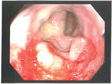 Figure 1: Endoscopic Image of Pseudotumor. View Figure 1
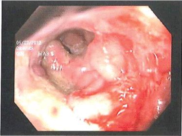 Figure 2: Endoscopic Image of Tumor. View Figure 2
The patient was then referred to colorectal surgery for evaluation of this mass but was briefly lost to follow-up. In the interim, the patient was started on HAART for his poorly controlled HIV infection. Several months had elapsed between his initial evaluation and referral to colorectal surgery. He was taken for exam under anesthesia shortly thereafter his initial surgical consultation. No gross condylomatous lesions were identified and the perianal tissue was soft. Anoscopy revealed a plaque-like whitening in the anal canal more pronounced on the right side. On digital exam there was a scar-like contracture at approximately 6 cm from the anal verge, which was biopsied. This stricture was at the level of the prior-noted mass. Pathology demonstrated eroded mucosa with inflamed granulation tissue and abscess formation. Immunohistochemistry was positive for CMV and low-risk HPV. Serum CMV PCR was negative. He was treated with a three-week course of ganciclovir for presumed CMV colitis. Subsequent exams have not noted recurrence.
Cytomegalovirus (CMV) is a common viral pathogen affecting immunocompromised patients, including HIV-positive patients. A recent review of the English literature identified fourteen cases of CMV-induced pseudotumor of the gastrointestinal tract, found in the esophagus, stomach, duodenum, ileum, and colon [1]. To our knowledge this is the first described report of possible CMV-induced pseudotumor of the rectum.
Mass lesions in the gastrointestinal tract should be treated as malignancies until proven otherwise. However, awareness of the potential for CMV pseudotumor is relevant because these patients may be prone to overaggressive treatment including surgery based on a misdiagnosis of malignancy. A few case reports describe exophytic luminal masses which are characterized by an inflammatory infiltrate in the submucosa and mucosa with fibrosis and are associated with CMV infection but can mimic malignancy [1-3]. The diagnosis is made by demonstrating inclusion bodies on histologic exam or by immunocytochemistry staining that detects CMV antigen via nuclear and cytoplasmic staining. Serum PCR for CMV is not consistently associated with pseudotumor, suggesting an end-organ tissue-level effect in the absence of CMV viremia [1].
The case we describe herein did not originally demonstrate CMV positivity on biopsy specimen of the mass seen on endoscopy. On repeat endoscopy by a colorectal surgeon, the mass had resolved leaving a scarred area in the rectum, which was CMV positive on immunohistochemistry. It has been previously suggested in the literature that immune reconstitution upon initiation of antiretroviral therapy may contribute to spontaneous resolution of these pseudotumor [1]. We feel it is unlikely that the patient developed a de novo CMV infection after initiation of antiretroviral therapy given that CMV is associated with declining, not improving, immune function. Therefore, it is reasonable to suggest that the negative immunostaining on biopsy of the mass was a sampling error or false-negative test given a reported sensitivity of 75% for CMV immunohistochemistry [4]. Given the lack of data regarding the natural history of CMV-associated pseudotumor in immunocompromised individuals and this being the first possible report of rectal pseudotumor, we feel it is prudent to add our case report to the aggregate literature.