Rheumatoid Arthritis (RA) is a systemic inflammatory disease with an autoimmune feature and an etiology that is still unknown. It is characterized by a symmetrical peripheral polyarthritis that affects primarily the small joints of hands and feet, with several levels of extra-articular manifestation. Cervical spine involvement is frequent, being the 3rd most affected region following hands and feet, with prevalence ranging between 17% and 80% [1,2] according to literature. It is considered the most common inflammatory disease affecting the spine. It is a relatively common entity, with a global prevalence of approximately 1%, and it affects around 1.3 million adults in the USA [3]. Women are most commonly affected in comparison to men (3:1), although the latter are subject to a higher risk of advanced cervical involvement [4]. Cervical disease in RA usually presents itself as one of these three forms: atlantoaxial instability or subluxation, vertical instability of the axis or basilar invagination and subaxial subluxation, involved in this sequence, respectively.
Cervical involvement typically initiates early in RA pathologic process, and its progress is strictly correlated to the extent of peripheral disease activity [5]. In most cases, radiological abnormalities remain asymptomatic for years. When symptomatic, its clinical presentation varies, generally initiating with a cervical or occipital pain and nonspecific neurological alterations. It's worth noticing that the rheumatoid patient neurological exam is often hindered by the presence of peripheral osteoarticular contracture and malformation, which are typical of this disease. Patients with RA with confirmed osteopenia or osteoporosis, particularly those with lower BMI appear to be at increased risk of cervical instability [6]. Established mutilating changes, concomitant corticosteroid treatment, and previous joint surgery are relatively robust indicators for a poor prognosis of the cervical spine in patients with RA [7]. Cervical myelopathy manifests itself insidiously, and, once it is established, mortality is a common outcome if the pathology isn't treated [8]. In an autopsy study, Mikulowscki, et al. state that unknown brainstem or medullary compression was the cause of 10% of deaths in RA patients [9]. In the series published by Pellici, et al. [10], 80% of cervical RA patients showed radiographic progression, although only 36% presented neurological progression. Ranawat, et al. [11] created a classification scheme to measure functional incapacity, neurological deficit and pain in cervical RA patients, as shown in Table 1.
Table 1: Ranawat's neurological deficit classification in RA. View Table 1
RA pathologic process is caused by a lymphoproliferative inflammatory synovitis with an autoimmune feature, which progresses and causes erosion of the subchondral bone and cartilage, forming a local hypertrophic tissue, known as pannus. The joint dislocation secondary to erosive synovitis overloads local ligament structures, possibly leading to injure and rupture of those ligaments. High cervical spine and Craniovertebral Junction (CVJ) often become unstable due to dependency of those ligament structures on the stability of this region. Atlantoaxial and occipitoatlantal hypermobility occurs as a result of damage to cruciform-alar ligament complex, transverse ligament and joint capsules [12].
Atlantoaxial instability is the most common presentation, accounting for 60 to 65% of rheumatoid cervical subluxation. About 70% are anterior, 20% are lateral, and 10% are posterior [13]. The damage to transverse ligament alone allows an atlantoaxial subluxation only up to 3-4 mm (0.12-0.16 in). The increase of Atlantodental Interval (ADI) causes a secondary damage to alar-apical ligament complex and an early rise of the odontoid process towards the base of the skull [5]. Basilar invagination or vertical instability of the axis results in bone loss and erosive arthritis of lateral masses, especially of the atlas. This progressive superior migration of the odontoid process generally leads to reduction of ADI, possibly causing a false impression of ventral atlantoaxial instability improvement in imaging exams [14]. Posterior dislocation is unusual, typically associated to odontoid process fracture, and presents a higher risk of compressive medullary lesion. In comparison, in the subaxial cervical spine disease, inflammatory process is primarily osteoarticular, located in uncovertebral joints. The emergence of subaxial subluxation usually occurs in a later stage, and it affects multiple levels, producing the classical "staircase effect".
The radiologic evaluation of cervical spine in RA usually initiates with simple radiographs. The combination of Anterior-Posterior (AP) incidence, transoral, neutral profile and flexo extension allows the identification and initial evaluation of atlantoaxial instability, basilar invagination and subaxial subluxation, as well as the rough evaluation of bone quality and level of osteoporosis in patient. Atlantoaxial instability may be identified and measured through Anterior Atlantodental Interval (AADI) and Posterior Atlantodental Interval (PADI), being the latter more reliable in correlation to potential neurological damage, particularly when its value is less than 14 mm (0.55 in) [15]. Basilar invagination may be evaluated by countless methods, none of which are completely satisfactory. However, there is an almost universal consent that Redlund-Johnell method is the most accepted one [16]. Traditional methods by McGregor and McRae measure the relation between tooth apex and skull, but they don't reflect the actual extent of vertical translocation if the tooth apex is eroded and shortened [17]. Besides, the apex of an osteoporotic tooth might be difficult to visualize due to superposition of mastoid processes.
Computerized Tomography (CT) with sagittal and coronal reconstruction has been further used in initial evaluation of RA of CVJ, in preoperative planning, and post-operatory follow-up, providing a better bone visualization in comparison to Nuclear Magnetic Ressonance (NMR) and radiographs. The case illustrated below is of a patient with RA of CVJ, where the subject radiographic imaging didn't contribute for the diagnosis, but CT clearly showed atlantoaxial instability associated to basilar invagination (Figure 1). NMR is the best exam to evaluate soft tissues and neural elements, helping to trace the actual available space for spinal cord, the magnitude of pannus and the extent of soft parts destruction. This is an important matter because, in addition to bone compression, the pannus reduces even further the available space for the cord within vertebral canal. Dvorak, et al. [18] show that two thirds of rheumatoid patients with atlantoaxial subluxation have a pannus diameter over 3 mm (0.12 in), and recommend surgical treatment for those with Space Available for the Cord (SAC) < 6 mm. Basilar invagination may be measured in MRI through the Cervicobulbar Angle (CBA), which is measured by two lines tangent to the anterior faces of the bulb and cervical cord. Normal CBA ranges from 135° to 175°. Values lower than 135° are related to the odontoid vertical migration and are associated to myelopathy [19].
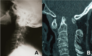 Figure 1: Lateral view radiography (A) of a patient with RA of CVJ, showing the difficulty to visualize CVJ. The diagnosis of pathologies in this region in simple radiographs might be difficult. CT with sagittal reconstruction (B) of the same patient, clearly showing the presence of atlantoaxial instability associated to basilar invagination. View Figure 1
The main objectives of the treatment for cervical spine RA are pain reduction and function restoration, as well as the prevention of progressive neurological deficit development, sudden death through unknown medullary compression, and unnecessary surgical intervention. The decision of when to seek surgical intervention is complex and should be individualized. It should also take into account the level of functional and neurological impairment, pathology natural history, and clinical alterations that might affect surgical risk.
The main controversies in surgical handling the RA of CVJ instability are related to opposite ends of clinical spectrum. In asymptomatic patients that show instability in radiographic exams (Ranawat I), is prophylactic surgery defensible? In non-ambulant quadriparetic patients (Ranawat IIIB), is there indication for surgery? A study developed by Tanaka, et al. compared the progress of asymptomatic patients with atlantoaxial instability radiologically demonstrated submitted to surgical and nonsurgical treatments. After 24 years of follow-up, best results were found in patients submitted to prophylactic surgery, with higher rates of survival, pain relief and functional recovery in comparison to those submitted to nonsurgical treatments [20]. Regarding those subjects with advanced functional impairment (ranawat IIIA and IIIB), Casey, et al. published an important study where they suggest that surgical treatment for terminal patients and patients with advanced disease is related to higher rates of morbimortality and a poor perspective for functional recovery [21]. On the other hand, Nannapaneni, et al. reported results for different types of treatment in those patients with advanced disease and showed that, with surgical treatment, 60% of them were capable of ambulating again and all of them experienced some level of cervical pain relief and quality of life improvement [22]. In this way, the answer to those dilemmas is not clear yet, and one must take into account the surgeon's experience and good judgement, as well as each patient's specificities.
Surgical approach for RA of CVJ instability basically consists in a stabilizing procedure associated or not to a decompression procedure. Currently, the posterior approach is the most commonly used, while the anterior approach is destined to specific cases.
Atlantoaxial fixation in RA is proposed to those patients with atlantoaxial instability that present an established or imminent neurological deficit, and for those with untreatable cervical or occipital pain. Atlantoaxial arthrodesis in patients with RA provides better results for cervical spine function, with an improvement in VAS scores for neck or shoulder pain or stiffness, but little improvement in pain or numbness of the lower extremities [23]. It is usually performed by a dorsal approach, and may use transarticular screws [24], sublaminar wiring or, more recently, the Harms-Goel technique, which associates screws in lateral masses of C1 to screws in C2 pedicle, connected by plates or rods. Goel, et al. introduced an innovative concept for surgically handling rheumatoid instability of CVJ [25]. Based on the principle that retro-odontoid pannus results of regional ligament slackness, they advocate that a bilateral facet distraction between C1-C2 associated to the placement of stainless steel joint spacers in distracted facets, with or without this segment instrumentation, allows the stabilization of atlantoaxial subluxation and, at the same time, the restoration of lateral masses height, providing simultaneous vertical stability and basilar invagination resolution. That way, by means of an atlantoaxial distractive arthrodesis, it's possible to treat atlantoaxial instability, basilar invagination and medullary compression simultaneously through retro-odontoid pannus. In early published papers, due to the use of stainless steel materials, the postoperative evaluation through MRI of pannus reduction and spinal cord alterations was difficult. By modifying the technique, we now came to use titanium materials for both lateral mass screws in C1 and pedicular screws in C2, as well as titanium mesh spacers, which provide a better radiologic control of soft parts through NMR [26]. Most recently, aiming to avoid subsidence due to the use of titanium mesh spacers and a better control of osseointegration, we've been using Polyetheretherketone (PEEK) cages in this technique, with consolidation rates in 100 percent of patients and absence of subsidence [27]. Figure 2 and Figure 3 illustrate the case of a patient with RA and basilar invagination associated to subluxation in C4-C5 and subaxial myelopathy. A posterior atlantoaxial distractive arthrodesis was performed with the Goel technique to treat basilar invagination and anterior corpectomy of C4 and C5 with C3-C6 arthrodesis (Figure 2 and Figure 3).
 Figure 2: Profile radiography (A), CT with sagittal reconstructions (B and C) and NMR sagittal acquisition (D) preoperative of a patient with RA Ranawat IIIa, illustrating the association of basilar invagination and instability with subaxial myelopathy. View Figure 2
 Figure 3: Profile radiography (A) CT sagittal reconstructions (B and C) and coronal (D) postoperative of the same patient, illustrating the surgical treatment performed with posterior atlantoaxial distractive arthrodesis through Goel technique for the treatment of basilar invagination, and anterior decompressive corpectomy with arthrodesis for the treatment of subaxial instability. View Figure 3
Occiptocervical arthrodesis in RA is traditionally proposed for patients with basilar invagination associated to myelopathy or neurological deficit, for instability combined to atlantoaxial and subaxial, when it's impossible to obtain arthrodesis with atlas posterior arch (due to laminectomy or insufficient bone stock), after transoral decompression, and in pseudoarthrosis after atlantoaxial arthrodesis [28]. This procedure presents a higher morbidity rate in comparison to atlantoaxial arthrodesis, especially in terms of movement limitation, restricting an average of 30° of flexoextension, and 35° to 40° of neck lateral rotation [29]. Nowadays, the mostly used constructions for occipitocervical fixation combine plates and occipital screws incorporated to a cervical instrumentation with poliaxial screws and rods.
We've been using less and less transoral decompression to treat basilar invagination in RA, limiting its use to those cases where there is significant anterior bone compression and they don't present a good clinical response to posterior treatment only. Further evidence in literature show regression of the pannus, and vertical dislocation progress is only interrupted with isolated posterior arthrodesis [30]. It's important to note that transoral decompression must be always supplemented by occiptocervical stabilization.
The surgical handling of subaxial disease in RA is usually performed through anterior decompression and stabilization. In specific cases where there's mobile atlantoaxial instability associated to significant subaxial disease, anterior atlantoaxial fixation avoids the morbidity of a second posterior approach. Max Aebi, et al. [31] described the atlantoaxial fixation technique through the anterior insertion of transarticular screws, providing satisfactory biomechanical results. In that way, the use of this technique allows the treatment of atlantoaxial instability and subaxial disease through a single anterior route, thus avoiding the additional damage from a posterior approach, like prone position and posterior muscle-ligament lesion. (Figure 4 and Figure 5).
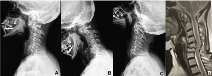 Figure 4: Profile radiography (A), flexoextention radiography (B and C) and sagittal acquisition of NMR weighed in T2 (D) preoperative of a patient with RA Ranawat I, showing mobile atlantoaxial instability associated to subaxial instability with typical "staircase" malformation. View Figure 4
 Figure 5: Profile radiography. (A) transoral radiography; (B) and TC with coronal reconstruction; (C) postoperative of the same patient, illustrating the surgical treatment performed with anterior trans articular atlantoaxial arthrodesis for the treatment of atlantoaxial instability associated to decompressive corpectomy with anterior arthrodesis for the treatment of subaxial instability. View Figure 5
Some algorithms were created to guide the therapeutic planning of these patients [32]; However, the emergence of new knowledge about the pathophysiology of the disease and the development of new surgical techniques such as those described above allowed different strategies, resulting in more benefits and less morbidity for the patients. Thus, we are proposing a new therapeutic algorithm, as illustrated in Figure 6.
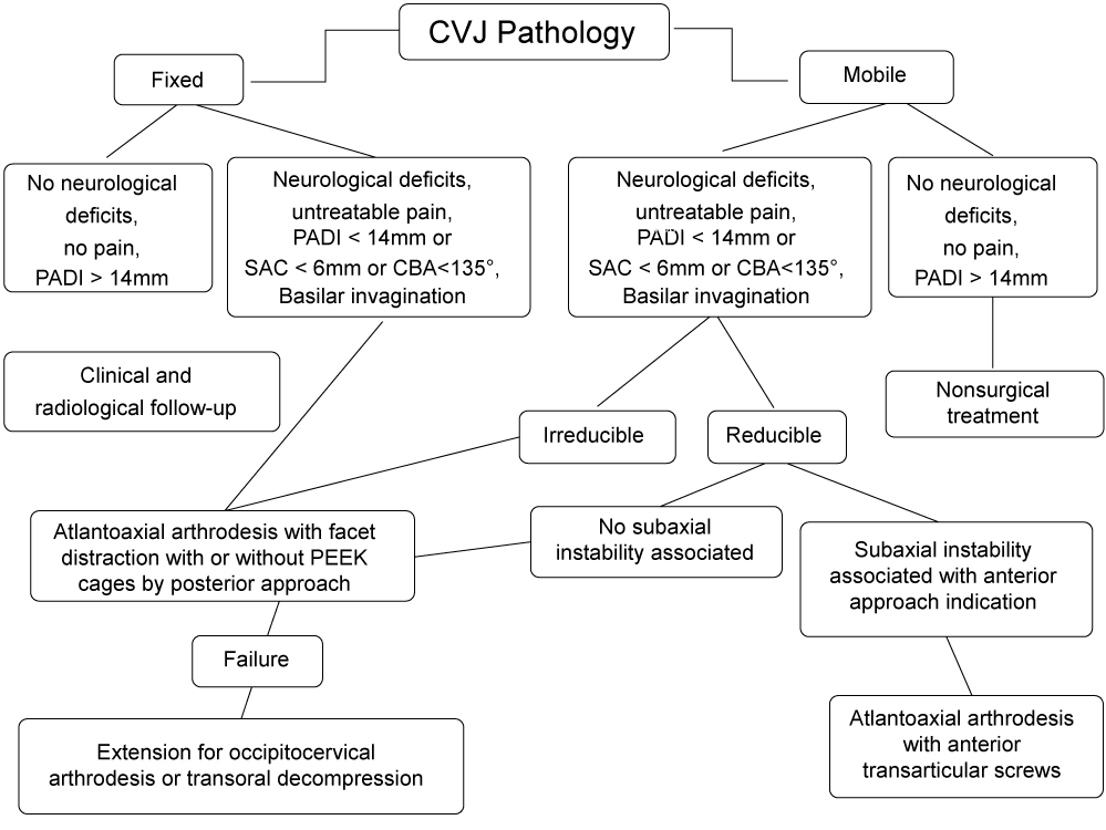 Figure 6: Therapeutic algorithm to RA of CVJ. View Figure 6
CVJ is commonly affected in RA. It might be asymptomatic, but it might cause cervical pain and neurological function deterioration. The presence of myelopathy is usually related to the advanced disease, being associated to an early death if a surgical intervention isn't performed. The surgical treatment is well indicated for patients with early neurological symptoms and for those that suffer from refractory pain to nonsurgical measures. There is still no clear answer to whether the prophylactic surgery for asymptomatic patients and surgical treatment for terminal patients are well indicated, so it is necessary to perform additional studies and to seek for better evidence. We presented a new therapeutic algorithm to handle CVJ impairment in RA.