Persistant disphagia, Arteria lusoria, Extrinsic compression
A 50-year-old woman referred to our internal medicine clinic complaining of recurrent solid food dysphagia at her upper-middle chest for a year. The patient’s history and laboratory test results were unremarkable. BE revealed a narrowing at the level of aortic arch depending on extrinsic compression. Upper gastrointestinal endoscopy confirmed a pulsatile extrinsic compression to esophagus. Unenhanced chest CT image showed the aberrant right subclavian artery origin from aortic arch and demonstrated esophageal compression.
Dysphagia lusoria is a rare diagnosis to describe association of dysphagia with vascular compression of the esophagus. The most frequent anomaly of aortic arch is aberrant right subclavian artery which appears in 0.5% to 1.8% of the population also known as arteria lusoria. Dysphagia rarely causes by extrinsic compression of the esophagus due to a vascular anomaly of the aortic arch. BE can show extrinsic compression on esophagus and excellent method to evaluate for possible dysphagia lusoria (Figure 1). Also CT and magnetic resonance imaging are great methods for evaluating the cause of extrinsic compression such as solid tumors and vascular anomalies (Figure 2). Medical treatment with proton pump inhibitor is the first choice for these patients. For patients with severe symptoms, surgical repair and reconstruction of the aberrant vessel should be considered [1-3].
The authors declare that there is no conflict of interest.
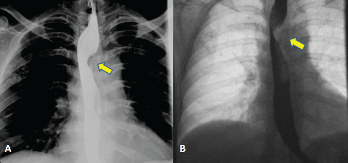
Figure 1: BE showed smooth extrinsic compression of esophagus at the level of the aortic arch (arrow).
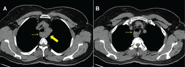
Figure 2: Axial unenhanced chest CT image (A) showed aberrant right subclavian artery origin from aortic arch (thick arrow); (A,B) esophageal compression (thin arrow).