A large free floating ball thrombus was diagnosed in an 8-year-old girl with severe restrictive cardiomyopathy following transient loss of vision. The thrombus developed despite therapeutic anticoagulation. The child died two days later with a massive embolic stroke.
Idiopathic restrictive cardiomyopathy is a rare disease in children and generally has poor prognosis. The severely dilated atia, along with high incidence of atrial fibrillation results in significant risk of intra-atrial thrombosis [1]. However, free floating ball thrombus remains very rare in children. It is more common in patients with mitral valve stenosis and usually requires urgent surgical treatment [2]. Development of free floating ball thrombus was rarely reported in patients with restrictive cardiomyopathy, even in sinus rhythm [3].
An 8-year-old girl who was diagnosed with idiopathic restrictive cardiomyopathy since the age of 4 years, and has a previous stroke was followed at our institution. She was on anticoagulation with war far in with a stable international normalized ratio of 2-3. She has severe ascites for which she was treated with abdominal paracentesis on a monthly basis. She presented with transient loss of vision. Her echocardiogram showed a large free floating left atrial thrombus (Figure 1). The thrombus was bouncing off the mitral valve with each ventricular systole in supplementary material (online movie). The patient had a fatal major thromboembolic event three days after the diagnosis of the ball thrombus.
None.
This research received no specific grant from any funding agency, commercial, or not-for-profit sectors.
None.
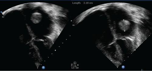
Figure 1: An echocardiographic image in apical view of an eight year old child with severe restrictive cardiomyopathy. There is severe dilatation of both atria, and a large ball thrombus in the left atrium measuring 2.2 cm in diameter.