

International Journal of Clinical Cardiology
Asymptomatic Left Atrial Appendage Aneurysm
Norliza Othman1,3, Alwi Mohamed Yunus2, Raad H. Mohiaddin3 and Masliza Mahmod4*
1Diagnostic Imaging Department, Serdang Hospital, Malaysia
2National Heart Institute, Malaysia
3Cardiovascular Magnetic Resonance Unit, Royal Brompton Hospital, UK
4Oxford Centre for Clinical Magnetic Resonance Research, Division of Cardiovascular Medicine, Radcliffe Department of Medicine, UK
*Corresponding author: Masliza Mahmod, Oxford Centre for Clinical Magnetic Resonance Research, Division of Cardiovascular Medicine, Radcliffe Department of Medicine, University of Oxford, John Radcliffe Hospital, Oxford OX3 9DU, UK, Tel: +44 1865 221868, Fax: +44 1865 740449, E-mail: masliza.mahmod@cardiov.ox.ac.uk
Int J Clin Cardiol, IJCC-2-027, (Volume 2, Issue 2), Case Report; ISSN: 2378-2951
Received: March 14, 2015 | Accepted: April 08, 2015 | Published: April 11, 2015
Citation: Othman N, Yunus AM, Mohiaddin RH, Mahmod M. (2015) Asymptomatic Left Atrial Appendage Aneurysm. Int J Clin Cardiol 2:027. 10.23937/2378-2951/1410027
Copyright: © 2015 Othman N, et al. This is an open-access article distributed under the terms of the Creative Commons Attribution License, which permits unrestricted use, distribution, and reproduction in any medium, provided the original author and source are credited.
Abstract
We report a patient with asymptomatic left atrial appendage aneurysm. Cardiac magnetic resonance was performed to further delineate the aneurysm, and the patient underwent successful surgical resection.
Case Report
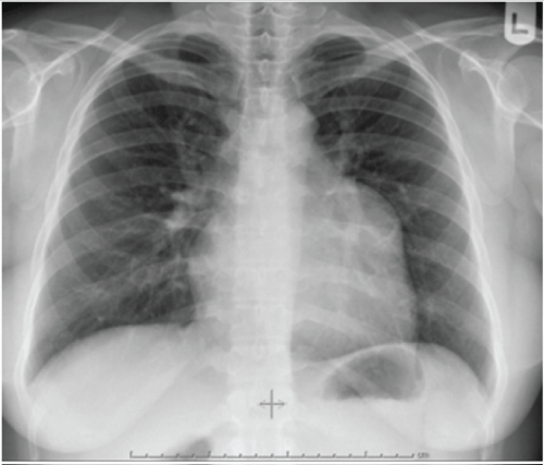
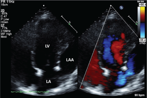
Asymptomatic healthy, 32-year-old woman was found to have enlarged left upper cardiac border on chest radiograph (CXR), undertaken as a routine health check-up (Figure 1). This was compared to a previous CXR taken 5 years earlier, which only showed a small bulging of the left upper cardiac border. Electrocardiogram showed normal sinus rhythm. A transthoracic two-dimensional echocardiogram showed left atrial appendage (LAA) aneurysm (Figure 2).
.
Figure 2: Transthoracic echocardiography of the apical 4-chamber views showing significantly dilated left atrial appendage. Colour Doppler (right) demonstrating flow between the left atrium and the dilated left atrial appendage. LA: Left Atrium, LAA: Left Atrial Appendage, LV: Left Ventricle
View Figure 2
To further delineate the aneurysm and the anatomical relationship with other structures, cardiac magnetic resonance (CMR) was performed. CMR confirmed large LAA aneurysm measuring 4.8 x 6.9 x 5.3 cm, communicating with the left atrium via a wide neck, compressing the lateral wall of the left ventricle, and without evidence of thrombus (Figure 3).

.
Figure 3: Cardiac magnetic resonance of the transverse (a) and coronal (b) views of the heart demonstrating a large LAA aneurysm communicating with the LA via a wide neck, without evidence of thrombus. Short axis views at the basal (c) and mid ventricular levels (d) showing mild compression of the lateral wall of the LV by the aneurysm. LA: Left Atrium, LAA: Left Atrial Appendage, LV: Left Ventricle, RV: Right Ventricle
View Figure 3
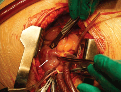
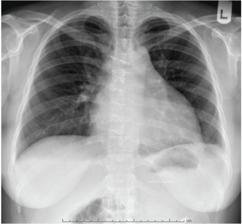
The left atrial size was normal, without mitral stenosis or regurgitation. Although asymptomatic, the patient underwent surgical resection of the LAA (Figure 4) in view of the risk of potential life-threatening complications. Macroscopic examination of the specimen revealed a partly open collapsed cyst-like structure with wall thickness between 2 and 4mm, smooth outer and inner surfaces, without blood clots. Microscopically, there were areas of muscular hypertrophy without evidence of tumour, inflammation, myxoid or cystic degeneration. She had uneventful postoperative recovery with normal post-operative CXR (Figure 5).
.
Figure 4: Intraoperative image showing the left aterial appendage aneurysm (white arrow)
View Figure 4
.
Figure 5: Post-operative chest radiograph showing normal left upper cardiac border
View Figure 5
LAA aneurysm is very rare with less than 100 cases have been reported in the literature [1]. An LAA aneurysm can be due to congenital or acquired causes. The congenital type is thought to be due to weakened appendage wall due to dysplasia of the pectinate muscles. The acquired form is usually associated with other cardiac abnormalities such as mitral valve disease, history of infection or other causes which can lead to raised left atrial and LAA pressures or weakened muscle wall [2,3].
Patients commonly present with palpitations, breathlessness and thromboembolic complications. Sixteen percent do not have symptoms and detected on routine medical assessment. LAA aneurysm can expand slowly over several years and have been reported to occur over 18 [4] and 30 years [5]. To our knowledge, this is the first report of significant progressive enlargement of LAA aneurysm in a relatively short time (5 years). Imaging modalities such as computed tomography scan and CMR are important techniques to further characterize the aneurysm [1]. However CMR is better suited in women of childbearing age without the risk of ionizing radiation, as was performed in our case. The most likely aetiology in our case was congenital as it occurred in isolation without known predisposing factors that could lead to the LAA enlargement. This was supported by CMR findings which revealed clear communication of the appendage with the normal-sized left atrium, no mitral valve stenosis or regurgitation. Surgery was performed in view of the risk of developing complications such as rupture, atrial tachyarrhythmias, thrombus formation and systemic embolism [1].
Acknowledgement
Masliza Mahmod acknowledges support from the National Institute for Health Research (NIHR) Oxford Biomedical Research Centre Programme. Raad Mohiaddin acknowledges support from the NIHR Cardiovascular Biomedical Research Unit of Royal Brompton and Harefield NHS Foundation Trust and Imperial College London.
References
-
Aryal MR, Hakim FA, Ghimire S, Ghimire S, Giri S, et al. (2014) Left atrial appendage aneurysm: A systematic review of 82 cases. Echocardiography 31: 1312-1318.
-
Culver DL, Bezante GP, Schwarz KQ, Meltzer RS (1993) Transesophageal echocardiography in the diagnosis of acquired aneurysms of the left atrial appendage. Clin Cardiol 16: 149-151.
-
Zimand S, Frand M, Hegesh J (1997) Congenital giant left atrial aneurysm in an infant. Eur Heart J 18: 1034-1035.
-
Park JS, Lee DH, Han SS, Kim MJ, Shin DG, et al. (2003) Incidentally found, growing congenital aneurysm of the left atrium. J Korean Med Sci 18: 262-266.
-
Pom� G, Pelenghi S, Grassi M, Vignati G, Pellegrini A (2000) Congenital intrapericardial aneurysm of the left atrial appendage. Ann Thorac Surg 69: 1569-1571.
